
Abstract
The present study was undertaken to investigate the gender-related liver injury induced by Dioscorea bulbifera L. (DB), a traditional medicinal plant, in mice, and further explored its hepatotoxic chemical compound. Serum and liver tissue samples were collected at 0, 4, 8, 12 h, after mice were administrated orally with 640 mg/kg ethyl acetate extracts (EF) isolated from DB. After treatments, serum alanine transaminase (ALT) and aspartate transaminase (AST) activities were both significantly elevated. Liver lipid peroxidation (LPO) level increased, while glutathione amounts, glutathione-S-transferase (GST), superoxide dismutase (SOD) and catalase (CAT) activities all decreased in the time-dependent manner. Further analysis demonstrated that ALT and AST activities in female mice were significantly lower than those in male. Meanwhile, liver glutathione amounts and CAT activity in female mice after giving EF for 12 h were both higher than those in male. Further, comparing the liver injury induced by Diosbulbin B isolated from DB with that induced by EF on the basis of chemical analysis for the amounts of Diosbulbin B in EF of DB, we found that Diosbulbin B could be the main hepatotoxic chemical compound in DB. Taken together, our results show that DB can induce gender-related liver oxidative stress injury in mice, and its main hepatotoxic chemical compound is Diosbulbin B, for the first time.
Introduction
The rhizome of Dioscorea bulbifera (DB) is widely distributed in Asia. It has been used to treat sore throat, struma and thyroma as a traditional Chinese medicine in ancient China. 1 Besides, it has also been traditionally used for treatment of leprosy and tumors at the northern districts in Bangladesh. 2 In the previous studies, the chloroform fraction of the methanol extract of DB was found to have caused liver injury in mice and rats. 3,4 However, up to now, the hepatotoxic chemical compounds and mechanism of DB are not clear.
There are many reports that oxidative stress plays key roles in liver injury induced by carbon tetrachloride, alcohol, acetaminophen, chemotherapeutic agents, chromium and so on. 5 –13 Reactive oxygen species (ROS) generated in the process of oxidative stress are extremely high-reactive, which may modify and inactivate lipids, proteins, DNA, and RNA, and thus induce cell dysfunctions. To prevent ROS-induced cell injury, the body has developed the antioxidant system, including low-molecular-mass antioxidants such as glutathione, alpha-tocopherol, ascorbate acid and antioxidant enzymes such as glutathione-S-transferase (GST), superoxide dismutase (SOD) and catalase (CAT). 14 Generally, cellular antioxidants maintain a point of balance between the oxidants and antioxidants, and thus prevent the organism from the injury induced by ROS. Up to now, there are many reports that such oxidative stress injury induced by various hepatotoxins has a gender-related difference. 15 –18 However, as for the liver injury induced by DB, there is no related report about whether its hepatotoxicity is gender-related.
The present study was designed to observe the liver oxidative stress injury induced by DB and whether the hepatotoxicity is gender-related. Further study investigated the potential hepatotoxic compound in DB.
Materials and methods
Animals
ICR female and male mice (18−22 g) reared in the animal house of Regional Center were purchased from Shanghai Slac Laboratory Animal Co. Ltd (Shanghai, China). Animals were given rodent laboratory chow and water ad libitum and maintained under controlled conditions with a temperature of 22 ± 1°C, relative humidity 65 ± 10 % and a 12/12 h light/dark cycle (lights on at 7:00 a.m.). All the procedures were in strict accordance with the P.R. China legislation on the use and care of laboratory animals and with the guidelines established by Institute for Experimental Animals of Shanghai University of Traditional Chinese Medicine and were approved by the university committee for animal experiments.
Reagents
Reduced glutathione (GSH), oxidized glutathione (GSSG) and NADPH were purchased from Roche Diagnostics GmbH (Mannheim, German). Unless indicated, other reagents were purchased from Sigma Chemical Co. (St Louis, Missouri, USA).
Plant material and preparation of samples
DB rhizomes were collected in Qingyang, Anhui province, and authenticated by Prof. Shou-Jin Liu (Anhui College of Traditional Chinese Medicine, Anhui, China). A voucher specimen was deposited in the herbarium of Institute of Traditional Chinese Medicine, Shanghai University of Traditional Chinese Medicine.
The dried rhizomes were crushed and the preparation of ethyl acetate extracts (EF) was described as follows. The powder (about 1 kg) was soaked in 80% ethanol (w/v = 1:10) and incubated at room temperature for 120 min. The mixture was extracted for consecutive three times at 85 ± 5°C with a rotary evaporator for 180 min at a time. The combined extraction was centrifuged at 800 × g for 10 min, and the supernatant was transferred to a glass container by decanting and concentrated under vacuum with a rotary evaporator under reduced pressure at /45 ± 5°C to about 78 g extracts. Then, the ethanol extract was extracted for eight times by ethyl acetate (v/v = 1: 1) in room temperature. Ethyl acetate layers were transferred to glass containers by decanting and concentrated under reduced temperature. Ethyl acetate layers were dried under vacuum with a rotary evaporator under reduced pressure at /45 ± 5°C, powered and decanted in a vacuum desiccator. The yield of EF of ethanol extract was 1.6% by DB raw medicinal materials.
Diosbulbin B was isolated from DB rhizomes in our laboratory according to the previous literature. 19 After the purification with silica gel column and gel chromatography, the purity of Diosbulbin B was more than 98% as determined by high-performance liquid chromatography (HPLC) with diode array detector (DAD).
Animal treatment protocol
Gender-related difference in the hepatotoxicity induced by DB
Female and male mice were divided into eight groups, including control (0), 4, 8, 12 h after administration of DB. Mice were administrated with 640 mg/kg EF of the ethanol extract of the DB or an equivalent volume of 0.5% sodium carboxymethyl cellulose (CMC-Na) by intragastric administration (ig). EF was dissolved in 0.5% CMC-Na and the volume was adjusted with 0.5% CMC-Na to the appropriate final concentration. Peripheral blood samples of groups were collected at 0, 4, 8, 12 h after the administration of DB. Livers of groups at the corresponding time were rapidly removed, washed in ice-cold saline and kept in ice. The lipid peroxidation (LPO) level, glutathione amount and antioxidant-related enzymes such as GST, SOD and CAT of mice hepatic tissues were assayed to evaluate oxidative injury, respectively.
Hepatotoxicity induced by Diosbulbin B isolated from DB
Male mice were randomly divided into four groups of 8 mice each. Mice of normal group received daily oral administration (po) of 0.5% CMC-Na (0.2 mL/10 g). The treated groups received indicated concentration of the Diosbulbin B at doses of 75, 150, 300 mg/kg po by ig. The peripheral blood samples of groups were collected at 24 h after the treatment for the determination of serum biomarkers for liver injury.
Alanine transaminase and aspartate transaminase assay
The blood samples were obtained from mice for the determination of serum biomarkers for liver injury. The serum alanine transaminase (ALT) and aspartate transaminase (AST) were assayed according to the reported method. 20
Assay for liver LPO level
Liver tissues were homogenized in cold phosphoric acid (phosphate buffered saline, PBS). LPO was determined by the previous reported method. 21 Malondialdehyde (MDA) formed as an end product of the LPO and served as an index of the intensity of LPO. MDA reacts with TBA to generate a pink-colored product, which has an absorbance at 532 nm. The standard curve of MDA was constructed over the concentration range of 0−40 μM. The LPO level was expressed as μM/mg protein based on tissue protein concentration.
Assay for liver glutathione amounts
The quantity of the glutathione amounts was measured immediately as described in the previous study. 22 The reaction mixture contained samples, 150 μL of a working solution (0.53 U/mL of glutathione reductase, 40.7 μg/mL of DTNB, 1 mM EDTA in 100 mM sodium phosphate buffer, pH 7.0) and 50 μL of 0.16 mg/mL of NADPH solution. The change in absorbance was determined at 412 nm against the reagent blank after standing in room temperature for 30 min and the glutathione amounts were determined in comparison with a standard curve. The glutathione amounts of mice livers were calculated based on tissue protein concentration.
Assay for liver GST activity
Tissues were homogenized in cold PBS and then centrifuged at 5000 × g for 5 min and the supernatant was transferred to new tubes for assay. GST activity was measured according to the previous reported method. 23 The activity of GST was calculated based on tissue protein concentration.
Assay for liver SOD and CAT activities
Tissues were homogenized in cold PBS, and then centrifuged at 5000 × g for 5 min and the supernatant was transferred to new tubes for assay. The liver activities of SOD and CAT were determined by the method of previous studies and calculated based on tissue protein concentration. 24,25
Assay for the amounts of Diosbulbin B in EF of DB
Amounts of Diosbulbin B in EF were measured by HPLC-DAD. The chromatography condition was shown as follows. The analysis was performed on a Prominence HPLC instrument (Shimadzu) equipped with quaternary pump, DAD, on-line degasser, autosampler and column heater compartment. The sample was separated on an ultimateTM XB-C18 column (4.6 × 250 mm, 5 µm). The mobile phase consisted of methanol and water containing 0.1% (v/v) acetic acid with the isocratic elution (v/v = 58: 42). The flow rate was 1.0 mL/min, and column temperature was set at 25°C. The DAD detector monitored in the range 190−400 nm, and the on-line UV spectra were recorded at 210 nm.
To obtain calibration curve of Diosbulbin B, purified Diosbulbin B (1.83 mg, weighed accurately) was dissolved in methanol in a 10-mL low-actinic volumetric flask. This solution was diluted with methanol to obtain standard solutions for the calibration curve in a range of 11.4–183 µg/mL on column, and a 10-µL aliquot was injected. EF was dissolved in methanol and an aliquot (20 µL) each was injected into the above HPLC-DAD system. The amounts of Diosbulbin B in EF were calculated using the calibration curve of Diosbulbin B.
Statistical analysis
All experimental data were expressed as mean ± standard error of mean (SEM). Significant differences between experimental groups were compared by one-way ANOVA (analysis of variance) followed by Least Significant Difference (LSD; p < 0.05) using the Statistics Package for Social Science (SPSS) program Version 11.5.
Results
Effects of EF of DB on serum biomarkers for liver injury
Serum ALT and AST activities are liver injury biomarkers and the conspicuous elevation of them often reflects liver injury. 20 In the present study, after giving EF of DB for 4−12 h, ALT and AST activities were both significantly elevated in serums of mice in the time-dependent manner (Figure 1). Further analysis showed that ALT and AST activities of the female mice were conspicuously lower than those in male after given EF for 12 h (Figure 1). All these results demonstrate that DB rhizome can significantly induce liver injury, and is gender-dependent.

Effects of ethyl acetate extracts (EF) of Dioscorea bulbifera L. (DB) on the activities of serum alanine transaminase (ALT) and aspartate transaminase (AST) in mice. Data are presented as mean ± SEM (n = 8). Significant differences compared with the Control (0 h) group under the same sex were designated as *p < 0.05 and **p < 0.01 and between the sexes at the same time point as # p < 0.05 and # # p < 0.01.
Effects of EF of DB on liver LPO level and glutathione amounts
MDA is one of the main end products of LPO. 26 As shown in Figure 2A , MDA amounts increased in livers of mice in a time-dependent manner, which demonstrated that EF of DB could induce LPO injury in mice livers. Through further comparison, we could see that the MDA amounts in livers of male mice were higher than those in female mice, especially after given EF of DB for 8 and 12 h, but there was no significant difference between them (Figure 2A). Glutathione, an antioxidant, protects cells against ROS such as free radicals and peroxides. 27 Excessive exhaustion of glutathione may lead to the damage on cellular-reducing environment. 27 The present study indicated that the glutathione amounts decreased in a time-dependent manner in livers of mice after giving EF of DB for 4−12 h (Figure 2B). Further results showed that liver glutathione amounts of the female mice were conspicuously higher than those in the male, especially after given EF of DB for 12 h (Figure 2B). Our results demonstrate that EF of DB can lead to the damage on the balance between cellular oxidants and antioxidants. Furthermore, there was gender-related difference in this change after given EF of DB for 12 h in mice livers.

Effects of ethyl acetate extracts (EF) of Dioscorea bulbifera L. (DB) on the malondialdehyde (MDA) level and glutathione amounts in mice liver tissues. Data are presented as mean ± SEM (n = 8). Significant differences compared with the Control (0 h) group under the same sex were designated as *p < 0.05 and **p < 0.01 and between the sexes at the same time point as # p < 0.05 and # # p < 0.01.
Effects of EF of DB on liver GST, SOD and CAT activities
GST, CAT and SOD are all intracellular antioxidant enzymes, participating in the process of oxidative stress. 23 –25 Our results showed that GST, CAT and SOD activities all decreased in livers of mice in the time-dependent manner (Figures 3 and 4). Further analysis showed that CAT activity in the female mice was conspicuously higher than that in the male after given EF for 12 h (Figure 4B). Although it seemed that GST and SOD activities were both higher in female mice livers than those in male, there was no significant difference (Figure 3 and 4A). Our results further confirmed the oxidative stress injury induced by DB. Meanwhile, to some content, the difference in the CAT activity in male and female livers reflected the different defensive capacity of them to the hepatotoxicity induced by DB.
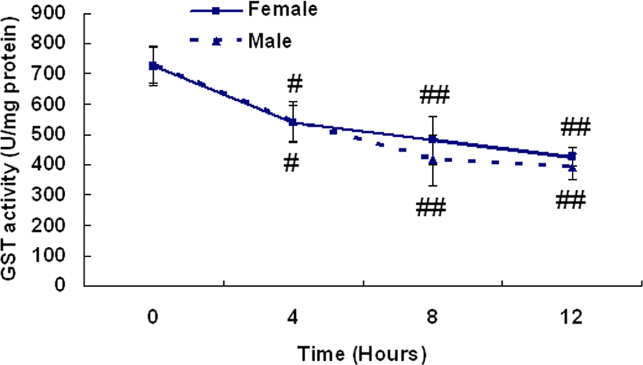
Effects of ethyl acetate extracts (EF) of Dioscorea bulbifera L. (DB) on the GST activity in mice liver tissues. Data are presented as mean ± SEM (n = 8). Significant differences compared with the Control (0 h) group under the same sex were designated as *p < 0.05 and **p < 0.01 and between the sexes at the same time point as # p < 0.05 and ## p < 0.01.
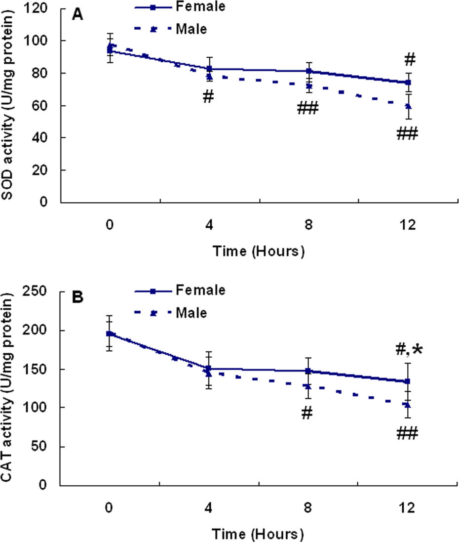
Effects of ethyl acetate extracts (EF) of Dioscorea bulbifera L. (DB) on the superoxide dismutase (SOD) and catalase (CAT) activities in mice liver tissues. Data are presented as mean ± SEM (n = 8). Significant differences compared with the Control (0 h) group under the same sex were designated as *p < 0.05 and **p < 0.01 and between the sexes at the same time point as # p < 0.05 and ## p < 0.01.
Effects of Diosbulbin B isolated from DB on serum biomarkers for liver injury
Diosbulbins as furanoid norditerpenes are abundant in DB. 19 Of them, Diosbulbin B is the major chemical compound. 19 Further to demonstrate the hepatotoxic compound in DB, we observed the effects of diosbulbin B on serum ALT and AST activities. In the present study, Diosbulbin B had no obvious effect on either serum ALT or AST at the dose of 75 mg/kg (Figure 5). However, when the dose of Diosbulbin B was more than 150 mg/kg, ALT and AST activities were both significantly elevated (p < 0.05) in a dose-dependent manner (Figure 5).
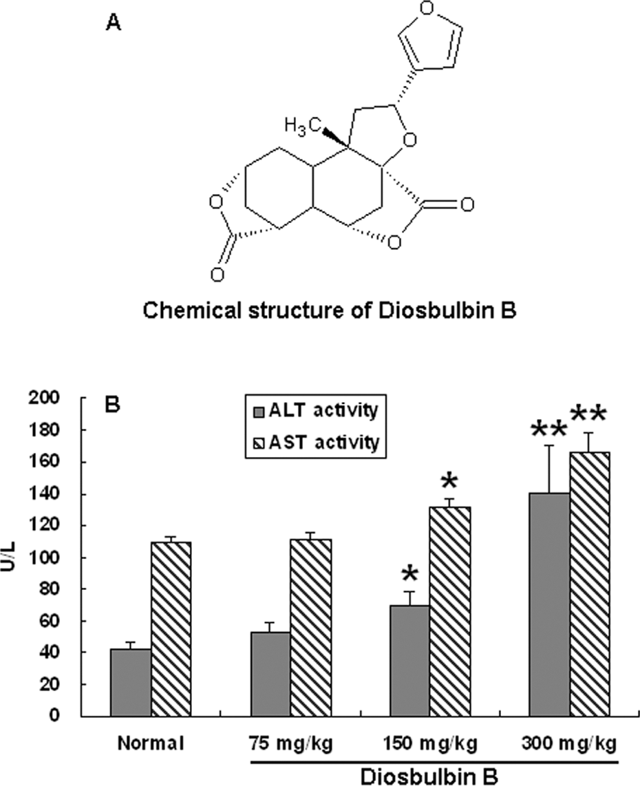
Chemical structure of Diosbulbin B and effects of it on the activities of serum alanine transaminase (ALT) and aspartate transaminase (AST) in mice. Data are presented as mean ± SEM (n = 8). Significant differences compared with the normal group were designated as *p < 0.05 and **p < 0.01.
Analysis of whether Diobulbin B is the main hepatotoxic compound in EF of DB
The amount of Diosbulbin B in EF of DB was 13.99% by HPLC-DAD analysis. The chemical structure, HPLC chromatograms and calibration curve of Diosbulbin B are shown in Figures 5A and 6, respectively.
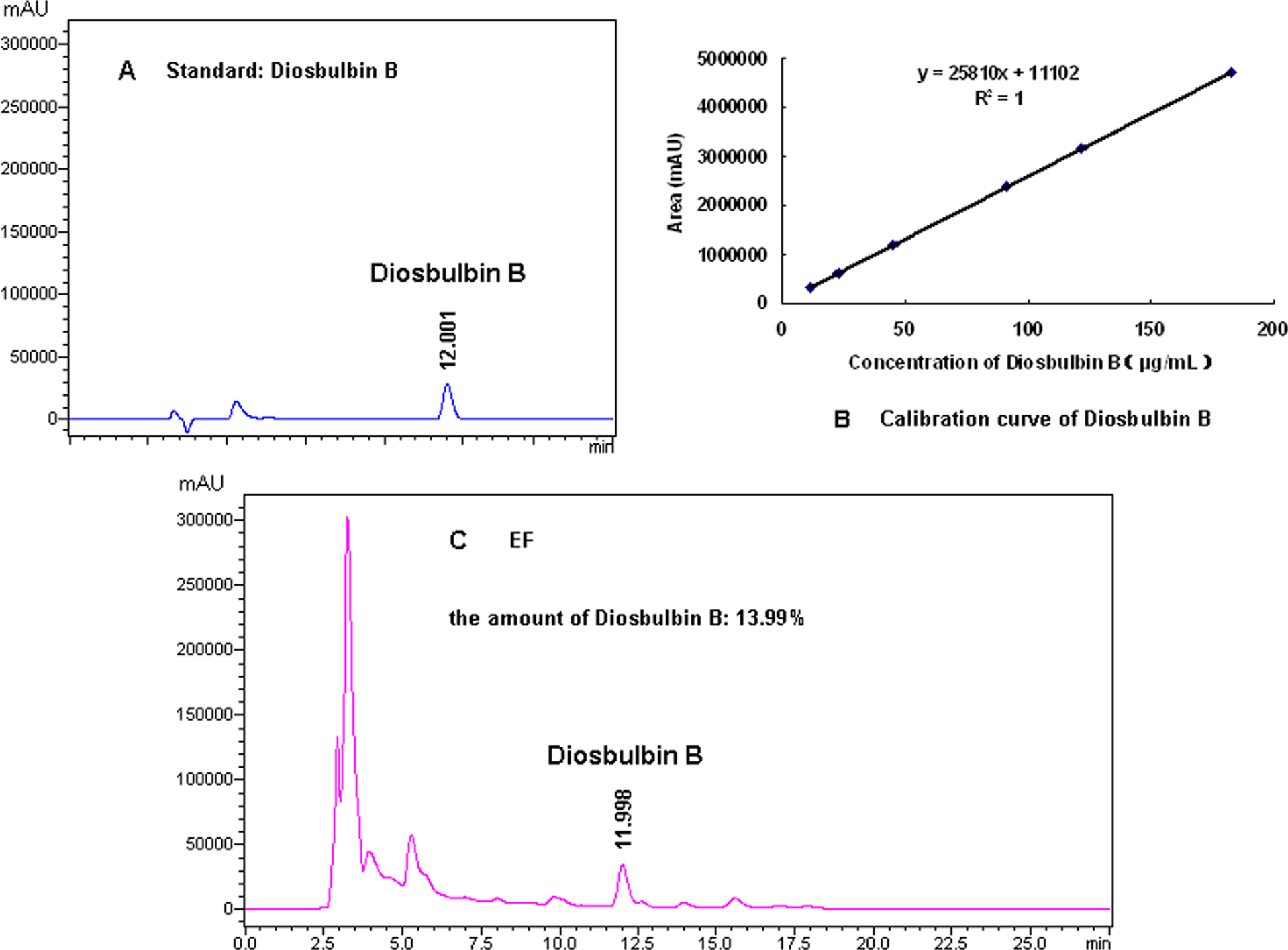
High-performance liquid chromatography (HPLC) chromatograms and calibration curve of Diosbulbin B. The general high-performance liquid chromatography with diode array detector (HPLC-DAD) condition was as described in Materials and methods.
Further to confirm whether Diosbulbin B was the main hepatotoxic compound in DB, we converted the hepatotoxic dose of EF to that of Diosbulbin B, and then compared this converted dose with the actual one. According to the above amounts of Diosbulbin B in the EF of DB, we could get about 90 mg of Diosbulbin B from 640 mg of EF through calculation. So the converted dose of Diosbulbin B was determined as 90 mg/kg (between 75 mg/kg and 150 mg/kg), equivalent to that of 640 mg/kg of EF of DB. Therefore, we could get a conclusion that the converted dose was exactly in the range of the actual one and Diosbulbin B was determined as one of the main hepatotoxic chemical compounds of DB.
Discussion
It has already been reported that liver is the major target organ for toxicity induced by DB. 3 When liver is damaged by hepatotoxins, the enzymes such as ALT and AST can leak from the damaged liver to the serum, and thus the conspicuous elevation of serum ALT and AST generally indicates the liver injury. 20 In the present study, ALT and AST activities were both significantly elevated in serums of mice after giving EF of DB for 4−12 h (Figure 1), demonstrating that DB can induce mice liver injury. Further through comparison, we found that the elevated ALT and AST activities in male livers were higher than those in female. All these results fully demonstrated that the liver toxicity induced by DB was gender-dependent, and male mice were more sensitive to its toxicity, which reminds us to consider the gender-related liver injury in the clinical administration of it.
Hepatic cellular oxidative stress happens due to the imbalance of oxidants and antioxidants, moreover the activities of many antioxidant-related enzymes and non-enzymatic antioxidants may be changed during this process. 28 –31 Among them, LPO is a free-radical-related process. 32 One of the main end products of LPO is MDA, which has the characterization of cross-linking cellular macromolecules such as protein or DNA and induces widespread cellular damage. 26 The results in Figure 2A showed that EF of DB significantly increased the MDA amounts, indicating it induced LPO injury in liver.
CAT mainly exists in the peroxisomes of all aerobic cells and serves to protect the cells against the toxicity of hydrogen peroxide by catalyzing it into molecular oxygen and water without generating toxic free radicals. 33,34 As peroxisomes are abundant in proteins, where oxidative stress always takes place, thus CAT is a classical oxidative biomarker. In the present study, CAT activity conspicuously decreased in livers of mice when given EF of DB for 4–12 h, demonstrating that it could induce the damage on CAT enzyme. Furthermore, our results also showed that the decreased CAT activity in male mice livers was higher than that in female, which indicated that CAT could contribute to the gender-related hepatotoxicity induced by DB. SOD is a metalloenzyme and can convert O2, which is produced during the oxidative stress, to hydrogen peroxide. 33 Our results showed that SOD activity significantly decreased in livers of mice when given EF of DB for 4–12 h, while there was no obvious difference in liver SOD activity between male and female. Our results indicate that SOD participates in DB-induced oxidative stress liver injury, while it may not be involved in DB-induced gender-dependant liver injury.
Glutathione plays an important role in protecting hepatocytes against exogenous toxins, and there are lots of reports that depletion of cellular glutathione is related with oxidative damage. 35,36 Our results showed that DB significantly decreased liver glutathione amounts, moreover such decrease was higher in male than in female. The results further confirmed the damage of liver normal antioxidants-oxidants balance and the gender-related oxidative stress injury induced by DB. The cytosolic GSTs are found in almost all of the aerobic species and have the capacity to catalyze the conjugation of electrophilic compounds produced in the process of oxidative stress with the glutathione, which plays vital roles in the detoxification of xenobiotics. 27,37 In the present study, the GST activity significantly decreased in livers of mice when given EF of DB for 4–12 h, while there was no obvious difference in liver GST activity between male and female. Our results demonstrate that GST is involved in DB-induced oxidative stress liver injury, while it may not participate in DB-induced gender-dependant liver injury.
Clerodane diterpenoids have been reported to have many bioactivities including antitumor, 38 antifeedant, 39 anti-inflammation, 40 anti-salmonellal, 41 cytotoxicity 42,43 and so on. The chemical constituents in DB mainly include clerodane diterpenoids, 44,45 steroids, 46 flavonoids 47 etc. As for the clerodane diterpenoids in DB, Diosbulbins as furanoid norditerpenes are abundant. Diosbulbin B is the major chemical compound of Diosbulbins in DB 19 and has been found to have antitumor activity in the previous study. 44 However, it is still not clear up to now on whether Diosbulbin B is the main hepatotoxic chemical compound of DB. In the present study, we found that Diosbulbin B could induce mice liver injury (as was shown in Figure 5B). Further, we carried out the correlated analysis between the hepatotoxicity and amounts of Diosbulbin B of DB. The result indicated that the converted amounts of Diosbulbin B were equivalent to the actual ones of it in EF of DB. (Figure 6), suggesting that Diosbulbin B could be the main potential hepatotoxic compound of DB.
In conclusion, the present study shows that DB can induce liver oxidative stress injury in a conspicuous time-dependent manner in mice. Moreover, it also demonstrates that such oxidative stress injury induced by DB is gender-related, for the first time. These results remind us to pay attention to the gender-related liver injury induced by DB in clinic. Finally, our results also indicate that Diosbulbin B is the main hepatotoxic chemical compound of DB. Further studies are in progress in our laboratory to explore the mechanisms of liver injury induced by Diosbulbin B isolated from DB.
Footnotes
Acknowledgements
The authors thank Tianyu Liu, KaiKai Shen and Ying Chen for their kind assistances during the assays for the antioxidant enzymes.
This work was financially supported by the National Basic Research Program of China (No. 2006CB504704), Shanghai Science and Technology Committee Grants (No.08DZ1972300), National Natural Science Foundation of China (No. 30701082) and Innovation Program of Shanghai Municipal Education Commission (09ZZ125).