
Abstract
Polymethylmethacrylate (PMMA) in powder form is broadly used as bone cement in orthopedic applications due to its expanded mechanical, physical, and chemical properties. In this study, a hybrid PMMA biopolymer nanocomposite is developed by the supplement of graphene nanoplatelets (GnP) and hydroxyapatite (HA) powders of nano-size with combined loadings ranging from 0.5 to 2.5 weight %. Both materials were applied uniformly to reinforce commercial bone cement made of polymethylmethacrylate. The findings showed that adding 1.5 wt.% of combined HA and GnP nanoparticles to the powder of PMMA bone cement resulted in the expansion of flexural strength by 49.28%, the flexural modulus by 30.80%, the compression strength by 31.7%, and the compression modulus by 57.41%. The nanocomposite was characterized using Energy Dispersive X-Ray Analysis (EDS), Fourier transforms infrared (FTIR) spectroscopy, and X-ray diffraction (XRD) to study the distribution of reinforced nanoparticles. Scanning Electron Microscopy (SEM) analysis of the prepared samples and fractured surface shows the proper dispersion of nanofillers into the matrix phase and possible reasons behind fracture. The inclusion of GnP and HA in the PMMA enhances the mechanical performances required for biomedical components. Also, the SEM findings of the mechanically tested broken surface of the polymer nanocomposite samples demonstrated the feasibility of the proposed material for joint replacement surgical procedures and orthopedic implants.
Introduction
Polymethylmethacrylate (PMMA) based bone cement has retained its acceptance as biomedical material since its discovery in 1960, and it is primarily being used to complete joint replacements anchor. 1 Implants use PMMA bone cement as a gap filler for retaining mechanical anti-bone interlocking. Besides biocompatibility, its biomechanical factors are explicit considerations for bone cement properties. 2 There are two components of PMMA bone cement: powder and liquid. The powder component comprises PMMA powder and benzoyl peroxide (BPO) initiator. N, N-dimethyl-P-toluidine is used as a reaction promoter in the liquid portion, which is made up of methyl methacrylate monomer. A paste-like cement is produced when these two pieces are combined, and after a few minutes, it gets converted to bone cement. 3 Over the last 30 years, PMMA bone cement has seen an increase in the use of the medical sector for bone-related surgical operations.4,5 Clinical experience clearly emphasizes high biocompatibility, bioactivity, biodegradation potential, and mechanical properties.6,7 The most recent biomaterials, bio-ceramics, and metallic compounds ensure biocompatibility and mechanical properties similar to bone and teeth. 8 Mainly PMMA bone cement is utilized as a technique for grouting during joint substitution. 9 The number of prosthesis replacements conducted in the west over the most recent decade has increased significantly. 10 A few considerations have shown the rise in the number of cases of traditional substitution techniques, like aging, has prompted this increment.11,12 PMMA bone cement stays the supporting material for full and incomplete removable prostheses. Due to its physical and esthetic properties, PMMA has been generally utilized in polymer-based prostheses. PMMA resins have positive results and have high flexural fatigue relative to other polymers.13,14 After 16 years, the failure rate has been reported to be as high as 67% in patients less than 45 years old. 15 PMMA bone cement failure is its weaker mechanical characteristics at continuous load applications. 16 Acrylic bone cements do not adhere directly to the bone. The indirect surface adhesion raises the chance of a gap forming between the bone/cement and cement/implant, resulting in the implant becoming loose and providing ideal conditions for bacteria to colonize. 17 Previous techniques have emphasized using extra additives and bioactive ceramics such as hydroxyapatite. 18 However, introducing additives could significantly alter the composite mechanical performances essential for load-bearing applications. 19
For strengthening PMMA, a few filler forms are used, glass fibers, carbon fibers, bamboo fiber, reduced graphene oxide, barium, zirconium, and particles of ceramics.20–22 Studies show that PMMA’s mechanical, thermal, and rheological properties can be improved with hydroxyapatite (HA) additive. HA (Ca10(PO4)6(OH)2) comes under the category of naturally occurring calcium apatite. It is the favored reinforcement due to its stiffness, density, and bioactivity.23,24 Higher HA volumes promote higher levels of adhesion plaque expression, pro-liberation, and alkaline phosphatase (ALP) activity.25,26 The tractability and compression properties of PMMA based cement have been observed to remain stable at 15 wt.% of HA. It raises the flexural module’s value by up to 25% from 2 to 2.5 GPa. 27 The essential advantage of the excellent surface area to volume proportion of nano-sized materials gives interesting data and improved mechanical properties at the limited scope of fixation. Carbon nanotubes (CNT), graphene (G), graphene oxide (GO), reduced graphene oxide (rGO), carbon fibers, and carbon black have all been incorporated into various polymeric matrixes to improve the mechanical properties.28–31 Carbon-based nanomaterials (CBNs) like graphene derivatives, including GO and rGO, offer multiple technological applications. It is due to CBNs superior thermal, electrical, optical, and mechanical capabilities.32,33 The homogeneous dispersal of graphene-based nano reinforcement inside the polymeric matrix is critical for achieving an effective interfacial connection between the reinforcement and matrix phase to optimize the mechanical performances. 34 MWCNT-PMMA bone cement has been broadly examined in several biomedical studies. These investigations demonstrate the utilization and potential of carbon nanomaterials like graphene, GnP, and GO in boosting the mechanical properties of polymer structures.35–37 MWCNT and GO are used in bone-based hydroxyapatite (HA)-PMMA cement to reduce hydroxyapatite’s adverse impact on mechanical properties. When graphene oxide particles and carbon nanotubes were added to bone cement made of PMMA and hydroxyapatite (HA), it was discovered that the elastic modulus and bending resistance of the cement were significantly enhanced by adding GO particles while adding CNTs to this cement had no impact.38,39 Different commercial PMMA bone cement properties were investigated using a GnP nanopowder that showed increased compressive strength by 12.6% and enhanced bone cement’s biological and physical properties. 40
Prior work on different wt.% of graphene reinforced polymer composites for biomedical applications.
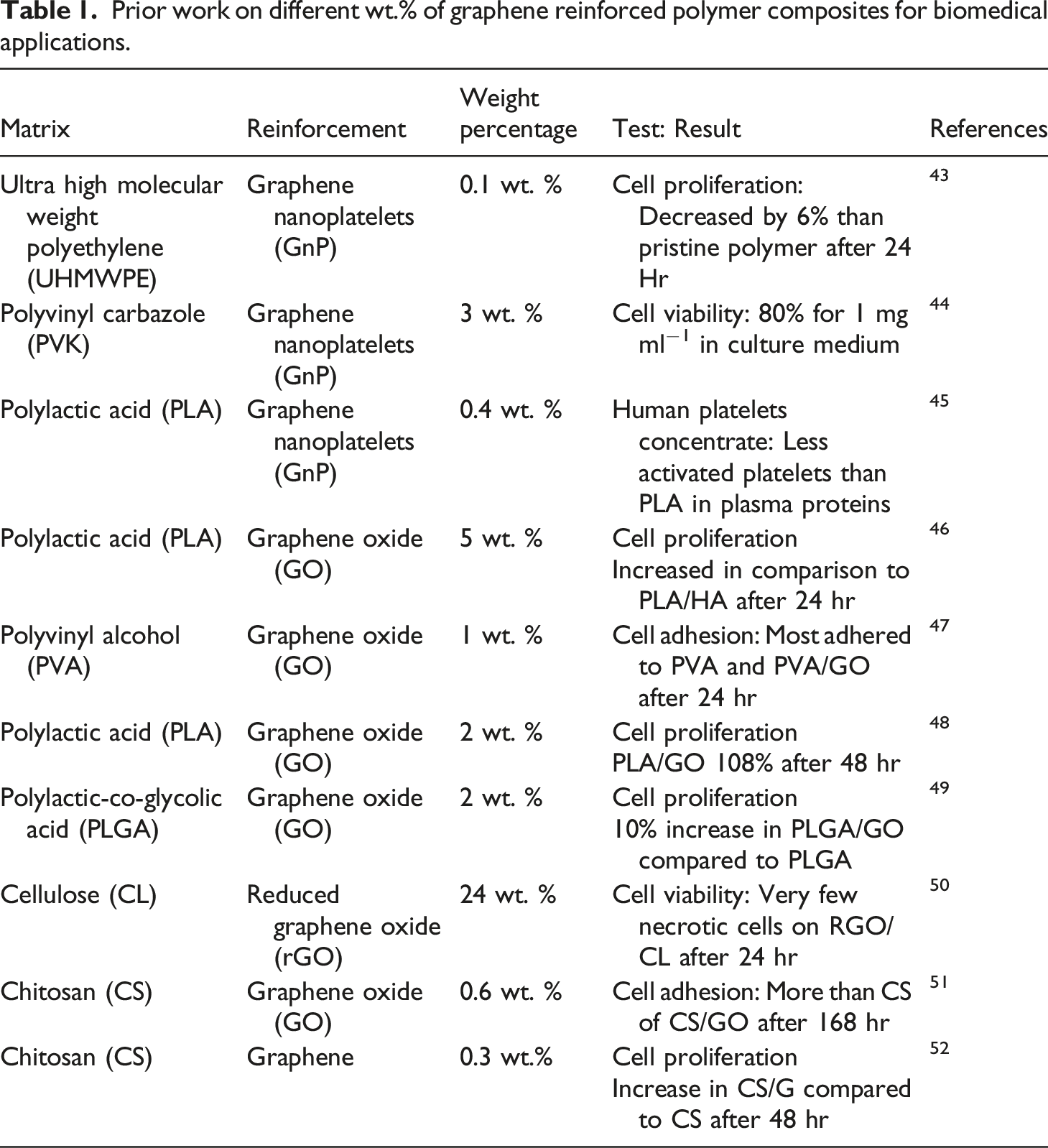
It has been remarked that very limited work exists on supplementing graphene nanoplatelets (GnP) in the PMMA bone cement. The investigation on the combined effect of hydroxyapatite and GnP on mechanical properties are also passing through the opening phase. It requires more attention from the researcher’s community, biomedical society, and manufacturing sectors. A few of them have explored incorporating carbon-based materials with bio-ceramic nanoparticles in PMMA based cement. This paper aims to expand the physio-mechanical performances of PMMA bone cement utilizing hybrid biocompatible nanofillers such as graphene nanoplatelets (GnP) and hydroxyapatite (HA). Most work exists to supplement single nanofiller materials in the matrix phase, as described in the literature review. The author tries to overcome the mechanical performance limitation of the PMMA bone cement by adding dual combination of nanofillers (GnP and HA).
Accordingly, the present article is mainly proposed to develop a PMMA bone cement-based hybrid reinforcing materials. HA and GnP were evaluated as potential reinforcement for PMMA bone cement. Mainly, three types of HA-GnP-PMMA bone cement nanocomposites with 0.5 wt.%, 1.5 wt.%, and 2.5 wt. % of HA and GnP were fabricated. Also, efforts have been made to select an optimized range for nanofiller addition in the matrix. The nanocomposite was characterized using EDX, FTIR spectroscopy, and X-ray diffraction (XRD) to examine the distribution of reinforced nanoparticles. The findings of SEM of the broken surface of the nanocomposite samples demonstrated the higher application potential for orthopedic surgery and implant fixing procedures. This provides the expected augmentation of mechanical performances for biomedical applications. The current research work could be an excellent contribution to biomaterials synthesis, development, and characterization. The developed PMMA based biopolymer nanocomposite can be cost-effective when produced at the mass production level. The findings show that it possesses excellent compressive and flexural strength performances. The chemical characterization of the synthesized material states the integrity of infused nanomaterials with the base matrix material. It can be endorsed to develop prosthesis and implants required for higher mechanical performance and biocompatibility objectives.
Materials and methods
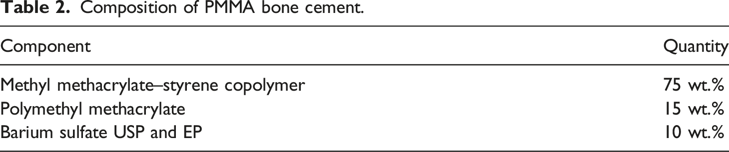
Composition of PMMA bone cement.
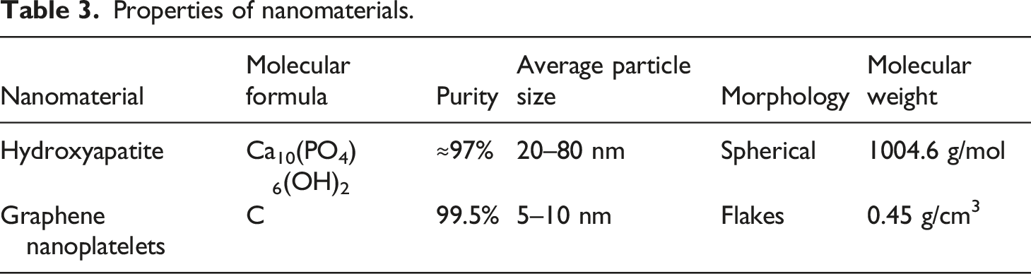
Properties of nanomaterials.
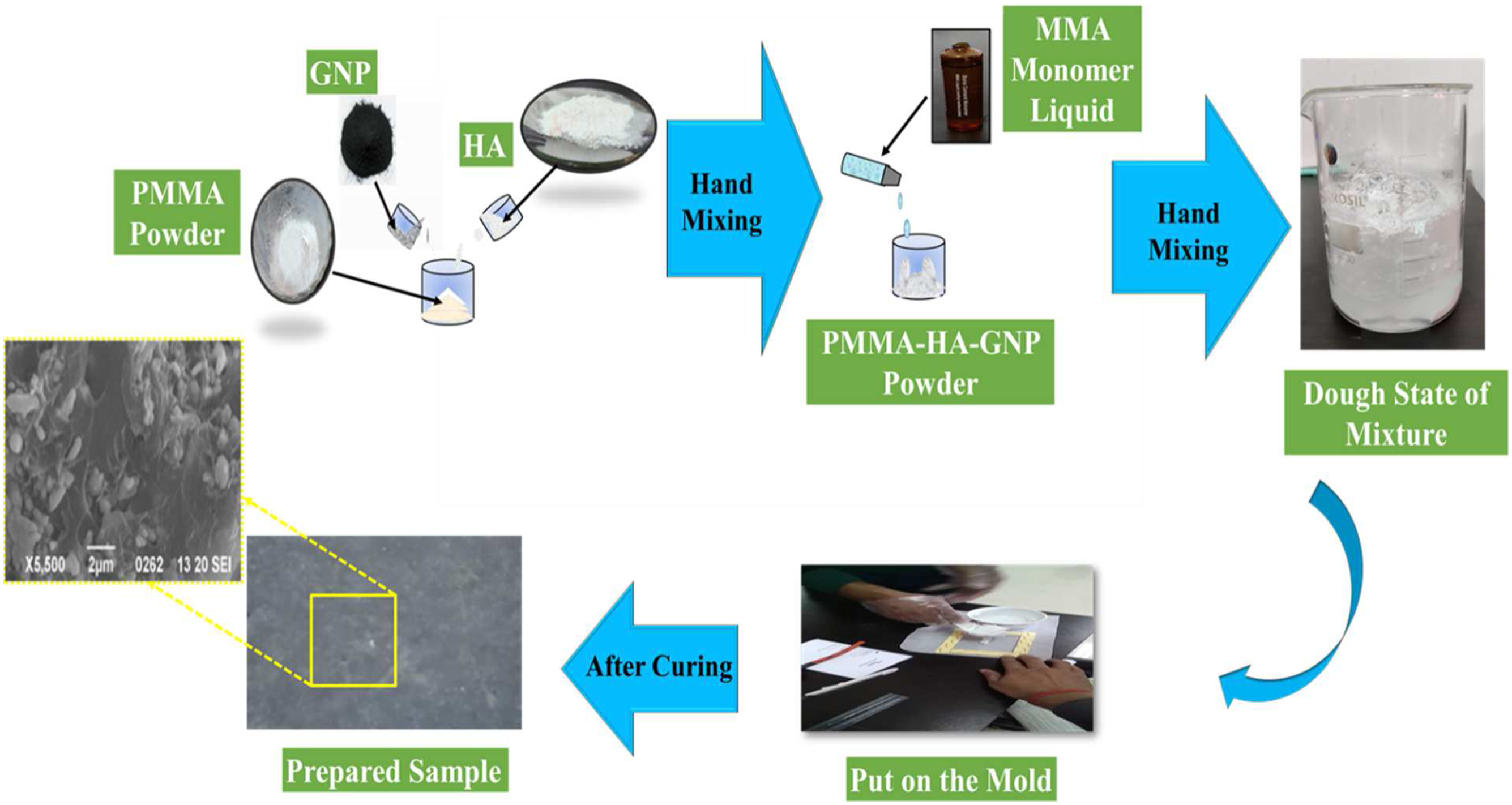
Nanocomposite fabrication procedure.
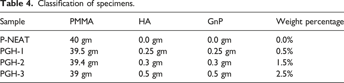
Classification of specimens.
Characterization techniques and mechanical testing
HR-SEM and EDS analysis
Scanning electron microscope (SEM) is used to analyze the prepared samples' surface morphology. The HR-SEM test was primarily performed before mechanical testing of biopolymer nanocomposite samples. It is performed to investigate the dispersion, surface morphology, and interface aspects of the proposed biopolymer nanocomposite samples. It helps to analyze the fracture property of bone and similar composites like PMMA bone cement. 54 Also, an analysis of the fractured surface was evaluated for observing the reason for the mechanical failure in samples. It becomes essential to investigate the composition of the developed nanocomposite and nanoparticles. Energy Dispersive X-Ray Analysis, referred to as EDS or EDAX, is an X-Ray technique used to identify the elemental composition of materials. EDS analysis produces spectra with peaks corresponding to the components that make up the actual composition of the material being analyzed. The samples were coated with Au-Pd, and a Philips XL-30 ESEM model high-resolution electron microscope (HR-SEM) was utilized for EDS and SEM examination.
X-Ray diffraction (XRD)
XRD is used to confirm the chemical composition and homogeneous dispersion of nanomaterials in the prepared composite. 55 The phase composition and crystal structure of the prepared nanocomposite, hydroxyapatite, graphene nanoplatelets, and PMMA bone cement were examined through XRD. The diffractometer used was a Raguku Model Ultima IV. 3 KW sealed X-Ray tube D/teX Ultra silicon strip detector, working at 200V and 30 A. At a scanning rate of 28 per minute, 508 datasets were obtained.
Fourier Transform Infrared Spectroscopy (FTIR)
FTIR evaluates the effect of functional groups, nanostructure, and elemental analyses of the prepared modified PMMA nanocomposite. 56 The FTIR spectrometer used was a BOMEM DA8 spectrometer, a testing-oriented FTIR spectrometer that allows the user to customize the standard configuration, including detectors, light sources, and beam splitters. To achieve the highest signal-to-noise ratio in the desired spectral regions, apodization parameters, beam aperture (radiation source), and several scans were used. The criterion was determined by measuring the sample transmittance phase. Bands in 2000–1800, 1550–1400, and 750–500 cm−1 ranges were selected for analysis.
Determination of compressive strength and modulus
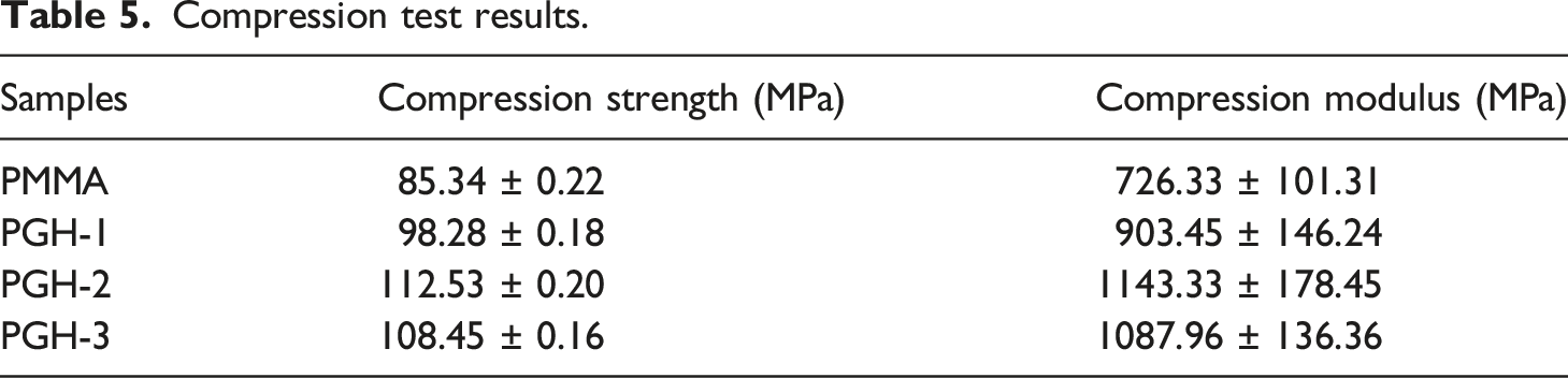
The compressive strength of bone cement is essential biomedical domains like tooth implantation and prosthesis replacement. 57 Therefore, it is vital to analyze the prepared nanocomposites’ compressive strength and compressive modulus. Also, compare it with pristine PMMA bone cement to get the optimum reinforcement quantity of the nanomaterials. Compressive strength was measured on HA and GnP-based bone cement as well as unfilled bone cement. Compression tests on solid specimens were conducted at room temperature on a Universal Testing Machine (UTM) Model H-75 KS equipped with a 50 kN load cell. A constant strain rate of 0.05 min−1 was used. ISO5833-2002 was used to perform the compression test. 58 The measurements of the specimens for this research were 12 ± 0.1 mm in length and 6 ± 0.1 mm in diameter. Compression modulus can be defined as the ratio of mechanical stress to strain in an elastic material when subjected to compression. The value of stress and strain were obtained with the help of the available stress–strain curve obtained from the UTM. Three replications of compressive testing were considered for each PMMA nanocomposite configuration, as described in Table 4.
Determination of flexural strength and modulus
Flexural strength becomes a vital property for applications like complete hip joint replacement and bolt fixing in the spine. 59 Flexural strength was measured on HA and GnP-based bone cement as well as unfilled bone cement. Universal Testing Machine Model H-75 KS was used for three points flexural test. The force applied to the specimens was 5 mm/min. The span to depth ratio was 16:1. At laboratory temperature (24 ± 1°C), the samples were incubated. ISO5833-2002 was used to perform the flexural test. 60 The measurements of the specimens for this research were 75 ± 0.1 mm in length, 10 ± 0.1 mm in width, and 3.3 ± 0.1 mm in thickness. In mechanics, the flexural modulus or bending modulus is an intensive property calculated as the ratio of stress to strain in flexural deformation or the tendency for a material to resist bending. 61 The value of stress and strain were obtained with the help of the available stress–strain curve obtained from the UTM.
For a three-point bend test specimen, the flexural strength is given by equation (1)
In order to obtain more consistency and accuracy in findings, statistical analysis was performed on these observed values. Means and standard deviations (SD values) were remarked as satisfactory for the conducted test. The results were deemed statistically significant under the p-value limits tend to be less than 0.05. The values are written as Mean ± SD.
Result and discussion
Chemical structure analysis
HR-SEM and EDS Analysis of prepared specimens
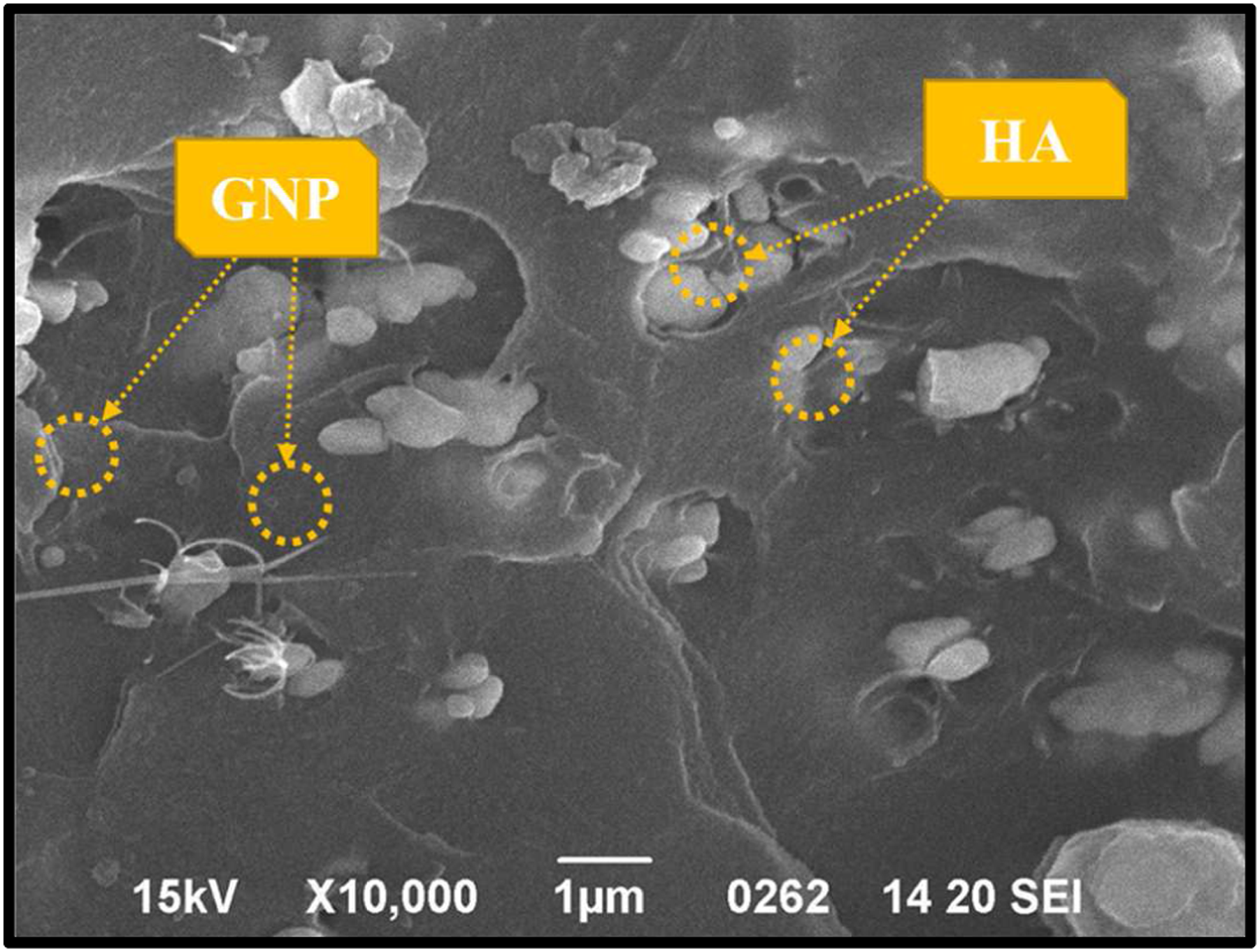
The high-resolution SEM test was primarily performed before mechanical testing of biopolymer nanocomposite samples. It is performed to examine the dispersion of nanomaterials in the matrix phase (Figure 2). The P-NEAT sample shows a bright image that indicates only white PMMA bone cement in the sample. The PGH-1 sample shows a dark surface, indicating the presence of graphene nanoplatelets (GnP) and some white particles, which indicates HA distribution. PGH-2 sample shows a smooth dark surface, with a tendency of homogeneous dispersion of both nanoparticles. Although PGH-3 sample appears to be slightly less even and smooth surface at 20 μm resolution than the PGH-2 sample.40,53 Figure 3 reveals the dispersion of nanomaterials in the prepared nanocomposite PGH-2. At 2 μm spherical shaped nanoparticles of HA, as well as GnP nanoflakes, could be detected within the matrix region of PMMA based modified nanocomposite. The foretasted higher results of mechanical properties of the PGH-2 sample could be explained by the dispersion uniformity, which can be observed in SEM results. SEM images of prepared samples at 10 μm resolution. Dispersed HA and GnP in PMMA bone cement nanocomposite.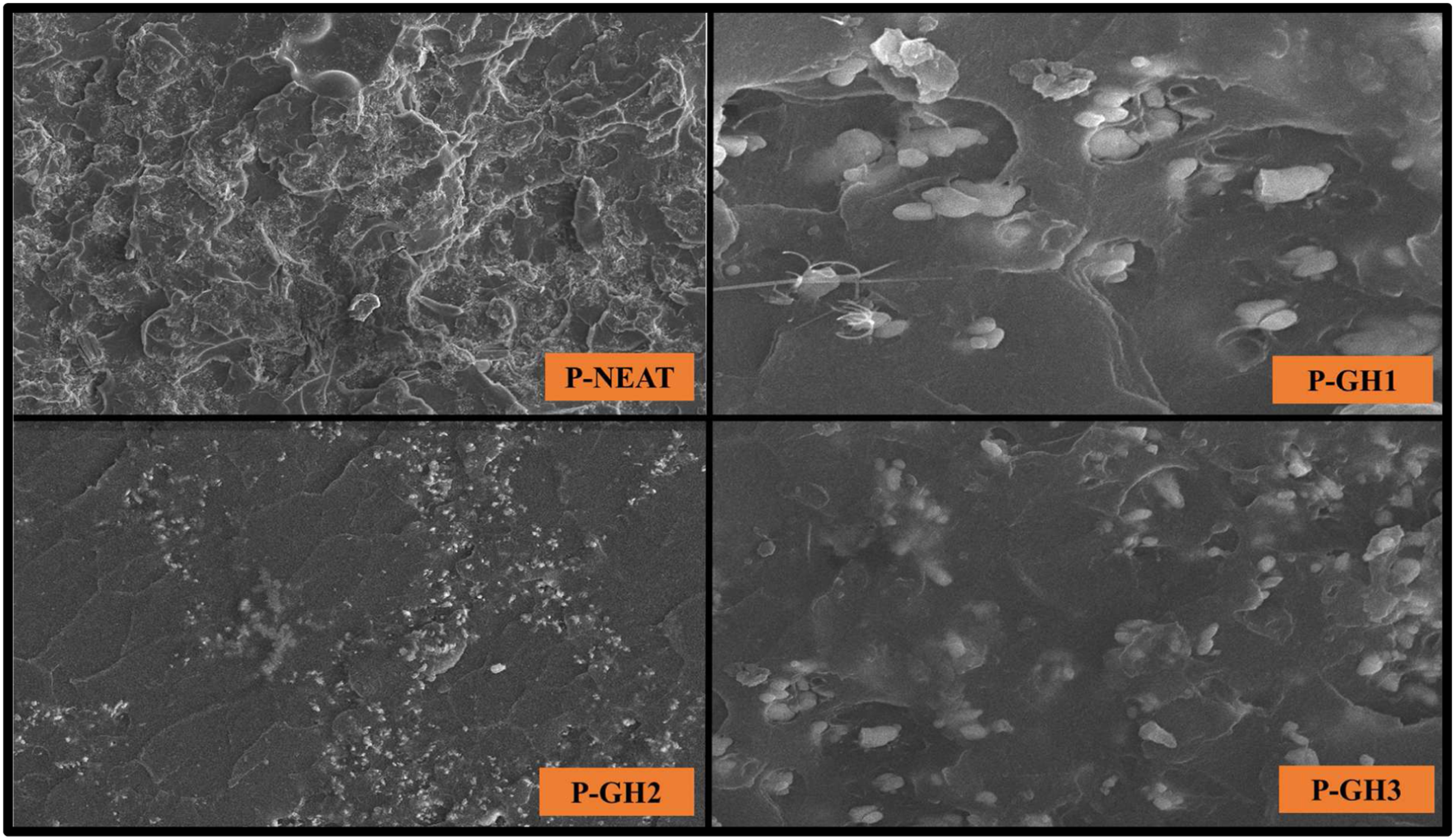
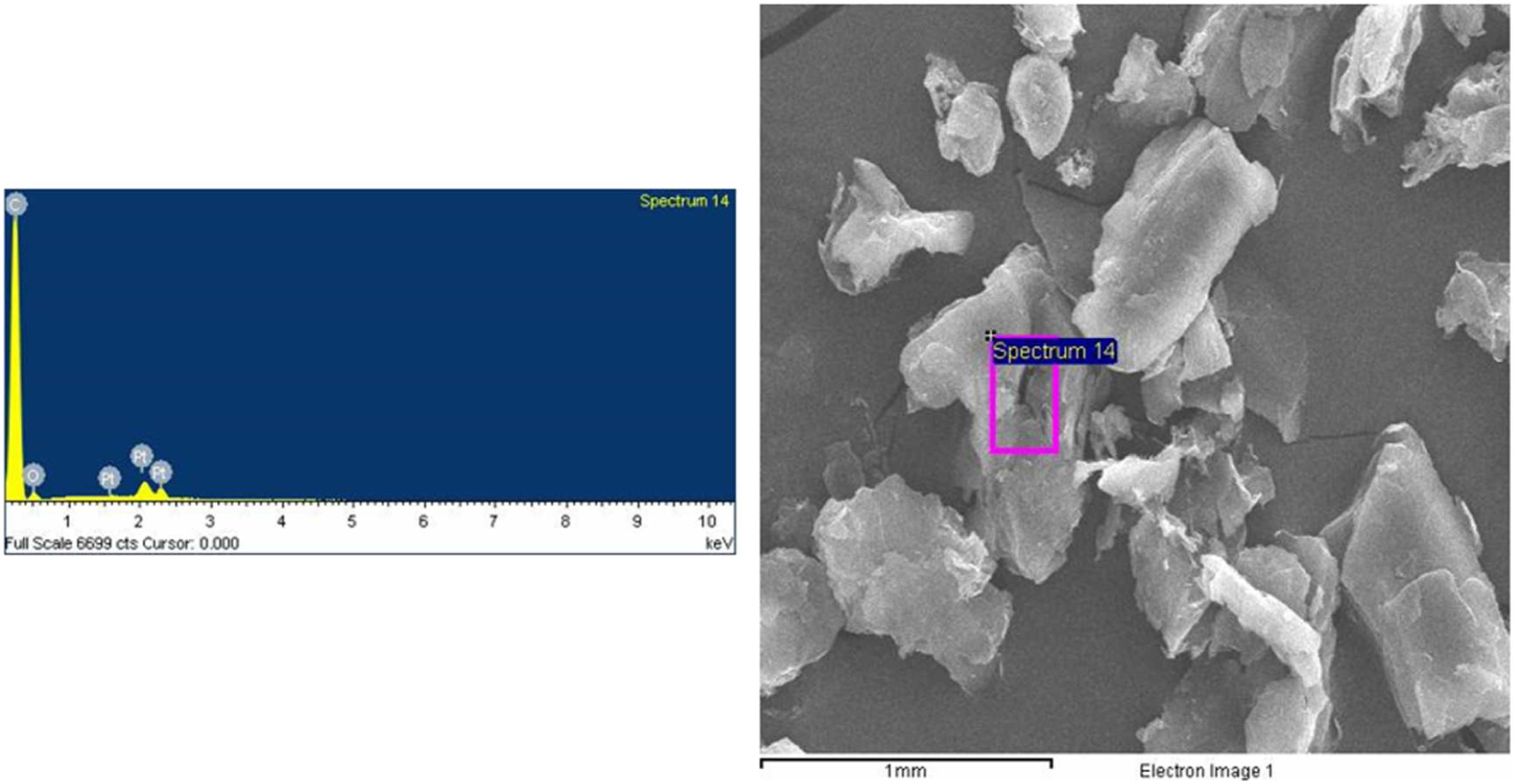
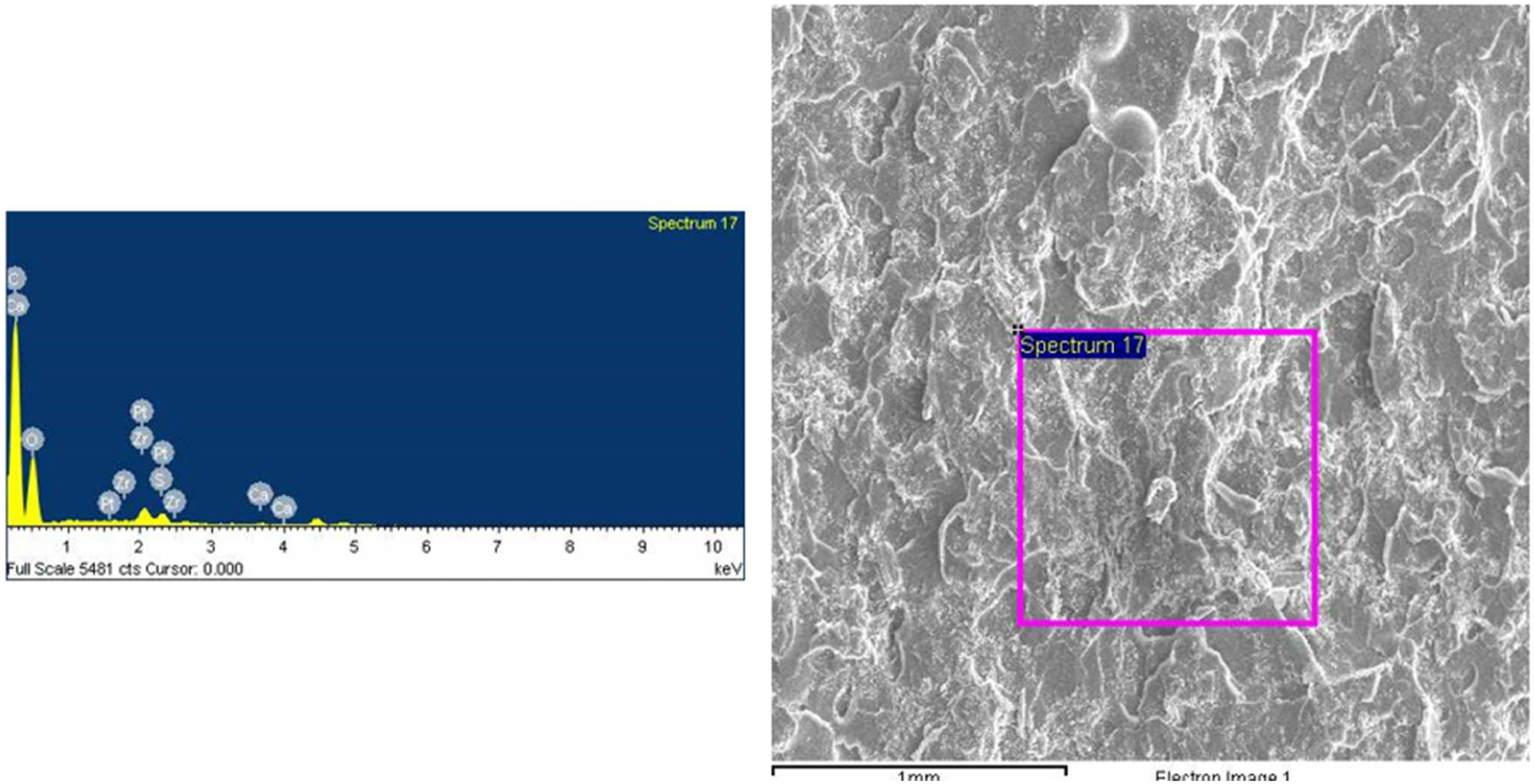
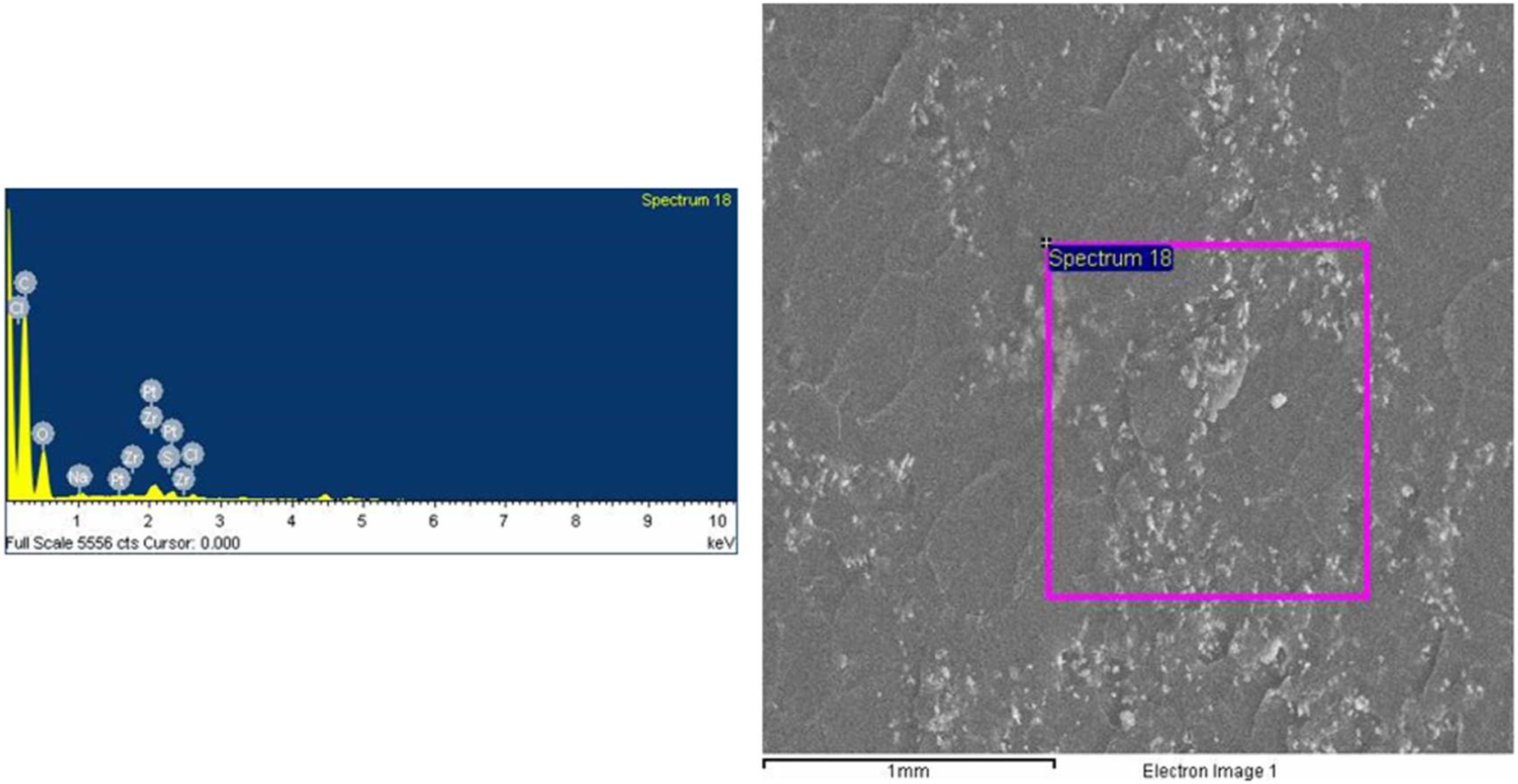
The primary use of PMMA bone cement is inside the living body. Hence, the PMMA based nanocomposite should consist of those elements which should be compatible with a living body. Therefore, to confirm the elemental composition of the prepared nanocomposite and the nanoparticles, Energy disperses X-ray spectroscopy (EDS) analysis of utilized hydroxyapatite, graphene nanoplatelets, neat PMMA bone cement, and developed PMMA-HA-GnP nanocomposites is performed. As revealed by EDS spectroscopy, the elemental composition of hydroxyapatite demonstrates the presence of Ca, P, and O in the nanoparticles, as shown in Figure 4. The elemental composition of GnP, as revealed by EDS, shows C, O in the nanoparticles in Figure 5. The elemental composition of PMMA bone cement as shown by EDS spectroscopy shows the presence of Ca, O, and P in the nanoparticles in Figure 6. The elemental composition of PMMA-HA- GnP nanocomposite shows the presence of Ca, O, and P in the nanoparticles in Figure 7. EDS spectrum of hydroxyapatite (HA). EDS spectrum of graphene nanoplatelets (GnP). EDS spectrum of PMMA bone cement. EDS spectrum of PMMA-HA-GnP nanocomposite.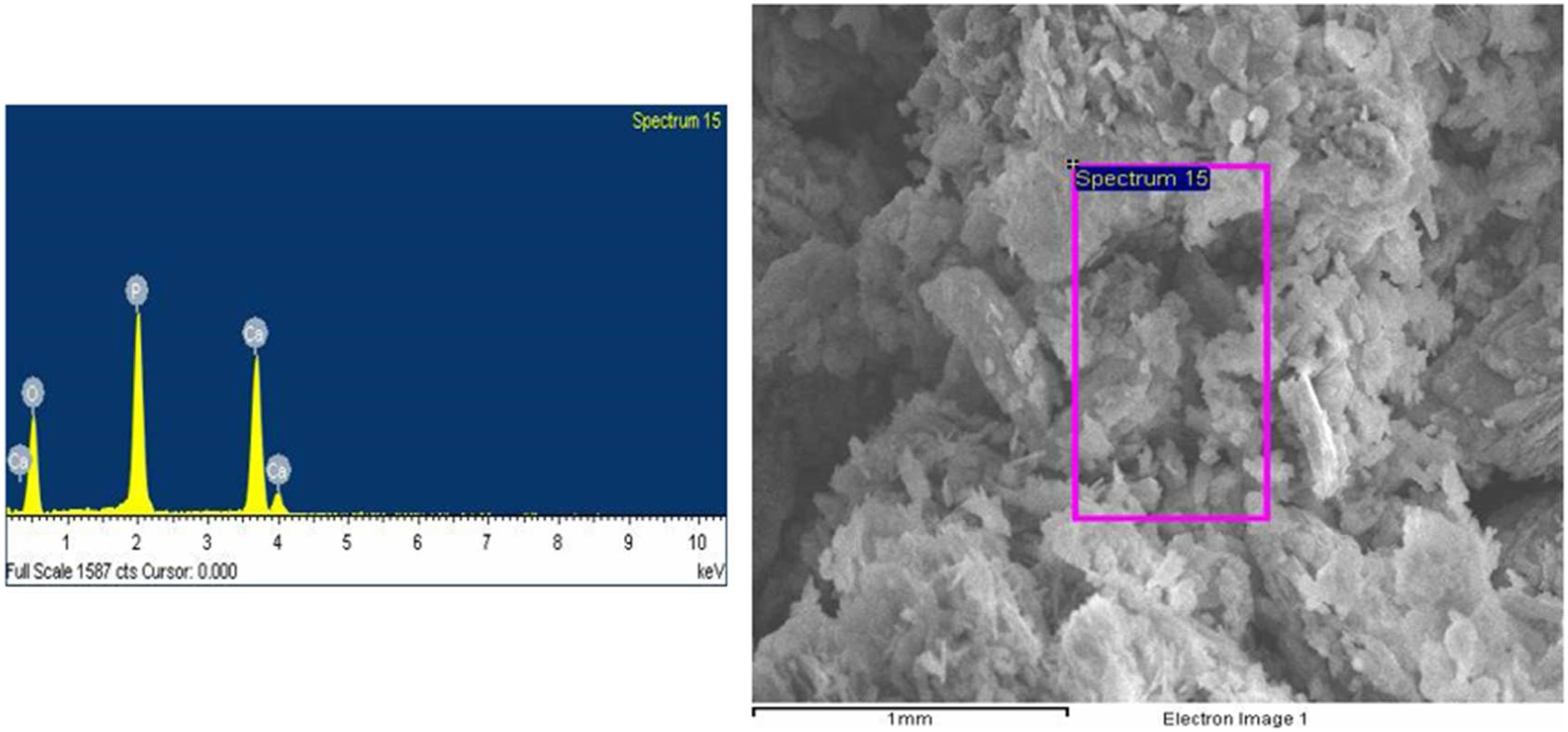
Fourier transform infrared spectroscopy (FTIR)
The FTIR reveals the various functional group in the PMMA bone cement, GnP, HA, and PGH-2 specimen (Figure 8). The transmission peaks at 1065 cm−1 and 837 cm−1 belong to PMMA bone cement.
16
The characteristic band of the C=O group at 1720 cm−1 peaks in PMMA bone cement shows band shift to 1730 cm−1 and broadening in the curve of the PGH-2 sample, representing the interactions in the mixture. An intense PO43- peak showing phosphate group appeared at 1048 cm−1, which was also appeared in previous studies.
8
Phosphate-related infrared bands were observed at 1097 and 962 cm−1, as shown in Figure 8. At 810 cm−1 and 757 cm−1 also, phosphate member bands are present. The spectrum for GnP revealed the characteristic vibration of the C=C at 1633 cm−1, which was due to graphene’s aromatic carbon composition.
61
The prominent bands appeared at 1140 cm−1 and 1730 cm−1, corresponding to an O–C–C stretching and a C=O stretching. These bands are also observed in previous studies having GnP and HA as reinforcement in PMMA.8,39 With the help of obtained transmission peaks and compared with earlier studies, the required functional group of nanomaterials is confirmed in the prepared nanocomposite. F spectrum of PMMA and PGH-2.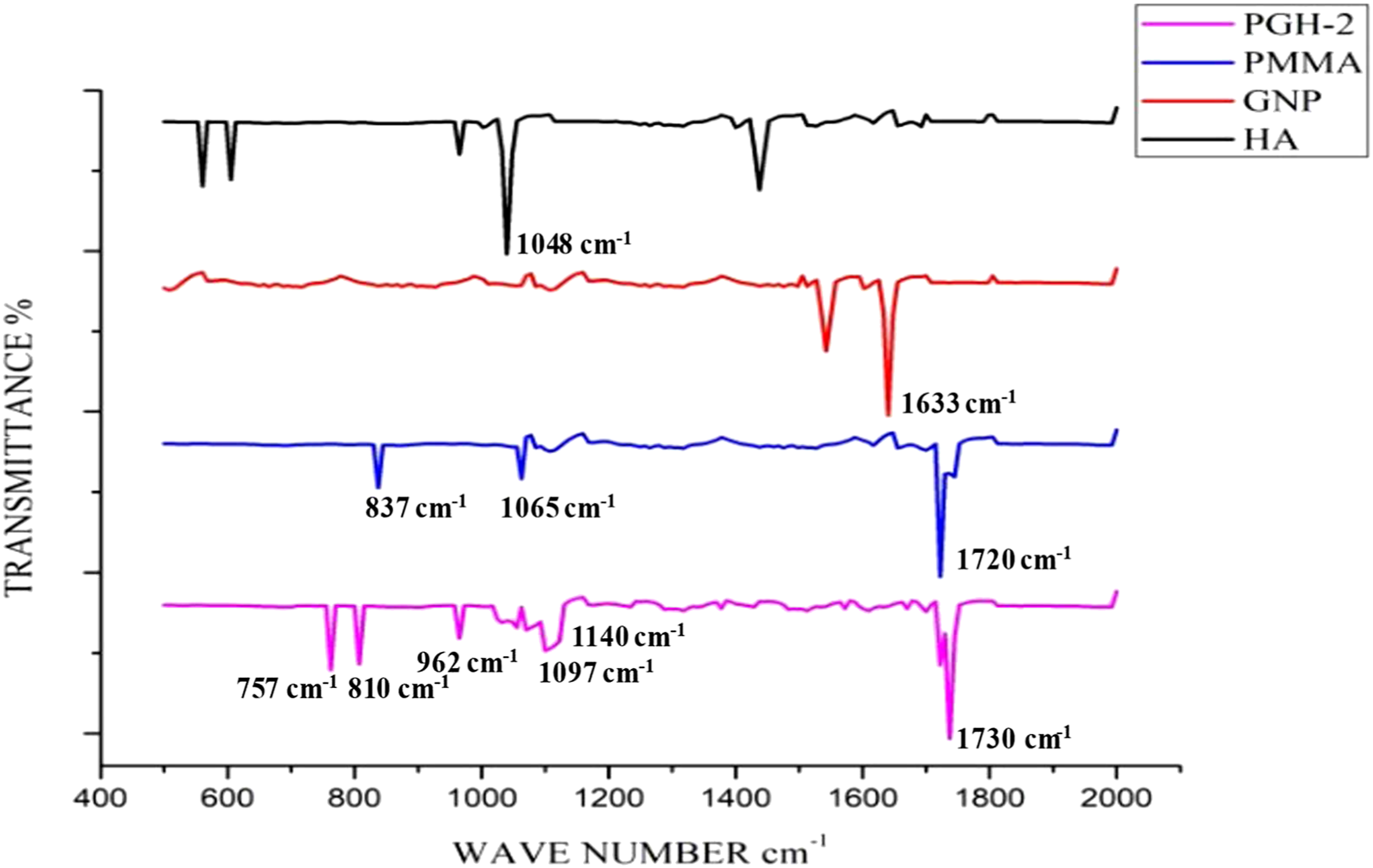
X-Ray Diffraction (XRD)
XRD reveals the diffraction peaks present in the PMMA bone cement, GnP, HA and PGH-2 sample as observed in a previous study.
62
The test showed the existence of hydroxyapatite on a polymeric matrix-like amorphous basis (Figure 9). These results indicate that when hydroxyapatite comes into contact with these acidic or essential functionalities, it does not alter structurally, preserving its ability to promote bone formation. Hydroxyapatite in XRD analysis shows peaks at 2θ = 28°, 31.4°, and 49° in various studies .63,64 The diffraction peak of GnP was found to be at 2θ = 25.64°, which was similar to previous studies.65,66 As per observations, the peak at 25.64° corresponds to an interlayer spacing of approximately 0.336 nm, which is extremely comparable in size to the d-spacing of graphite .
67
The needed crystal structure of nanoparticles in the developed nanocomposite is validated using the acquired diffraction peaks and compared to earlier investigations.8,68 As can be seen from the XRD pattern, PMMA bone cement was grafted on HA and GnP. X-Ray diffraction of PMMA and PGH-2.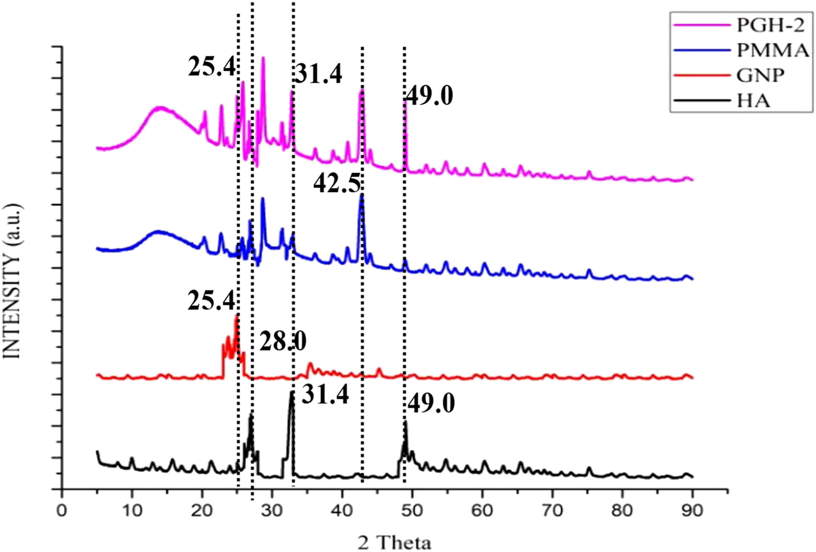
Mechanical Properties Analysis
Effect of reinforcement on compressive strength
Compression strength, compression modulus, and stress–strain curve are obtained while performing the compression test. Figure 10 depicts the stress–strain curve of the performed compression strength test. The addition of different pre-defined amounts of nanomaterials substantially increased compression strength and compression modulus, as seen in Figure 11 and Figure 12, respectively. The PGH-2 sample showed the highest value of compression strength. The compressive strength of the PGH-2 sample was increased by 31.7% (from 85.34 ± 0.22 MPa to 112.53 ± 0.20 MPa), and the compression modulus of the PGH-2 sample increased by 57.41% (from 726.33 ± 101.31 MPa to 1143.33 ± 178.45 MPa) in comparison to the commercially available PMMA bone cement. In a related analysis, applying 0.5% by weight GO to PMMA bone cement improved compressive strength by just 8.4% in comparison to the commercially available PMMA bone cement, whereas in the present study on adding an equal amount of HA with GnP in 0.5 wt.% reinforcement improved compression strength by 15.16%.
40
Similar experimental values of 16.2% improvement were found in compression strength when Chitosan (CS) and GO was used at a high concentration of 25 wt.%, which is also less than the present result.
37
Only 14% improvement is observed in compression strength when HA is alone loaded to PMMA, but in this study with hybrid reinforcement, 31.4% improvement is observed.
69
As the weight % of GnP and HA increased, compressive strength and modulus increased significantly up to 1.5 wt.% and decreased when loaded above 1.5 wt.%, as shown in Table 5. Earlier studies show that the decrement in compression strength could be observed possibly at high loading due to the higher agglomeration rate of carbon-based nanoparticles in matrix material and formulation of internal voids during fabrication procedure of modified PMMA based nanocomposite.37, 70 Stress v/s Strain curve for compression strength. Compressive strength of all the tested samples. Compression modulus of all the tested samples. Compression test results.
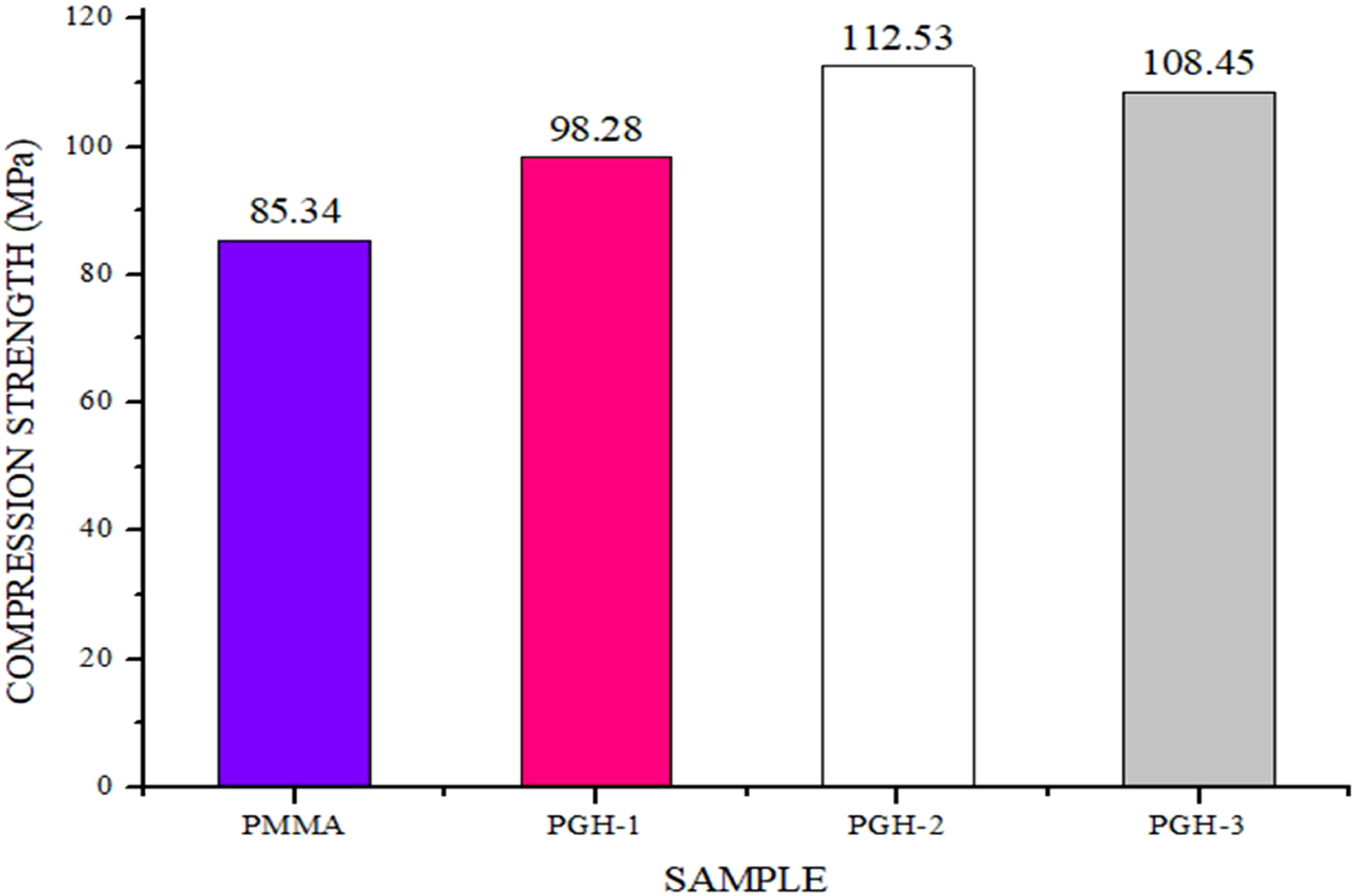
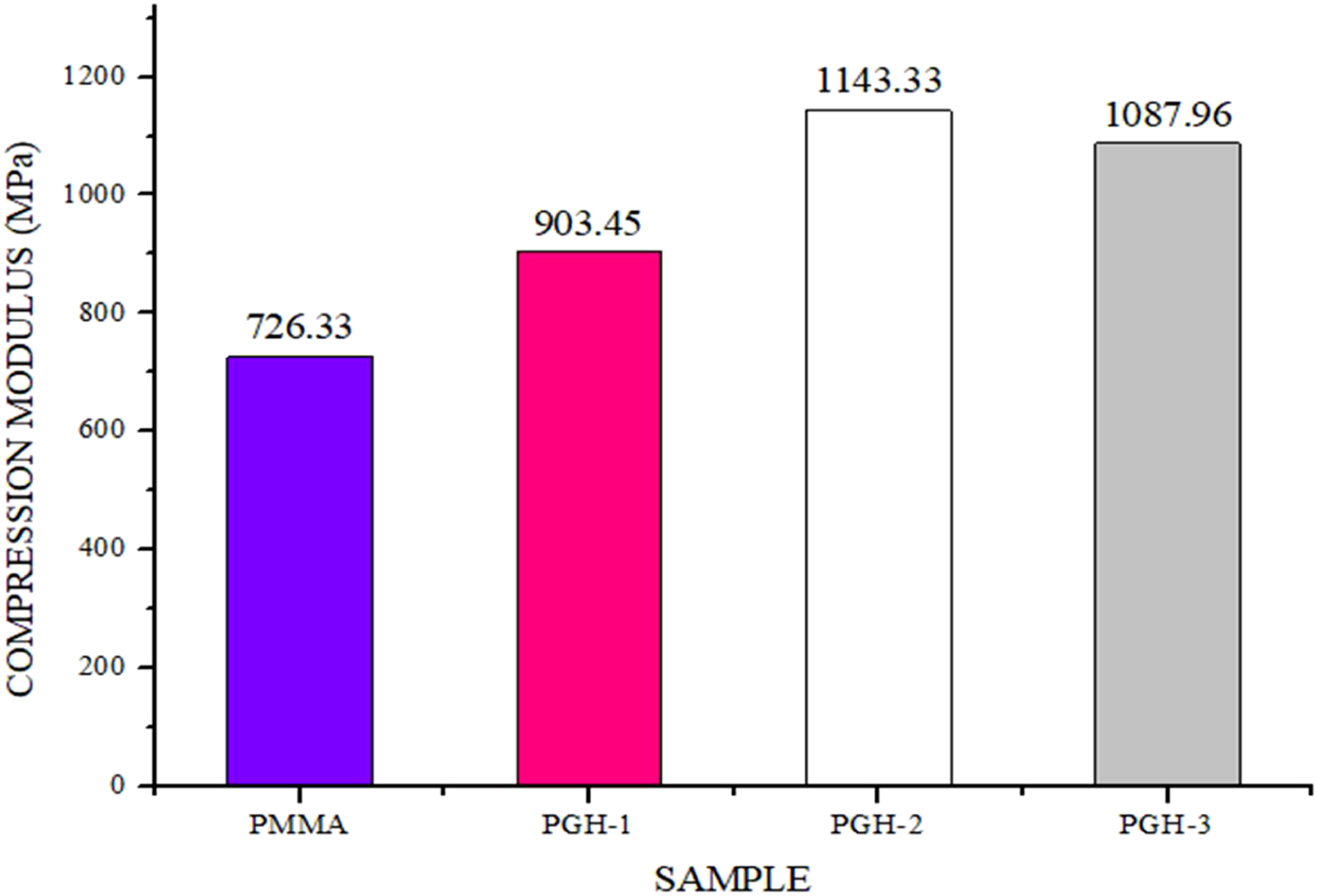
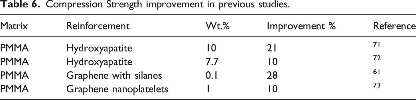
Figures 13 and 14 show the error bars of the obtained values of compression strength and compression modulus. Table 6 shows more studies showing improvement in percentage in compression strength of PMMA bone cement on adding different nanomaterials. The PGH-2 sample was chosen as the best mechanical property sample for future experiments, according to the findings. Error bar of compression strength. Error bar of compression modulus. Compression Strength improvement in previous studies.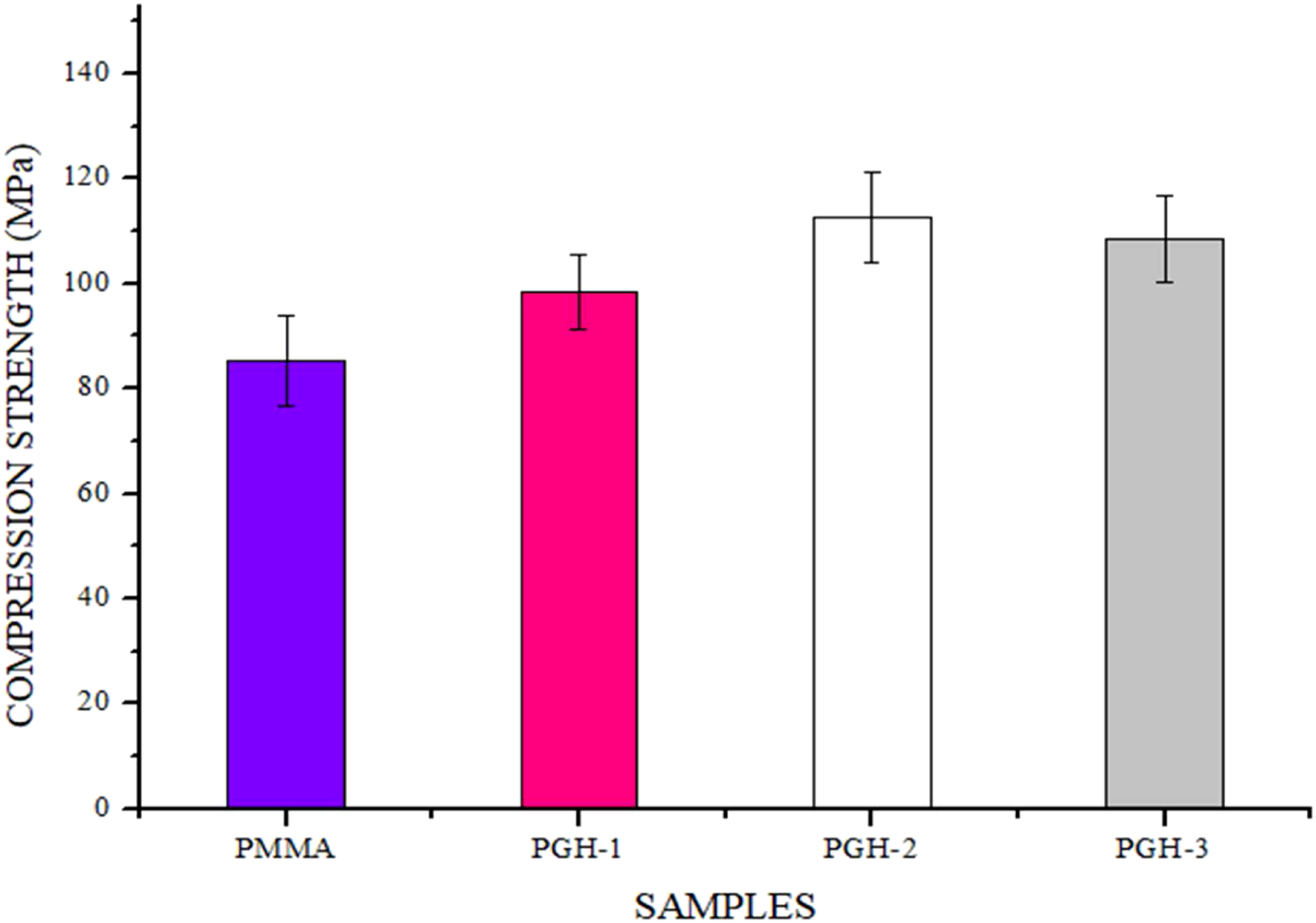

Effect of reinforcement on flexural strength
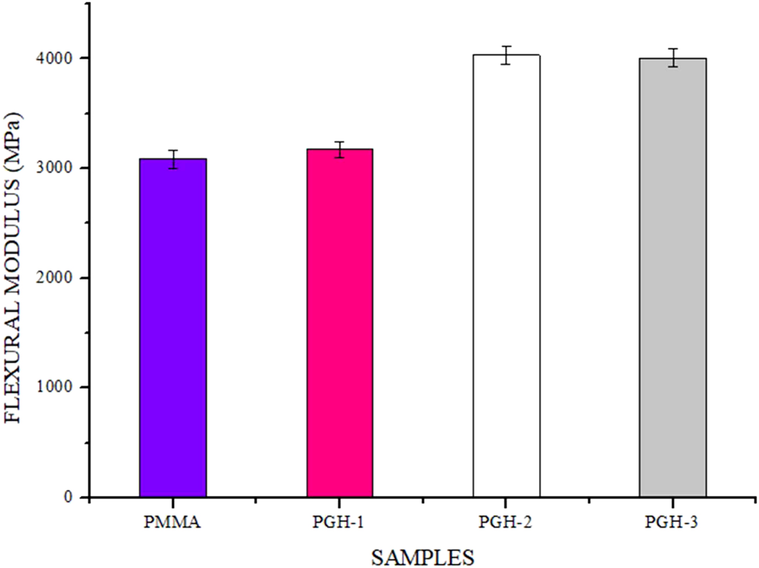
Flexural strength, flexural modulus, and stress–strain curve are described in Figure 15. The addition of nanomaterial substantially increased flexural strength and flexural modulus, as seen in Figure 16 and Figure 17, respectively. The PGH-2 sample showed the highest value of flexural strength. The flexural strength of the PGH-2 sample was increased by 49.28% (from 63.32 ± 0.43 MPa to 94.54 ± 0.38 MPa), and the flexural modulus of the PGH-2 sample increased by 30.8% (from 3085.21 ± 351.53 MPa to 4035.70 ± 378.86 MPa) in comparison to the pristine PMMA. The flexural strength of the PGH-1 sample has 0.25 wt.% of GnP and 0.25 wt. % of HA was increased by 24.0% (from 63.32 ± 0.43 MPa to 78.43 ± 0.47 MPa) in comparison to the neat sample; however, in a similar study at 0.25 wt. % GO powder loading, the best bending properties were improved by just 13%.
40
Similar research found a 24.0% improvement in flexural strength when GO and chitosan (CS) nanomaterial was used at a concentration of 25 wt.%, which is still less improved than the present study analysis.
37
The addition of GnP in up to 5 wt. % in polymer matrix composite results in 45.2% improvement in flexural strength, whereas in the present study with hybrid reinforcement only 1.5 wt. % GnP improved flexural strength by 49.2%.
74
As the weight % of GnP and HA increased, flexural strength and modulus increased significantly up to 1.5 wt.% and decreased when loaded above 1.5 wt.%, as shown in Table 7. Earlier studies show that the decrement in flexural strength could be observed possibly at high loading due to the higher agglomeration rate of carbon-based nanoparticles in matrix material and formulation of internal voids during fabrication procedure of modified PMMA based nanocomposite.37, 70 Stress v/s Strain curve for flexural strength. Flexural strength of all the tested samples. Flexural modulus of all the tested samples. Flexural test results.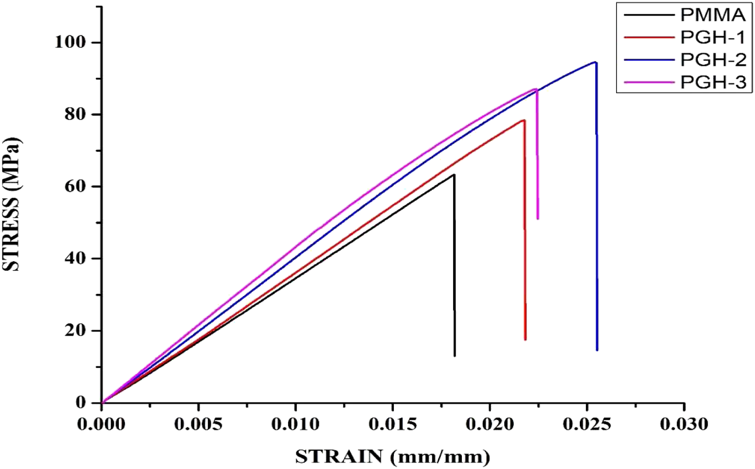
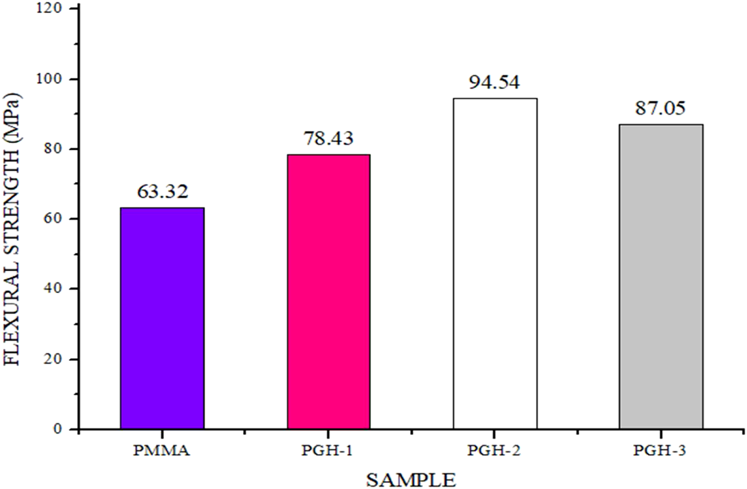
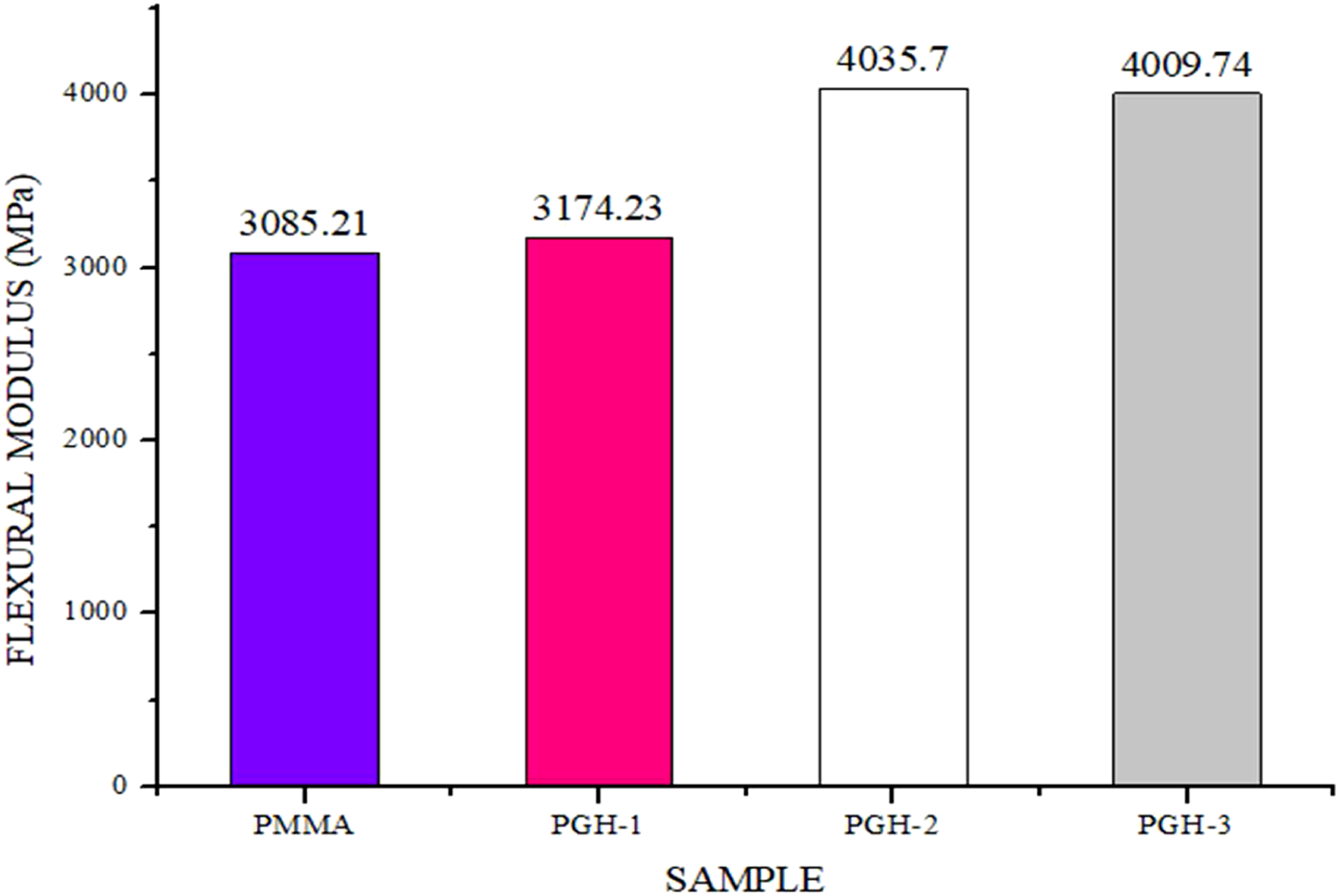
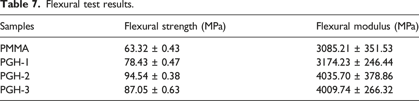
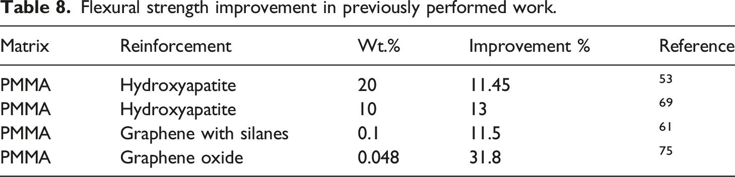
Figure 18 and Figure 19 show the error bars of the obtained values of flexural strength, and flexural modulus. Table 8 shows improvement in percentage in flexural strength of PMMA bone cement on adding different nanomaterials. The PGH-2 sample was chosen as the best mechanical property sample for future experiments. Error bar of flexural strength. Error bar of flexural modulus. Flexural strength improvement in previously performed work.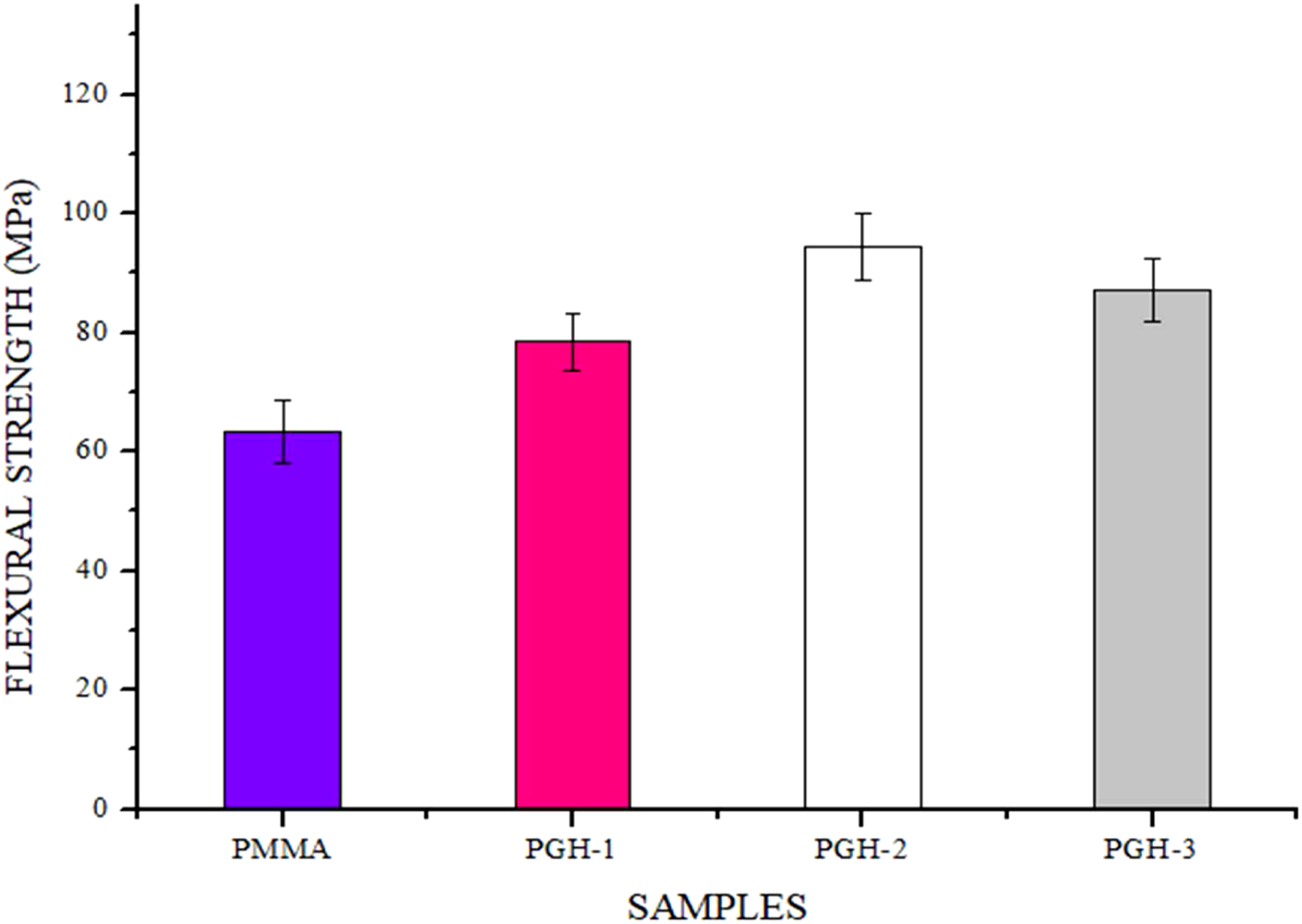
HR-SEM analysis of the fractured sample
The finding of mechanical tests demonstrated a desired improvement in the prepared bio-composite samples. The broken surface analysis at high-resolution microscopy is essential to investigate the surface morphology. The SEM images of all four samples are shown in Figure 20 at 200 μm resolution. The P-NEAT sample shows a bright image that indicates only white PMMA bone cement in the sample. The PGH-1 sample shows a rough surface, indicating the poor distribution of nanoparticles. In the PGH-2 sample, a smooth surface is seen, indicating fine dispersion of nanoparticles, as reported in the SEM examination of the composite bone cement’s fracture surface supported this claim in previous work.
53
The presence of agglomerates is a deciding factor for the mechanical performance of the HA-GnP-PMMA nanocomposite, as suggested in previous studies.
40
As the HA content rose to 1.5 wt.%, the dispersion of unadulterated GnP and HA in the HA-GnP-PMMA cement’s cross-section is standardized, as seen in Figure 20. As a result, the material was more uniform and had better mechanical properties, as the modified HA and GnP dispersed much smoother in the PMMA matrix.
76
Voids are visible in the PGH-3 sample, shown in Figure 20. A large content of graphene results in the agglomeration and improper diffusion in polymer matrix due to the structural property of graphene resulting in void formation.
77
Void formation in some other hybrid nanocomposites of HA is also seen at a high loading of reinforcement.71,78 Due to void formation, stress concentration occurs in the sample, resulting in fracture of the sample. SEM images of all four samples at 200 μm resolution.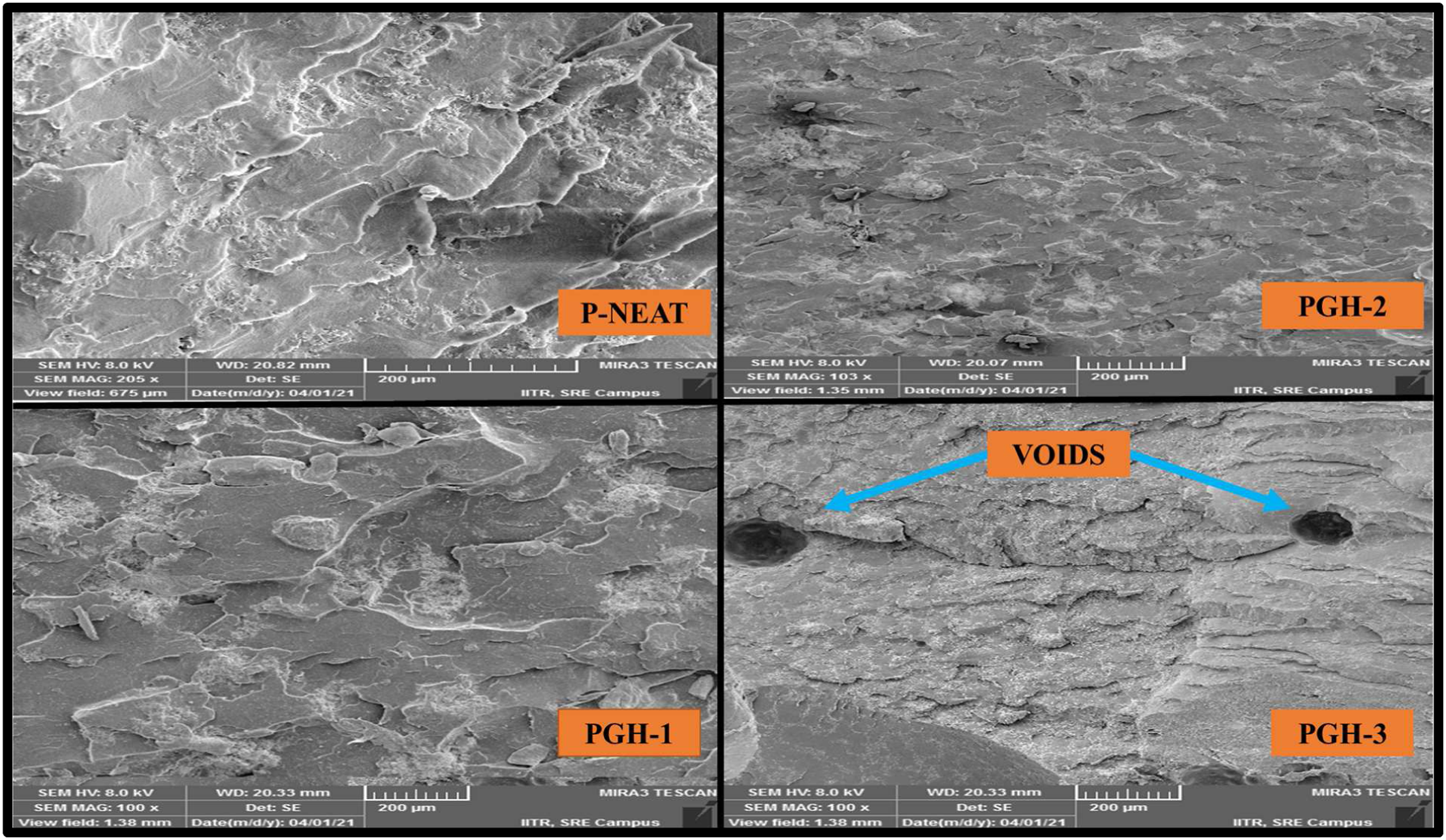
The samples with a small amount of GnP resulted in no agglomerates, as can be seen in PGH-1 and PGH-2 samples images as observed in the earlier investigation also of polymer matrix nanocomposite.73,79 The PGH-3 sample has the highest GnP and HA content among all four samples, showing weak dispersion inside the PMMA matrix, resulting in voids and poor dispersion. With high HA content, the HA dispersion in the HA-GnP-PMMA cement cross-section became less uniform, as shown in the PGH-3 sample. In the case of the PGH-2 sample, the uniform distribution is visible, resulting in high mechanical strength. 80 All these observations suggest using hybrid reinforcement of HA and GnP up to 1.5 wt.% only for maximum mechanical strength in the PMMA bone cement matrix.
Discussion
PMMA bone cement is used widely as a biomaterial, with a huge domain in orthopedic application. It still holds some disadvantages with such great potential, one among them being due to the weak mechanical property. The only possible solution is the use of nanoparticles. The present study observed a significant improvement in the mechanical property by chemically bonding of HA and GnP in the PMMA matrix. This study revealed that adding only a small amount of HA with GnP in the PMMA bone cement can significantly change mechanical properties. In many investigations, a minor change in bending strength was produced with a large quantity of nanoparticle reinforcement. 81 During the application of flexural test on any specimen, some part specimen tends to compressive loading while others are put in tensile, whereas the compression test applies only the compressive stresses, which motivates yielding and fracture. Thus, both types of testing might be utilized to measure the strength and fracture strengths of different materials. It is commonly established that brittle materials are significantly stronger in compression than flexural. A transverse fracture will seek to close up and hence cannot propagate under compressive stress. The flexural strength increased from 63.32 ± 0.43 MPa to 94.54 ± 0.38 MPa and compression strength increased from 85.34 ± 0.22 MPa to 112.53 ± 0.20 MPa, representing a major improvement in mechanical properties due to the loading of GnP and HA at wt.% of 1.5. In previous research, increasing GO above 1 wt. % diminishes mechanical properties due to a significant number of voids and pores, which can also be observed in the present study as in the PGH-3 sample the wt. % of GnP is 1.25 resulted in void formation as seen in SEM image analysis at 200 μm resolution, which reduced the mechanical properties. 40 Earlier research has also shown an increase in bending strength up to 25% on adding HA by 15 wt. % and increase in compressive strength up to 4% on adding HA by 2.5 wt. % whereas in the current research article, with hybrid reinforcement, 49.28% increment in flexural strength and 31.7% increment in compressive strength is achieved. 25 Also, when 8 wt.% HA powder was applied to the bone cement, the result was the compressive strength improved from 110 MPa to 122 MPa, that is, 10.9% only, which is less than the present study result. 72 In recent studies, it has been observed that the hybrid reinforcement in PMMA bone cement nanocomposite is utilized to modify the mechanical properties of the base matrix. It was found that the improvement percentage in mechanical properties was not as high as it was seen in our study. Tavakoli et al. 37 utilized chitosan and graphene oxide (GO) in PMMA bone cement and found 16% improvement in compression strength and 23% improvement in flexural strength. At the same time, Pahlevanzadeh et al. 70 utilized reduced graphene oxide (rGO) and carbon nanotube (CNT) in PMMA bone cement and observed an improvement of 34% in bending strength as compared to neat. The mechanical analysis results show that the mechanical properties and the weight percent of HA and GnP used in PMMA are directly related. It is worth noting that strong interconnection between the GnP and HA nanoparticles and the PMMA bone cement matrix is needed to improve mechanical behavior. There are two possible reasons for the decrement in mechanical properties. The nano-sized powder sheets caused variations in the propagation crack fronts, resulting in off-plane loading and new fracture surfaces. As a result, the requisite strain energy for the fracture to continue increases, resulting in a decrement in mechanical properties at high loading.82,83 Also, if the HA particles are in small numbers and are evenly distributed, they act as load carriers and enhance the mechanical aspects. As the percentage of HA particles increases, a non-uniform distribution can develop, which causes particle aggregation. This phenomenon could also lead to weak matrix adhesion and a reduction in mechanical performances.84,85 In the present study also, it is observed that the PGH-3 sample with the highest wt. % of HA resulted in a decline in mechanical properties. Graphene nanoplatelets (GnP) were more helpful in improving flexural strength and modulus of PMMA as GnP has exceptional mechanical properties.74,83 In the field of polymer composites, GnP was found to be a promising member for improving mechanical properties.83,84 However, to realize this potential, the flakes must be well-dispersed in the polymer matrix, and the interactions between the two components must be strong. GnP was reinforced in PMMA by enhancing the interfaces between the filler and polymer. The “grafted” polymer chains serve as “bridges,” powerfully connecting the two components of the composite and producing a “continuity” between the matrix and nanofiller.85,86 The enhanced dispersions and strong filler-polymer interfaces created by grafting polymer chains onto reinforcements resulted in large amounts of reinforcement, indicating that chemically altering the interface between filler and host polymer is critical for achieving superior mechanical performance in composite materials. In prior state-of-art investigations, the aforesaid outcomes were noticed.18,86,87 brittle compatibility between bone and prosthetic cement. 18 Furthermore, hydroxyapatite has also been widely accepted as a commercial additive in PMMA bone cement for improving other highly requisite properties like radiopacity, biocompatibility, and osteoconductive. 87
The presence of different functional groups in the developed nanocomposite is best analyzed with the help of FTIR. The FTIR peaks confirm the proper dispersion and presence of both HA and GnP. The FTIR data show similar spectra among the evaluated groups, with no major differences in the functional groups’ positions. PMMA has distinctive peaks for C=O at 1725 cm−1. 75 The FTIR result showed the identical spectra as demonstrated by other researchers on adding HA and GnP.8,37 When XRD analysis was conducted to analyze crystal structure on pure PMMA bone cement, three large peaks were observed at 2θ =14.1°, 2θ = 29.0°, and 2θ = 42.5°. These patterns point to the PMMA’s usual amorphous form, and the findings seem to be in line with previous research82,88 With the help of results obtained through FTIR and XRD analysis, it is confirmed that both the reinforcements are properly dispersed in the PMMA bone cement matrix. The developed HA-GnP-PMMA bone cement nanocomposite shows the ability to be utilized as possible bone cement and fillers for covering micro-fractures and filling voids in the bone in the event of damage or injury. With minimal experience and supplies, this HA-GnP-PMMA nanocomposite bone cement can be made in a short period. Also, the HA-GnP-PMMA bone cement nanocomposite may be used to stabilize implants as bone screws. However, for such an application, additional manufacturing processes will be needed. The current results are clinically relevant because they shed light on how the actions of bone cement can change as a result of GnP and HA incorporation. Although, the findings provide knowledge on the physiochemical and experimental composite’s mechanical operation. It is critical to understand the biological behavior of materials containing GnP and HA fillers. A more thorough study into the HA-GnP-PMMA bone cement nanocomposite microstructure is also needed to resolve the GnP and HA nanoparticles reinforcing process complications.
Conclusion
The present work demonstrates the feasibility of the proposed biopolymer nanocomposites in orthopedic function. The findings of this study could pave the way for a new approach to commercial PMMA bone cement production with an improvement in mechanical properties. The developed PMMA based biopolymer nanocomposite can be very cost-effective when produced at the mass production level. The findings show that it possesses excellent compressive and flexural strength performance. The effect of wt.% of GnP and HA and the limit of the reinforcement are investigated here. The chemical characterization of the synthesized material states the integrity of infused nanomaterials with the base matrix material. Based on the result of mechanical test and advanced characterization, the following conclusion can be drawn: (1) The preparation of HA-GnP-PMMA bone cement nanocomposite was accomplished by employing a conventional and cost-effective fabrication method. The prepared PMMA bone cement-based nanocomposites with varying weight % of HA and GnP were evaluated for morphological as well as mechanical properties. (2) High-resolution SEM and EDS analysis of the prepared PMMA bone cement-based nanocomposites was evaluated for nanoparticles' uniform dispersion in the samples. SEM analysis reflected the homogeneity in dispersion, while EDS analysis of PMMA bone cement composites showed all the vital elements that confirm the prepared nanocomposite’s chemical composition and validate the SEM results. (3) XRD and FTIR spectroscopy illustrated a proper dispersal of nano reinforcements of HA and GnP in PMMA bone cement by assessing the obtained pattern peaks in XRD and FTIR as compared to neat PMMA bone cement. (4) It is observed that the PMMA bone cements mechanical properties were enhanced by the inclusion of 1.5 weight% HA and GnP in modified PMMA bone cement nanocomposite. It is observed that during mechanical evaluation, in contrast to the neat PMMA bone cement, the integration of HA and GnP of 0.75 wt.% each, that is, 1.5 wt.% of hybrid reinforcement, resulted in a significant change in compression strength and compression modulus by 31.7 and 57.41%, respectively. (5) At 1.5 wt.% of HA and GnP hybrid reinforcement in PMMA based bone cement, flexural properties of PMMA based bone cement also improved by 49.28% in flexural strength and 30.80% in flexural modulus. (6) SEM image of fractured segment reveals the distribution of HA and GnP in PMMA bone cement matrix at varying weight% in PMMA bone cement. Furthermore, voids due to GnP and inhomogeneous distribution of HA are seen in the PGH-3 specimen due to high reinforcement resulting in a decrement in mechanical property.
The present work demonstrates the practicability of the recommended biopolymer nanocomposites in orthopedic functions. It could be used in other potential fields like food preservation, tissue engineering, and gene therapy applications. The finding of improved properties like high compressive strength and flexural strength is highly required for prosthesis and implant’s long-term life. The researchers can utilize the obtained results to further investigate other affected properties due to increased mechanical properties like physical, thermal, and biocompatibility. Industrialists can develop more efficient PMMA bone cement by introducing these reinforcements once they get clinical approval. The current research work could be an excellent contribution to biomaterials synthesis, development, and characterization. The different wt. % of graphene derivatives could be a part of further analysis to explore the best possible use in society and trade interest. Additional chemical analysis is also needed in the future for the complete utilization of proposed polymer nanocomposite in society and trade interest.
Footnotes
Acknowledgment
The authors would also like to thank to Indian Institute of Technology, Roorkee (Saharanpur campus) India, for providing testing facilities and Ministry of Textiles, Government of India, New Delhi, India for their kind support during this study.
Declaration of conflicting interests
The author(s) declared no potential conflicts of interest with respect to the research, authorship, and/or publication of this article.
Funding
The author(s) disclosed receipt of the following financial support for the research, authorship, and/or publication of this article: This work was supported by the o/O DC (HC) New Delhi, Ministry of Textiles, Govt. of India (Project ID: K-12012/4/19/2020-21/R&D/ST).