
Abstract
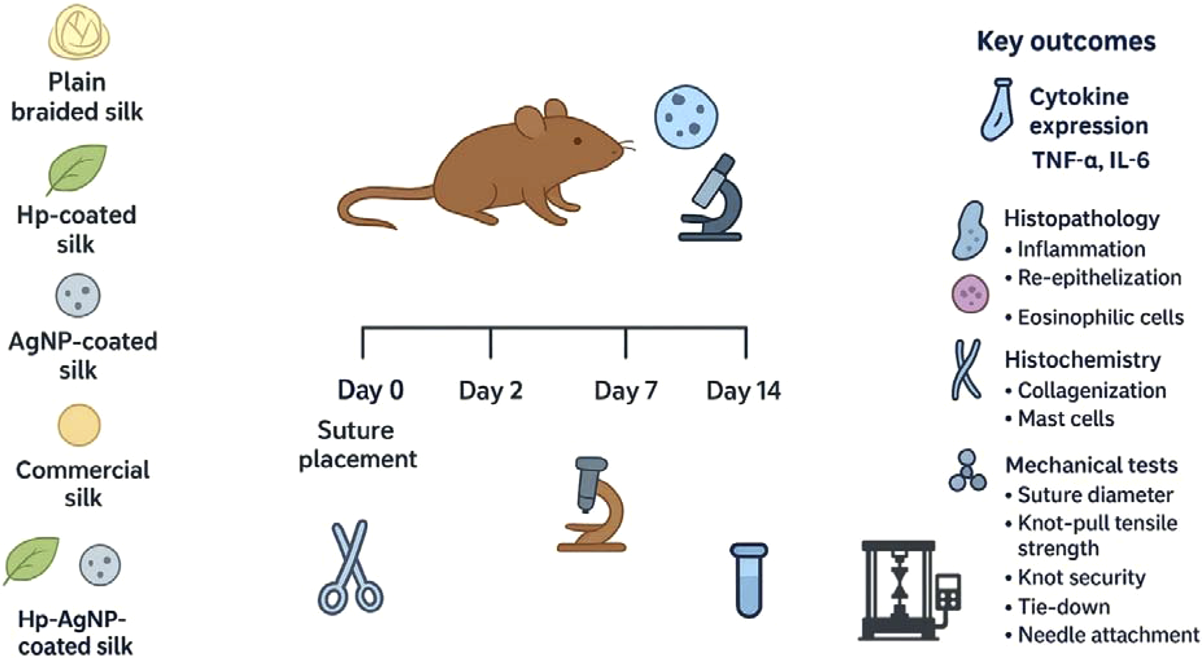
Development of surgical sutures coated with antimicrobial agents is a promising strategy to minimize surgical site infection (SSI) and improve wound healing. The antimicrobial features of Hypericum Perforatum and biogenic silver nanoparticles (AgNPs) have arised an increasing demand for processing surgical sutures. Herein the results of the animal experiments and mechanical tests of a novel antimicrobial silk suture coated with H. perforatum extract (Hp) and biogenic AgNPs (Hp-AgNP) are reported. The study used in vivo histological, histochemical, and immunohistochemical techniques to illustrate the variations in inflammatory response, re-epithelialization, and collagenization of the coated silk sutures in a rat buccal mucosa incision model. Diameter, knot-pull tensile strength, knot security, tie-down, and needle attachment tests were carried out for evaluating the effects of the coating process on mechanical and handling properties. Histopathological and immunohistochemical evaluations revealed progressive healing in all groups, with variations in wound closure, inflammation, and cytokine expression. Hp-AgNP-coated sutures showed significant improvements in re-epithelialization and reduced TNF-α and IL-6 levels over time, highlighting their potential benefits in enhancing wound healing compared to other materials. The coating process had a remarkable effect on the mechanical and handling properties. Coated sutures exhibited higher values than control groups. Suture diameter, knot-pull tensile strength and knot security revealed the highest values for Hp-AgNP-coated suture. The Hp-AgNP coating on the silk suture significantly improves wound healing, mechanical and handling properties. This implies that it has the potential to be a feasible substitute for commercially available silk sutures in surgical interventions. (Scheme 1).
Introduction
Surgical site infection (SSI) remains a major complication occurring after surgical procedures, caused by bacterial contamination at the surgical site.1,2 SSIs result in additional surgical interventions, prolonged length of hospital stays and significant postoperative morbidity and mortality.2,3 Several factors are implicated in the onset of SSIs, ranging from surgical issues to patient-derived factors. 4 Of these factors, surgical sutures are one of the most susceptible materials associated to the occurrence of SSI. 5 Acting as a foreign body, sutures promote bacterial adherence and lead to microbial colonization on the surgical sites. Attachment of bacteria on the suture surfaces follows colonization, resulting in biofilm formation.1,6 Once the biofilm has developed, it shields bacteria from the host immune response as well as systemic and local antibiotic therapy, thus the risk of SSI is higher. 3
Silk suture has been widely used due to its ease of use, excellent physical and handling properties, and ideal knot security. It has been commonly utilized for general soft tissue closure and ligation in dental, ocular, neural, and cardiovascular surgery.5,7,8 However, the braided and multifilamentous nature of silk suture facilitates bacterial attacment, as a consequence may lead to SSI.5,8 To solve this problem silk sutures have been coated with antimicrobial agents including berberine, 9 artemisinin, 10 chitosan, 11 aloe vera, 12 neem oil, 13 ciprofloxacin, 14 levofloxacin, 1 and metallic nanoparticles.15–17
The development of nanotechnology has been shown as a promising strategy to produce new biomedical products with antimicrobial properties. 17 Silver nanoparticles (AgNPs) have been extensively studied in recent years among other metallic nanoparticles.15,16 AgNPs have potent antibacterial and antifungal properties over Gram–positive and Gram-negative bacteria, as well as pathogenic and conditionally pathogenic microorganisms. 18 Studies have evaluated the efficacy of AgNP-based antimicrobial sutures in eradicating various pathogenic microorganisms. Baygar et al. investigated the in vitro antimicrobial characteristics and biocompatibility of silk sutures coated with biogenic AgNPs. 15 In another study Baygar coated silk sutures with biogenic AgNPs and reported enhanced antimicrobial and wound healing effects. 16 Zhang et al. impregnated AgNPs on the surface of absorbable sutures and observed reduced tissue inflammation and improved wound mechanical strength in a mouse model of intestinal anastomosis. 19 The research conducted by Syukri et al. showed that nylon sutures coated with AgNP had remarkable antibacterial and wound healing characteristics. 20
Hypericum perforatum (H. perforatum), also referred to as St. John’s wort, has a long history of being used as a traditional herbal remedy for many ailments such as burns, stomach ulcers, colds, migraines, headaches, diabetes mellitus, and depression. 21 H. perforatum is recognised for its antibacterial and anti-inflammatory properties, as well as its ability to enhance wound healing. 22
Sutures are characterized by measurable traits including mechanical and handling properties. These properties are associated with physical characteristics and can be evaluated in experimental studies. 23 A review of the existing literature reveals that the mechanical and handling properties of suture materials have mostly been studied in the fields general surgery, obstetrics, orthopedics, and veterinary surgery.24,25 Oral and maxillofacial surgery has unique characteristics in comparison with other specialties, due to the presence of saliva, high vascularization, masticatory functions and smaller suture materials and sizes. Hence, it is crucial to examine the mechanical and handling properties of sutures during oral and maxillofacial surgery.
In a previous study we developed a novel suture coated with biogenic AgNPs and H. perforatum extract and evaluated its synergistic in vitro antimicrobial, antioxidative and wound healing effects. 26 H. perforatum extract (Hp) and AgNPs were combined and incorporated directly into the USP size 3.0 plain braided silk sutures by using dip coating process. The antimicrobial activity was evaluated against common oral pathogens (S. aureus, E. faecalis, C. albicans, and S. mutans) using the standard agar plate method. All coated variants demonstrated activity against S. aureus, with Hp-AgNP-coated sutures exhibiting the highest inhibition. Partial antifungal and antibacterial effects were also observed against C. albicans and S. mutans, respectively. Antioxidant activity tests indicated a higher antioxidative potential than positive controls. In vitro wound healing assay demonstrated that the Hp-AgNP-coated sutures had higher wound healing potential in comparison with uncoated silk sutures. 26
In this study we explored in vivo anti-inflammatory and wound healing potential induced by Hp-AgNP-coated suture in Wistar albino rats. Additionally mechanical and handling properties were studied. Hp-coated, AgNP-coated and plain-braided silk sutures were also produced and in vivo tests were carried out. Furthermore we have compared this novel suture with the commercial silk suture, which served as the positive control group.
During antibacterial coating process, optimization of the modifications on braided silk sutures should be considered and these coatings should be compatible with mechanical and handling properties.8,27 Thus mechanical and handling properties including diameter, knot-pull tensile strength, knot security, tie-down, and needle attachment were evaluated and measured. Hp-coated, AgNP-coated, plain braided and commercial silk sutures were also used for mechanical tests and served as a reference for comparisons. To the authors’ best knowledge, there is no published data available on the needle attachment test of coated silk sutures. We believe that Hp-AgNP-coated antimicrobial suture offers a promising strategy for reducing SSIs and promoting wound healing.
Methods
Animal studies
The Local Ethics Committee of Gazi University for Animal Experiments (Code No: G.Ü.ET-18.011) approved all the in vivo experiments. The experiments were performed in accordance with the national guidelines and regulations. A total of 72 male Wistar Albino rats, aged between 10 and 12 weeks and weighing between 250 and 300 gr, were used for the buccal mucosa incision model. The rats were randomly divided into four groups (n = 18) in a random manner. These groups included the AgNP-coated group (rats stitched with AgNP-coated suture), the Hp-coated group (rats stitched with Hp-coated suture), the Hp-AgNP group (rats stitched with Hp-AgNP-coated suture), and the silk group (control group, rats stitched with either plain braided or commercial silk suture [Doğsan AŞ, Trabzon, Türkiye]). The four primary groups were each subdivided into three subgroups, categorised according to the time at which the rats were sacrificed on the 2nd, 7th, and 14th days. The number of experimental subgroups was determined based on similar studies in the literature,28–31 while ensuring adherence to the 3 Rs principles (Replacement, Reduction, Refinement) in animal research.
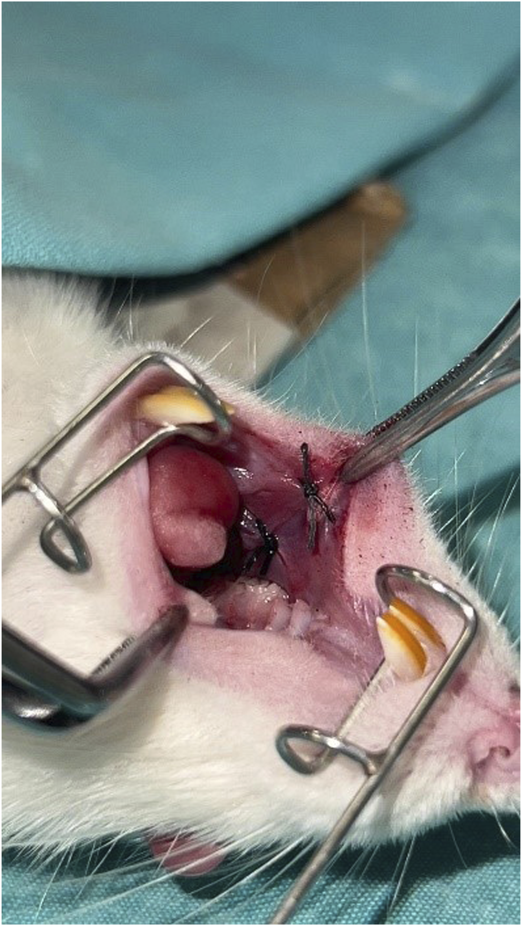
Rats were put under general anaesthesia by injecting 5 mg/kg of xylazine (Alfazyne 2%, EGE-VET) and 45 mg/kg of ketamine HCl (Alfamine 10%, EGE-VET) into their muscles. The surgical procedures were conducted in a sterile surgical environment. A 10 mm incision was made in the right buccal mucosa of each rat, running parallel to the occlusal plane and reaching the muscle tissue (Figure 1). The incision sites were sutured using two sutures that were spaced 5 mm apart (Figure 2). The rats were euthanised on the 2nd, 7th, and 14th days. The mucosal portions that had been treated were removed from the sacrificed subjects, together with the surrounding 2 mm of undamaged soft tissue. These samples were then preserved in a solution of 10% buffered formalin. The buccal mucosa incision. Sutures placed on the incision line.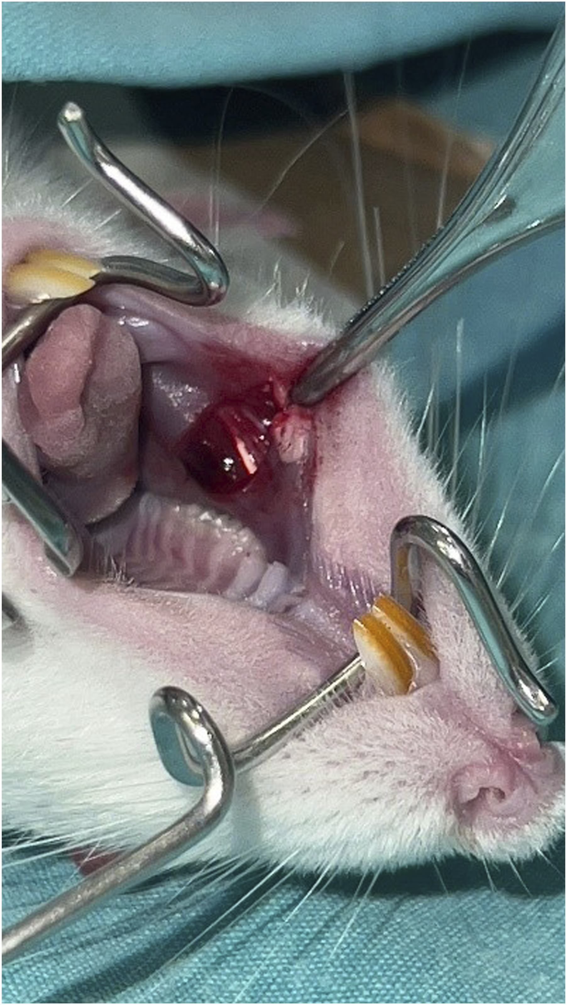
Histological methods
The tissue samples collected following sacrifice were preserved in a 10% neutral phosphate-buffered formalin solution for a period of 24–72 h. After standard tissue processing, slices with a thickness of approximately 4–5 μm were produced from paraffin blocks using adhesive slides (Surgipath, X-tra Adhesive Microslides, Illinois, USA). The sections were stained with conventional hematoxylin-eosin for histological evaluation. They were then analysed under a Leica DM 4000 B light microscope (Leica Microsystems GmbH, Wetzlar, Germany) to assess inflammation, re-epithelialization, and the presence of eosinophilic cells. Histochemically, collagenization was evaluated using Masson Trichrome (with Anilin Blue) and the presence of mast cells was evaluated using Dominichi stainings. The level of inflammation was evaluated using a four-point scoring method, ranging from 0 (no inflammation) to 3 (severe inflammation), in three randomly chosen x200 magnification fields observed under a light microscope. 32 The mast cells and eosinophils were listed individually in the lamina propria and submucosa. This was done in three randomly chosen areas at a magnification of x200. The counts from these fields were then averaged to obtain the final numbers.
Immunohistochemistry
The formalin-fixed paraffin embedded (FFPE) tissue samples were sectioned into 4µ slices and subsequently underwent immunostaining with the usual Streptavidin-Biotin complex procedure. After removing paraffin, the antigen retrieval process was conducted using sodium citrate buffer and TNF-α and IL-6 antibodies. In order to determine specific immunoreactivity, the samples were subjected to treatment with polyclonal antibodies against TNF-α (Biorbyt, rabbit polyclonal, Cambridge, UK) and IL-6 (Biorbyt, mouse polyclonal, Cambridge, UK), and subsequently analysed using immunohistochemistry. The optimal dilution for TNF-α was determined to be 1/50, and IL-6 was diluted at the same ratio of 1/50. Gill’s haematoxylin was used for counterstaining. The positive control for TNF-α was liver tissue, while skin was used as the positive control for IL-6. The examination was conducted using a light microscope and Leica Application Suite Version 4.13.0 (Leica Microsytems Switzerland, CMS GmbH). Scoring was conducted for cells that exhibited positive staining patterns in both the cell membrane and cytoplasm, as anticipated for the antibodies. The immunological scoring was performed and yielded a score of 0:0. 1: Less than 25% - 2: Between 26% and 50%, while the number three exceeds 50%.
Mechanical testing procedures
The testing standards for the mechanical and handling properties assessed in this study were established according to the guidelines outlined in the European Pharmacopoeia 9.0 for surgical sutures.
Suture diameter
The diameter was measured using five sutures. A mechanical instrument capable of measuring with an accuracy of at least 0.002 mm was used. The diameter was measured at intervals of 30 mm over the whole length of the suture. Two measurement were taken at each point; the second measurement was conducted after rotating the suture through 90°. The diameter of that point was calculated as the average of the two measurements.
Knot-pull tensile strength
The knot pull tensile strength test was performed using five sutures of 90 cm length. The minimum breaking load was assessed over a simple knot formed by placing one end of the suture held in the right hand over the other end held in the left hand, passing one end over the suture and through the loop so formed and pulling the knot tight. The test was performed using Instron tensilometer (Instron Corporation, Norwood, MA, USA), which was driven at a constant rate of 300 mm/min. The length of the suture between the clamps was 12.5–20 cm and the knot was placed midway between the clamps. The mobile clamp was set in motion and the force required to break the suture was noted.
Knot security
The knot security test was performed using 10 sutures, each measuring 75 mm in length. The knot security was determined by using an Instron tensilometer. The gauge length was set to 200 mm, and extention rate was set at 250 mm/min. The sutures were tied around a paper cylinder with a diameter of 10 cm using a (2 = 1 = 1) knot. The observation of knot breakage without slipping was recorded as an indication of knot safety.
Tie-down
The tie-down test was performed using 10 sutures of 75 mm in length. The suture was tied around a sponge tube, with a (1 = 1) knot, and placed in the clamps of the Instron tensilometer. The gauge length was 50 mm, and the extention rate was 20 mm/min. Upon analysing the graphical outcomes of the test, the distribution of the knot breaking strength corresponding to USP 3/0 below two-thirds (<0.96 kgf x 2∕3) was recorded as an indicator of appropriate tie-down.
Needle attachment
The Instron tensilometer was utilized for the test. The needle attachment test was conducted using five sutures. The needle and the suture (without knot) were fixed in the clamps. The mobile clamp was set in motion and the force needed to break the suture or to detach it from the needle was noted.
Statistical analysis
Statistical analysis was performed using IBM SPSS Statistics for Windows (Version 26.0; IBM SPSS Statistics, Chicago, Illionis). A 95% confidence interval was selected (p < 0.05). By using Bonferroni correction, p < 0.01 was taken as reference for intragroup comparisons. Frequency (n) and percentage (%) statistics were given for categorical variables, whereas mean ± standard deviation (Mean ± SD), maximum, minimum, and median (Max-Min (M)) statistics were presented for numerical variables. The sample size was determined according to the guidelines provided by the European Pharmacopoeia 9.0 for surgical sutures. The recorded values were given as mean with their SD. The Kruskal Wallis test was employed to compare categorical variable groups in terms of numerical variables. Cochrane Q test was used to compare categorical measurements according to different time intervals. The chi-square test was used to assess the relationship between two categorical variables. Friedman test was used to compare time-dependent measurement values.
Results
Histolopathological evaluations
Histopathological assessment mostly relied on the examination of sections stained with hematoxylin and eosin. Therefore, samples collected on the 2nd, 7th, and 14th days were examined to assess the characteristics of epithelium and connective tissue (Figure 3). Microscopic assessment was conducted by examining wound closure, re-epithelialization, and type of epithelium (para/orthokeratinized). The evaluation also included assessment of the presence of abscess foci and/or necrotic material. The histopathological representation of tissue samples of all groups from day 2,7 and 14 (Hematoxylin & eosin, x40 magnification).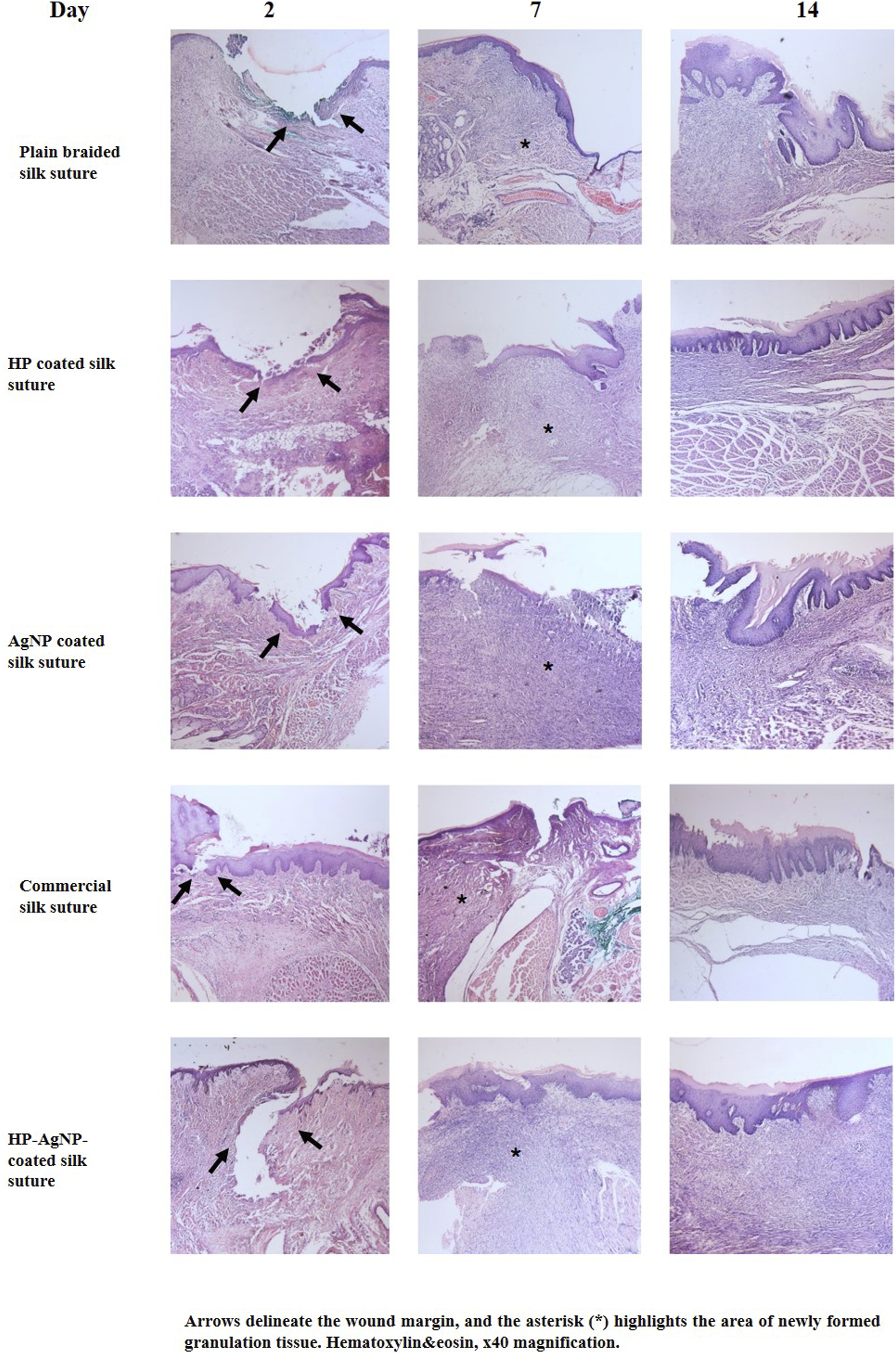
Masson’s trichrome staining was used to assess connective tissue structure (Figure 4). The composition of connective tissue, including collagen, fine fibrils, and fibrovascular elements, was evaluated. The histopathological characteristics of connective tissue healing from tissue samples of the control group (plain braided silk) and the Hp-AgNP-coated group (Masson Trichrome, x40 magnification).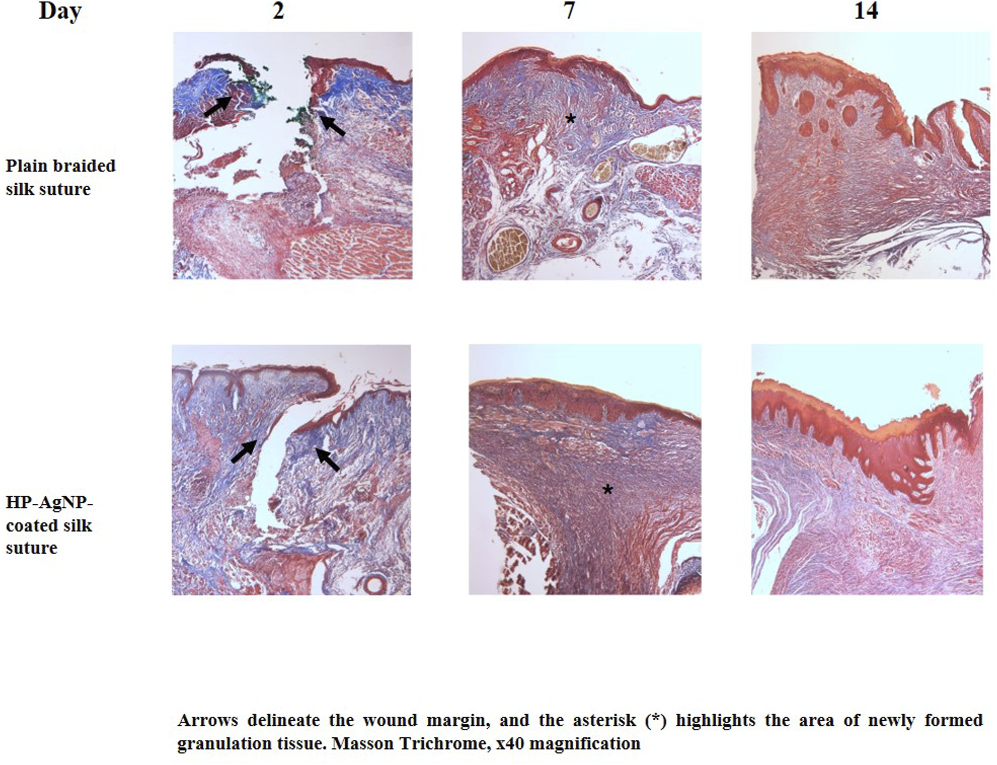
In addition, the assessment of inflammation was based on the severity of inflammation in the connective tissue, the composition of the inflammatory cells (mixed or mononuclear), and the location of the infiltration, whether within the epithelium or deep within the connective tissue. To assess the effect of the sutures on the tissue and level of inflammation in both the lamina propria and submucosa, we used specialized stain (Dominichi) to measure the number of mast cells. Immunohistochemistry was used to assess the proinflammatory cytokines IL-6 and TNF-α.
None of the groups exhibited re-epithelialization or wound closure on the 2nd day. However, on 7th day, the plain braided silk group achieved 100% closure, whereas the AgNP-coated and Hp-AgNP-coated groups achieved closure rates of 83.3% and 100%, respectively. On the 14th day, all groups showed the presence of epithelial lining, only plain braided silk group and Hp-AgNP-coated groups exhibited the development of a fully formed stratified squamous epithelium. The connective tissue was collagenized, similar to the normal healing process. On the 2nd day, a thin fibrillar collagenized structure was observed. On the 7th day, it resembled a granulation tissue. By day 14, the connective tissue had become fibrocellular mature structure in all groups.
Evaluation of mast cells
When analyzing the groups based on mast cell count in the lamina propria, a statistically significant difference was observed on the 2nd day (p < 0.05) (Figure 5(A)). Within the commercial silk group, the number of mast cells reached its peak on the 2nd day, with a count of 15.33. There was no statistically significant difference between the 7th and 14th days (p > 0.05). There was a significant difference in mast cell counts between the plain braided silk and Hp-AgNP-coated groups, based on the passage of time (p < 0.05). The difference in the other groups was not significant (p > 0.05) (Table 1). Representation of mast cells (A- Lamina propria, B- Submucosa, x100 magnification, Dominichi). Evaluation of mast cells in lamina proria and submucosa. *p < 0.05 significant difference; Kruskal Wallis, Friedman test.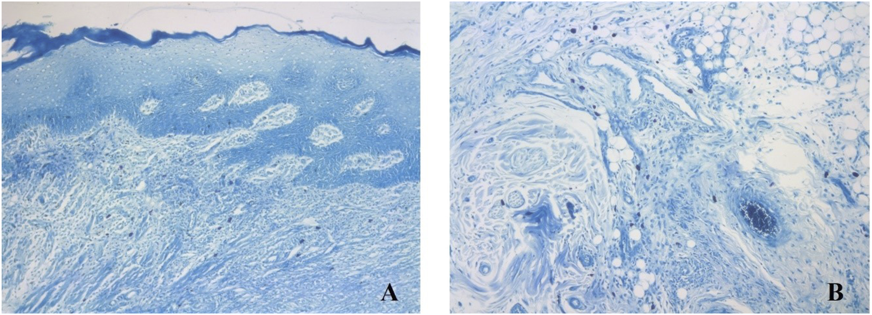
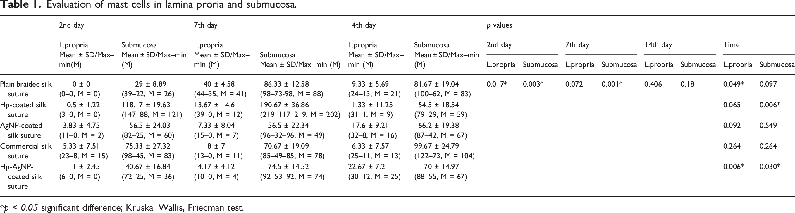
A statistically significant difference was observed between the groups in relation to the number of mast cells in the submucosa from the 2nd to the 7th days (p < 0.05) (Figure 5(B)). In the Hp-coated group, the number of mast cells in the submucosa peaked on the 2nd day (118.17) and 7th day (190.67). There was a statistically significant difference in the number of mast cells between the Hp-coated and AgNP-coated groups, as well as the Hp-AgNP-coated group, on the 2nd day. Additionally, a significant difference was observed between the Hp-coated and AgNP-coated groups on the 7th day (in-group p < 0.010). On the 14th day, there was no statistically significant changes (p > 0.05). There was a significant difference in the number of mast cells in the submucosa between the Hp-coated and Hp-AgNP-coated groups over time (p < 0.05). The difference in the other groups was not significant (p > 0.05) (Table 1).
Evaluation of inflammation and eosinophilic cells
Evaluation of eosinophilic cells in lamina propria and submucosa.
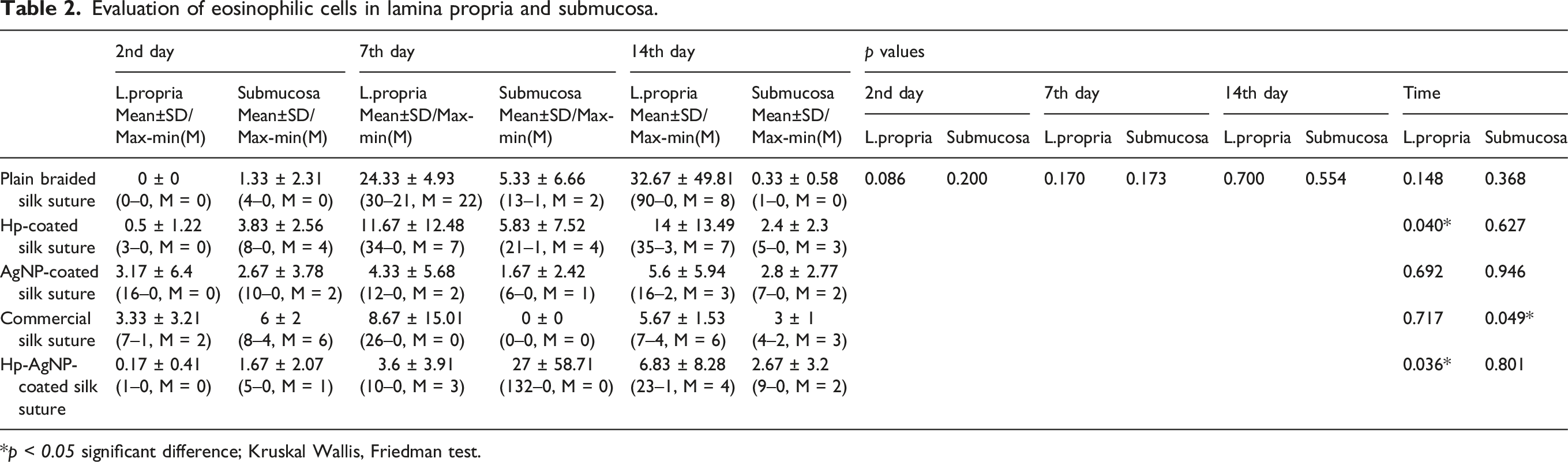
*p < 0.05 significant difference; Kruskal Wallis, Friedman test.
There was no statistically significant difference in the number of eosinophils in the submucosa across groups on the 2nd, 7th, and 14th days (p > 0.05). In the commercial silk group, there was a statistically significant change in the number of eosinophils in the submucosa with time (p < 0.05). The difference in the other categories was not significant (p > 0.05) (Table 2).
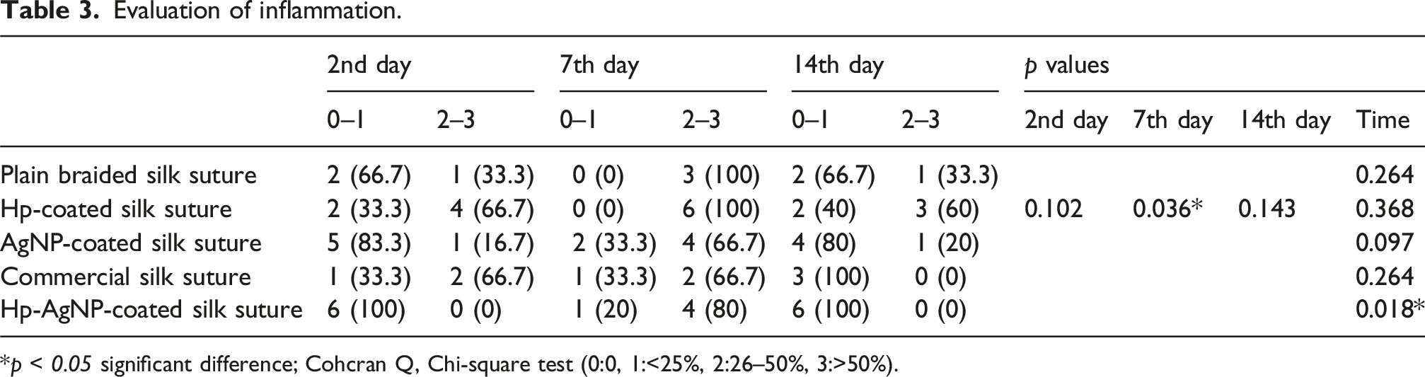
On the 7th day, there was a significant difference in the inflammation score between the groups (p < 0.05) (Figure 6 (A)–(B)). The inflammation score was the highest in the plain braided silk (100.0%) and Hp-coated (100.0%) groups. The difference between the 2nd and 14th day was not statistically significant (p > 0.05). The Hp-AgNP-coated group exhibited a significant change in the inflammation score over time (p < 0.05). The differences were not significant among the remaining groups (p > 0.05) (Table 3). Respresentive images of inflammation in A-Lamina propria, B-Submucosa, (Hematoxylin & eosin, x100 magnification). Evaluation of inflammation. *p < 0.05 significant difference; Cohcran Q, Chi-square test (0:0, 1:<25%, 2:26–50%, 3:>50%).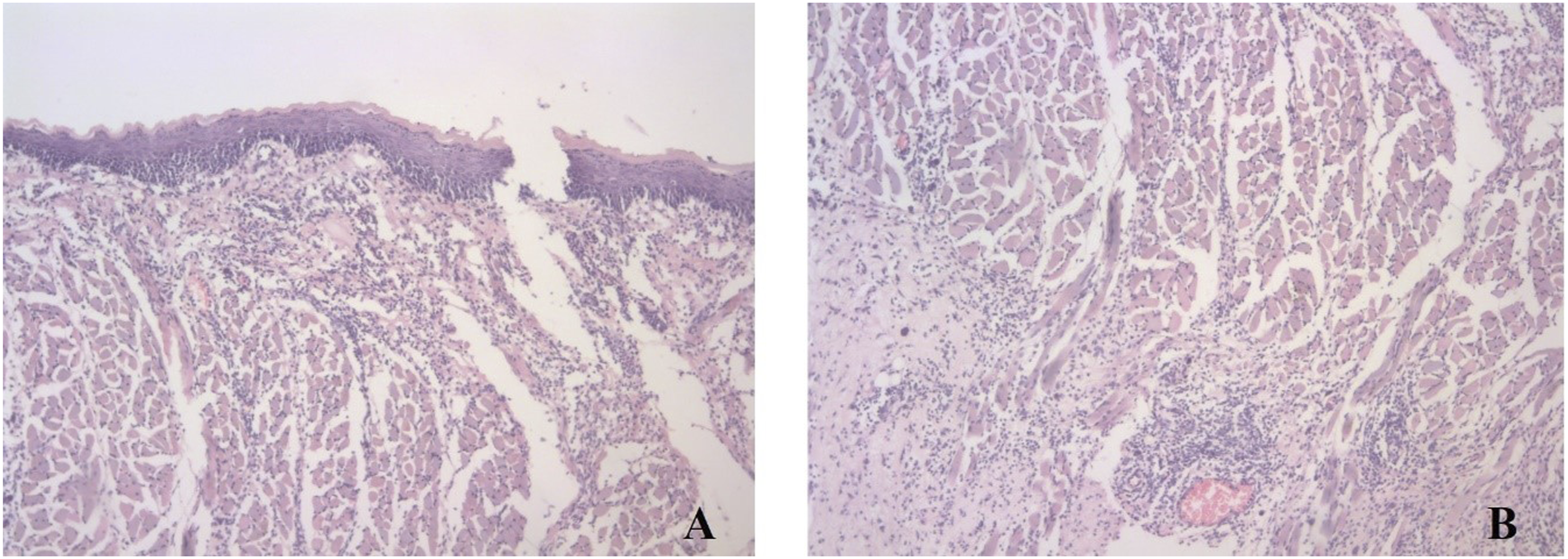
The connective tissue was characterized by the formation of fine fibrillar collagen structures on 2nd day, granulation tissue on 7th day and fibrocellular structures on 14th day in all groups, similar to the normal healing process. None of the groups showed re-epithelialization or wound closure on the 2nd day. However, on the 7th day, 100% of the blain-braided silk group and 83.3% of the AgNP-coated and Hp-AgNP-coated groups showed re-epithelialization and wound closure. On the 14th day, all wound sites had fully developed stratified squamous epithelium, except for the silk and Hp-AgNP-coated groups.
Immunohistochemical evaluations
TNF- α expression
Evaluation of TNF- α expression in lamina propria and submucosa.
*p < 0.05 significant difference; Cohcran Q, Chi-square test (0:0, 1:<25%, 2:26–50%, 3:>50%).
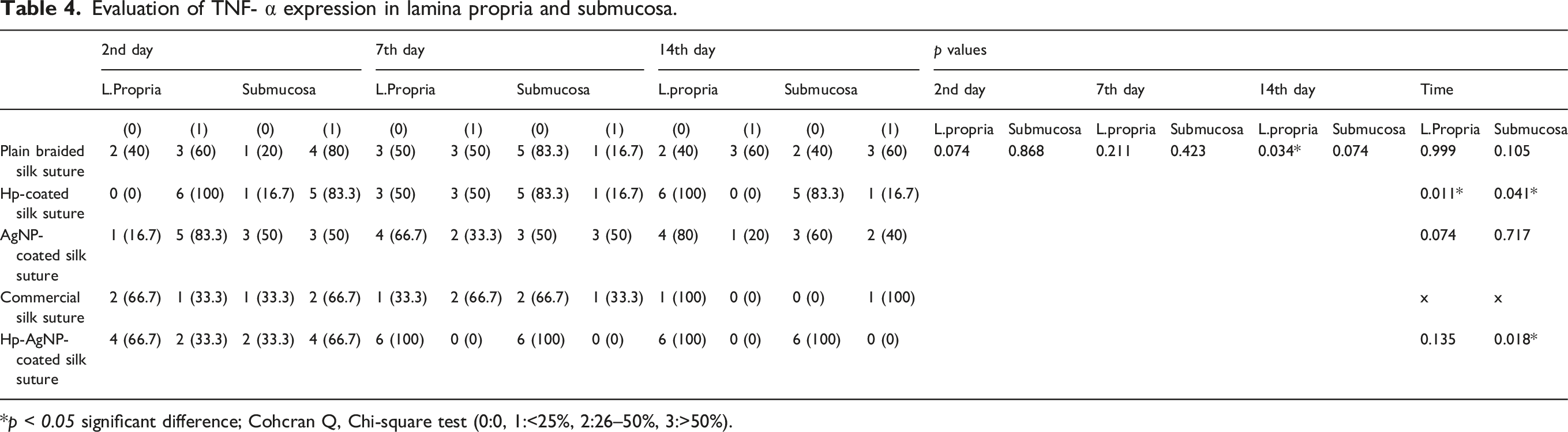
The submucosa was assessed for TNF-α scores, and the highest levels were found in the Hp-coated group (83.3%) on the 2nd day, the AgNP-coated group (50%) on the 7th day, and the commercial silk group (100%) on the 14th day. Nevertheless, there was no statistically significant difference between the groups (p > 0.05). When comparing the groups based on time, there was a significant difference in the submucosa total score between the Hp-coated and Hp-AgNP-coated groups (p < 0.05). These groups exhibited a notable decline in TNF-α expression over time (Table 4) (Figure 7). Demonstration of TNF-α expression in Hp-AgNP-coated group. Arrows indicate immunopositive inflammatory cells (x400 magnification, DAB).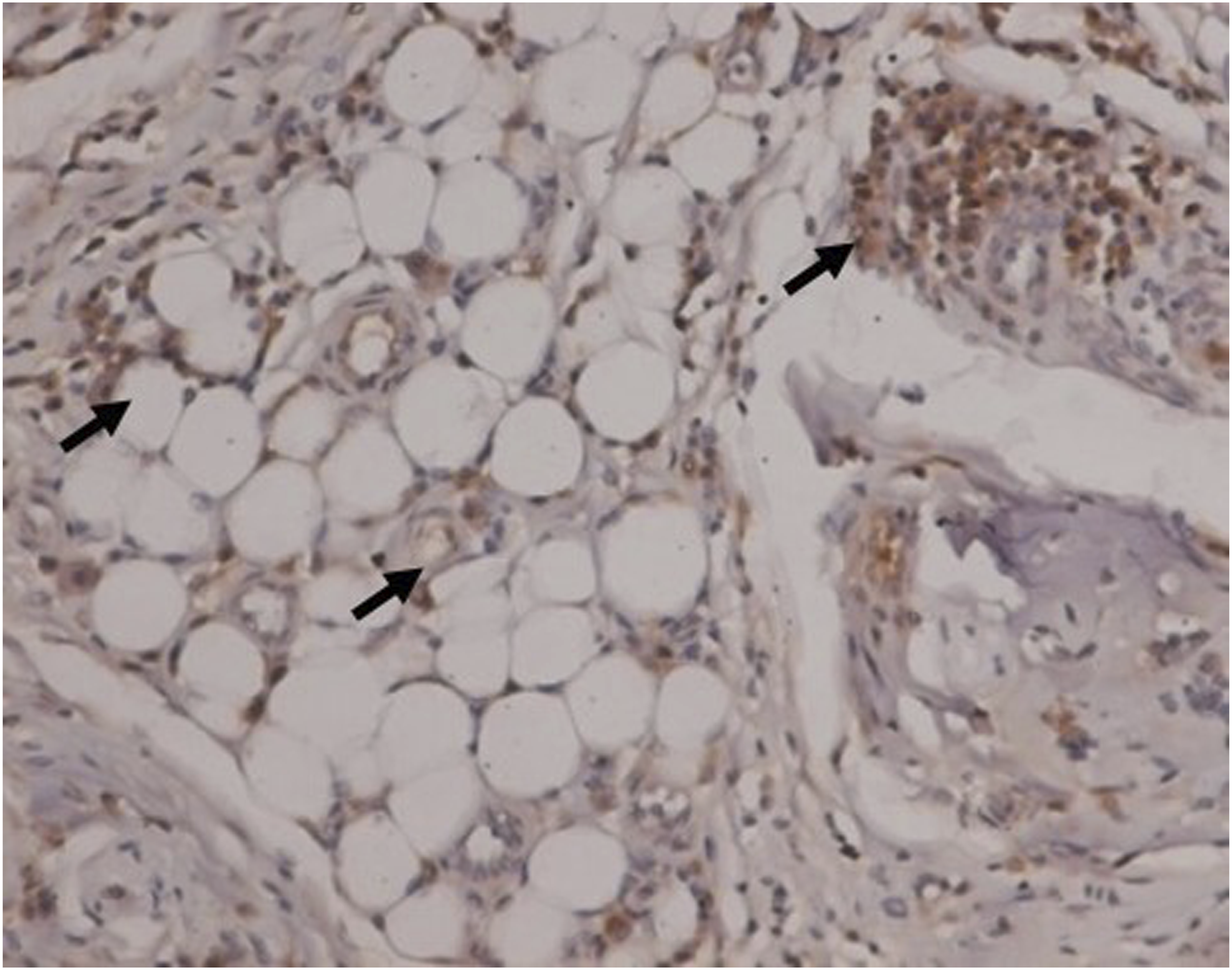
IL-6 expression
Evaluation of IL-6 expression in lamina propria and submucosa.
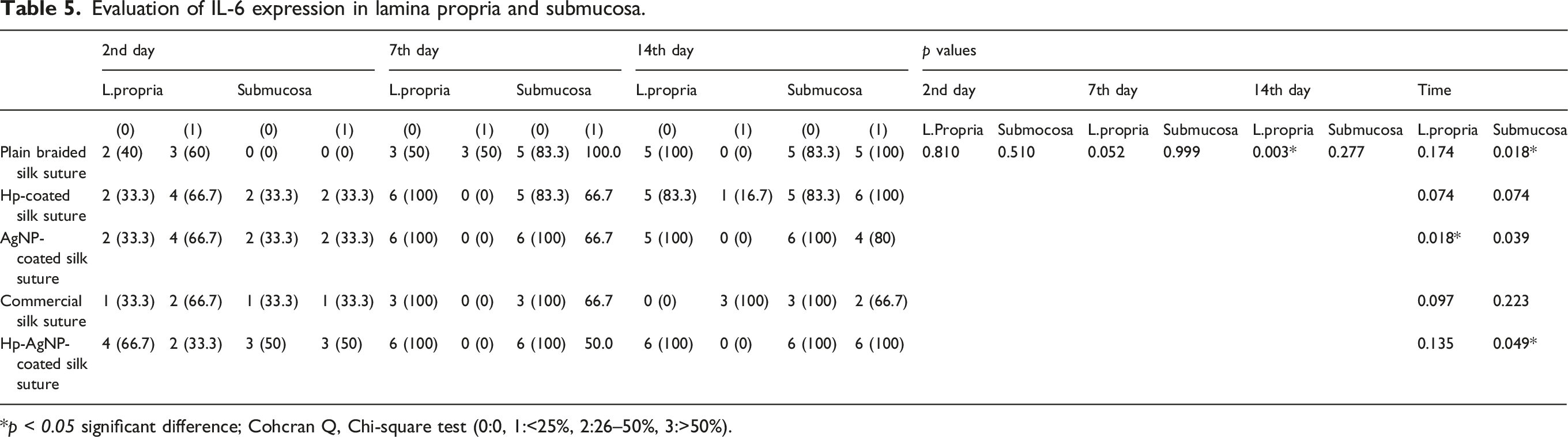
*p < 0.05 significant difference; Cohcran Q, Chi-square test (0:0, 1:<25%, 2:26–50%, 3:>50%).
When evaluating IL-6 expression in the submucosa, the Hp-AgNP-coated group (50%) exhibited the highest levels on the 2nd day. No statistically significant differences were identified between the groups. On the 7th day, the plain braided silk (100%) exhibited the highest values, and there was no statistically significant difference. On the 14th day, the Hp-coated (100%) and Hp-AgNP-coated (100%) groups showed the highest values, with no statistically significant difference between them. When comparing the groups based on time, there was a significant difference in the total submucosa score levels between the plain braided silk and Hp-AgNP-coated groups (p < 0.05) (Table 5) (Figure 8). Demonstration of IL-6 expression in the Hp-AgNP-coated group. Arrows indicate immunopositive inflammatory cells (x400 magnification, DAB).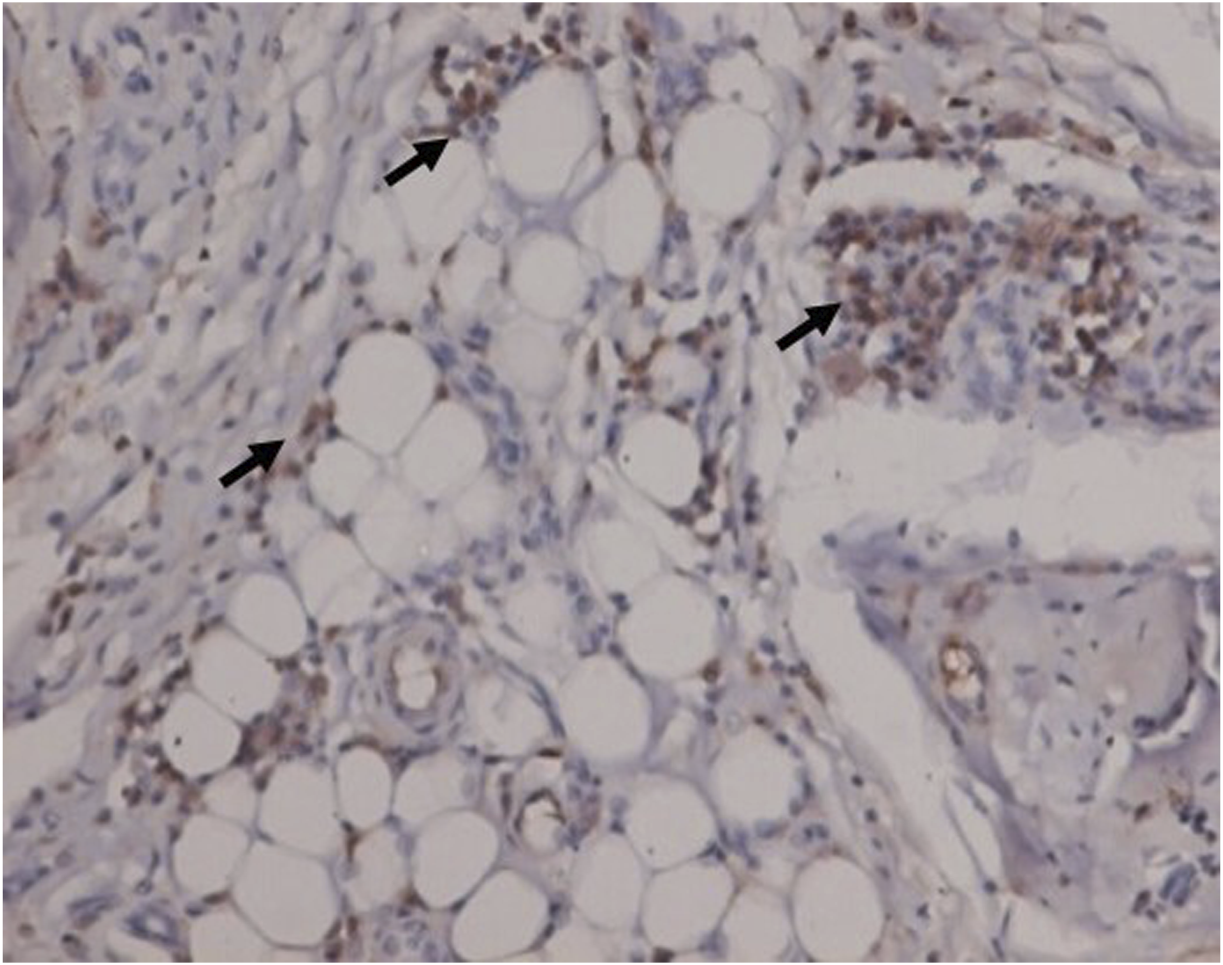
Mechanical tests
Mechanical and handling properties including diameter, knot-pull tensile strength, knot security, tie-down and needle attachment were evaluated. In the diameter test, the diameters of the coated sutures were measured following coating process. The average diameter of each suture was calculated and expressed in mm. Maximum load and elongation were recorded continuously using a specialized software for the knot-pull tensile strength, knot security, tie-down and needle attachment tests. Maximum load (kgf) and elongation (expressed in mm and %) at the time of failure were analyzed. Elongation was calculated as the displacement that a suture can withstand before breaking in tensile testing. The maximum load was noted as the maximum tension the material can experience without breakage. 33
Suture diameter
Maximum, minimum, mean and standard deviation values in the diameter test.
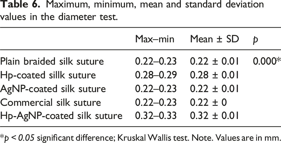
*p < 0.05 significant difference; Kruskal Wallis test. Note. Values are in mm.
Knot-pull tensile strength
Maximum, minimum, mean and standard deviation values for maximum load and elongation in the knot-pull tensile strength test.
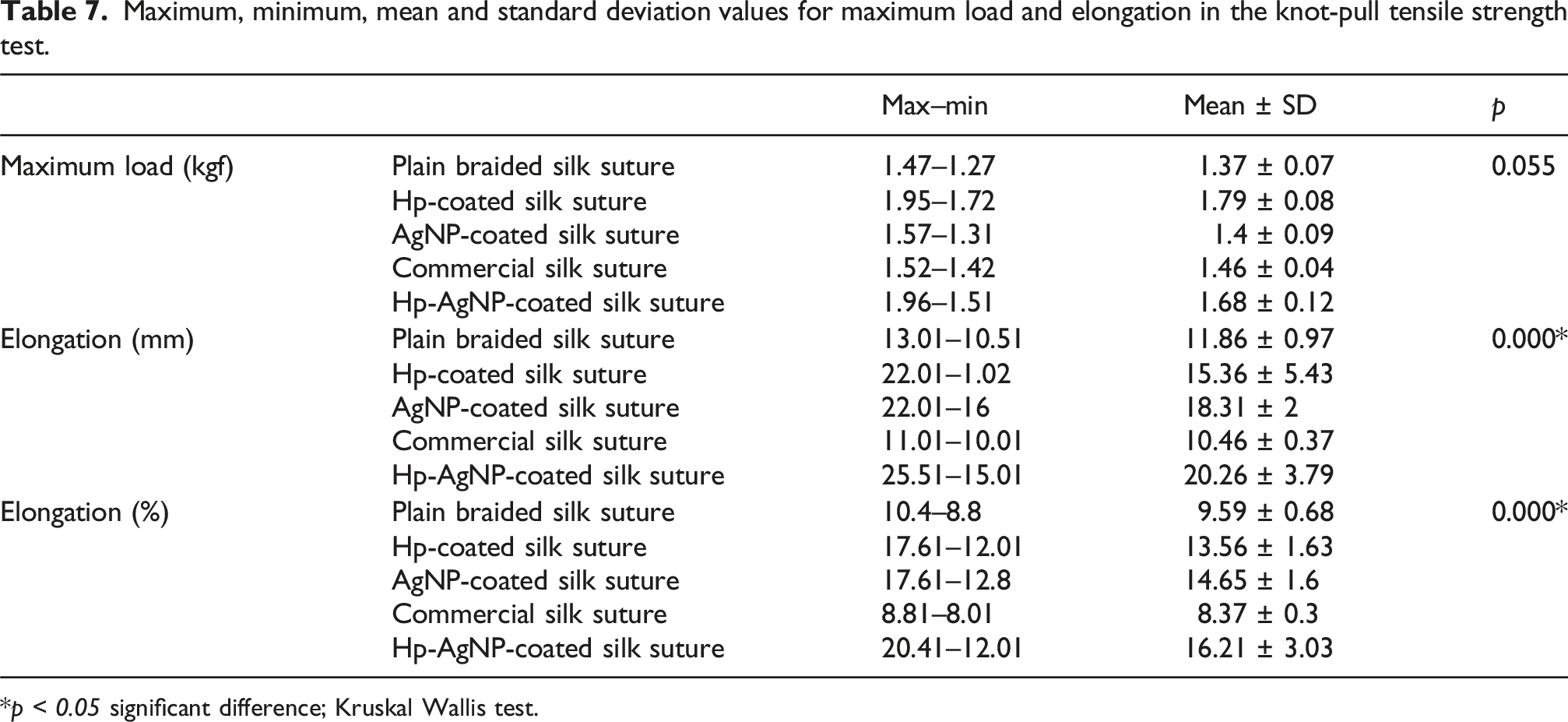
*p < 0.05 significant difference; Kruskal Wallis test.
The analysis of elongation revealed that the Hp-AgNP-coated sutures exhibited the highest values. The lowest values were observed in commercial silk sutures. The results were statistically significant (p < 0.05).
Knot security
Maximum, minimum, mean and standard deviation values for maximum load and elongation in the knot security test.

*p < 0.05 significant difference; Kruskal Wallis test.
Tie-down
Maximum, minimum, mean and standard deviation values for maximum load and elongation in the tie-down test.
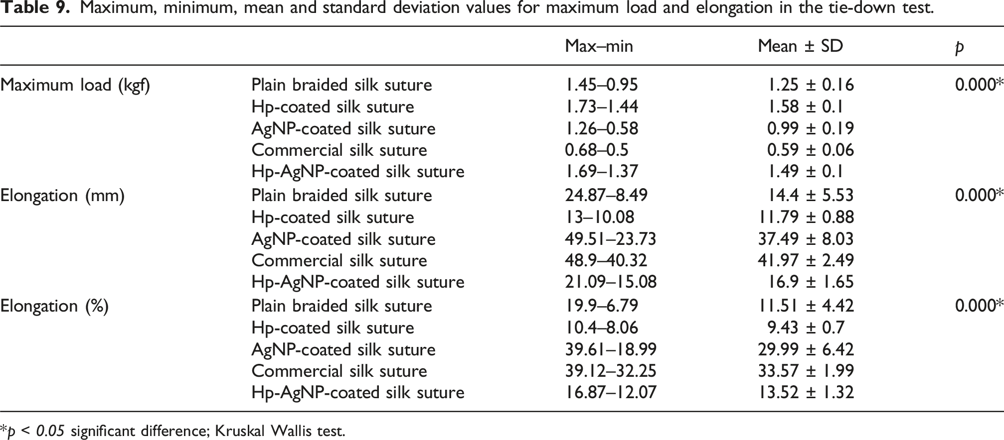
*p < 0.05 significant difference; Kruskal Wallis test.
Needle attachment
Maximum, minimum, mean and standard deviation values for maximum load and elongation in the needle attachment test.

*p < 0.05 significant difference; Kruskal Wallis test.
Discussion
In contrast to other areas of the body, the oral region is exposed to several factors that might affect the effectiveness and contamination of the suture materials. 35 Wound healing is a controlled process that involves certain cell types and signalling chemicals that repair damaged tissues. The wound healing process consists of four partly overlapping phases: haemostasis, inflammation, proliferation, and remodelling, regardless of the kind of wounded tissue. 36 When the wound is primarily closed, these activities accelerate; however, variations in processes and cell types can be observed depending on the type of suture used. 37
Research conducted in pigs has shown that mast cells emerge in the wound approximately 14 days after damage and eventually decrease in oral wounds. As a consequence, there was a significant decrease in the number of mast cells present in the area of the oral wound 60 days after the injury. 38 The commercial silk and Hp-coated groups exhibited the highest number of mast cells. These materials are believed to be associated with the presence of highly active mast cells in both the lamina propria and submucosa on the 2nd and 7th days. The increased number of mast cells indicated that the substances utilized caused notable allergic reactions in the tissues. However, in the Hp-AgNP-coated group, there was a significant decrease in the number of mast cells over time.
Eosinophils are the primary cells involved in type I hypersensitivity reactions and are seldom seen in normal oral wound healing processes, as shown by histological examination. 39 Eosinophilic cells are only identifiable in the tissue after an allergic reaction is initiated. The Hp-coated group showed a notable presence of eosinophilic cells, which corresponded to the infiltration of mast cells.
In this study, inflammation in the oral mucosa peaked on day 7, which is consistent with previous reports indicating that inflammatory activity typically subsides around the sixth day of the healing process. 40 Both TNF-α and IL-6 were markedly elevated during the early phase and subsequently declined over time, underscoring their critical roles in initiating inflammation and orchestrating the transition to tissue repair. Elevated levels of these cytokines at the lesion site, however, may prolong the inflammatory phase and thereby delay wound healing.41,42 Thus, maintaining TNF-α and IL-6 within an appropriate range appears essential for efficient mucosal regeneration.
Our findings also align with previous work by Liu et al., who demonstrated that AgNP-coated sutures reduced macrophage infiltration and lowered cytokine production compared to control sutures in a murine intestinal anastomosis model. 43 In the present study, plain braided silk and Hp-coated groups exhibited higher inflammatory scores, in agreement with the known irritative potential of silk sutures. Nevertheless, complete epithelialization by day 14 in these groups suggests that, despite their irritant properties, silk sutures may still support wound closure. By contrast, AgNP-coated sutures were associated with a progressive reduction in TNF-α and IL-6 expression, indicating their capacity to modulate the inflammatory response and improve tissue compatibility. Taken together, these results suggest that suture type exerts a significant influence on cytokine dynamics and inflammatory scores. It is noteworthy that AgNP-coated sutures may provide a promising alternative in oral surgery, as they appear to promote wound healing while minimizing excessive inflammation. After analyzing the inflammatory scores in the present study, it was determined that the plain braided silk and Hp-coated groups had the most pronounced inflammation scores. The plain braided silk material is known to induce irritation of the oral mucosa and is unsuitable for oral tissues. However, it is often utilized in oral surgery because of its cost-effectiveness, easy accessibility, and stabilizing characteristics. 44 The achievement of complete epithelialization by day 14 in the silk groups in our study serves as evidence for the advantageous effects of plain braided and commercial silk sutures in the process of wound healing.
H. Perforatum is thought to possess notable anti-inflammatory properties. These effects are partially linked to its capacity to stimulate the production of inducible nitric oxide synthase (iNOS) and COX-2. 45 In a study assessing the impact of H. perforatum on wound healing in rats, a notable enhancement in the rate of healing was reported in rats treated with H. perforatum in comparison to those treated with mupirocin and vaseline. 46 H. perforatum contains several constituents, such as naphthobacrons, flouroglusinol, flavonoids, bioflavonoids, and phenylpropanoids. 47 These components exhibit antifungal, anti-inflammatory, and antiviral properties that assist in the process of wound healing. Scientific research indicates that H. perforatum has the ability to accelerate the growth phase, stimulate collagen formation, and enhance the migration of fibroblasts to the injured area. 48 Furthermore, the long-lasting reduction in inflammation resulting from the application of sutures coated with AgNPs in the later stage was also confirmed by the suppression of inflammatory cytokines. The finding of substantial inflammation, together with increased levels of eosinophils and mast cells, caused by the use of Hp-coated suture, indicates that H. perforatum alone is not suitable for use on the oral mucosa. However, the combination of H. perforatum and AgNP has shown noteworthy results in terms of tissue regeneration. Over time, there was a decrease in the numbers of mast cells and eosinophils, while inflammation scores followed the expected progression in the typical healing process. Upon comparing this work with prior studies, we put forth two potential reasons for the prolonged anti-inflammatory effectiveness of AgNP and Hp-AgNP-coated sutures. Initially, the release of silver from the AgNP-coated suture is limited to the early stage of the healing process. However, this release significantly decreases local inflammation, therefore creating a more favorable local environment for tissue repair and regeneration. Another potential explanation is that the AgNP-coated suture consistently releases silver, which exerts a potent anti-inflammatory impact on the nearby mucosal tissue. Similarly, a study conducted on mice demonstrated that sutures coated with AgNP had a more potent and enduring anti-inflammatory effect while also enhancing tissue regeneration. 43 Our findings provide experimental support for these proposed mechanisms. Consistent with the controlled release profile of AgNPs from coated sutures, our immunohistochemical analyses revealed a time-dependent decline in TNF-α and IL-6 expression in both AgNP and Hp-AgNP-coated groups. This cytokine modulation was paralleled by lower inflammatory scores and more advanced epithelial and connective tissue regeneration compared with plain silk or commercial sutures. By day 14, AgNP-containing groups exhibited well-organized fibrocellular connective tissue and complete epithelial coverage, whereas persistent inflammation and delayed epithelial maturation remained evident in certain control groups. Collectively, these findings indicate that the sustained release of AgNPs dampens the early pro-inflammatory response while promoting tissue repair and remodeling, thereby establishing a mechanistic link between controlled AgNP delivery, cytokine regulation, and improved wound healing. Building on this evidence, the combination of AgNPs with H. perforatum appears to further enhance these beneficial effects through synergistic mechanisms, leading to improved wound healing and inflammation modulation. While H. perforatum accelerates healing through multiple mechanisms—such as enhancing fibroblast activity, collagen deposition, and immune modulation 22 —AgNPs promote tissue repair by reducing inflammation and fostering cell migration. 49 Together, these agents complement each other’s actions, leading to more effective tissue regeneration and faster wound closure.
Antimicrobial sutures incorporating antiseptic agents such as triclosan and chlorhexidine have been developed and are currently available for both medical and veterinary applications. The first such suture to gain clinical approval from the United States Food and Drug Administration (US FDA) was a braided polyglactin 910 coated with triclosan (Vicryl Plus, Ethicon Inc., Somerville, NJ, USA). Subsequently, additional triclosan-impregnated suture formulations have been commercialized, including monofilament poliglecaprone (Monocryl Plus, Ethicon Inc., Somerville, NJ, USA), monofilament polydioxanone (PDS Plus, Ethicon Inc., Somerville, NJ, USA), and multifilament polyglactin 910 (Petcryl Plus, Ethicon Inc., Somerville, NJ, USA). Chlorhexidine-coated sutures have also been developed, primarily for veterinary indications, offering alternative antiseptic profiles for infection control at the surgical site. 50
Previous studies evaluating the biological effects of triclosan-coated sutures reported variable outcomes. Gomez-Alonso et al. demonstrated that triclosan-coated sutures exhibited strong antibacterial efficacy both in vitro and in vivo, significantly reducing bacterial load and inflammatory markers without impairing wound healing mediators. 51 Similarly, our results showed that enhanced re-epithelialization and decreased TNF-α and IL-6 expression over time in the Hp-AgNP-coated suture group, indicating improved inflammatory modulation and wound healing. These findings suggest that the incorporation of antimicrobial agents, such as triclosan or Hp-AgNPs, can support infection control without impairing the physiological healing process. In contrast Storch et al. found no significant differences in wound strength or histopathological parameters between triclosan-coated and non-coated sutures in a guinea pig model, suggesting that the coating does not adversely affect wound integrity. 52 In addition, the study by Gartti-Jardim et al., which assessed various absorbable sutures, reported that poliglecaprone 25 elicited the least inflammatory response, while triclosan-coated polyglactin 910 exhibited moderate tissue compatibility. 53
The experimental conditions in this study were standardized based on the guidelines outlined in the European Pharmacopoeia 9.0 for surgical sutures. The testing requirements were executed according to these specified conditions.
The suture diameter is quantified in millimetres and denoted by a zero. The magnitude of the number preceding the zero is inversely correlated with the diameter. The measure of the diameter is indicated by USP size, which is associated with the type of the suture material being used. 54
The selection of suture size or diameter is a crucial consideration for the surgical area to be used. Increased suture diameter correlates with greater tensile strength. 55 The diameter of the silk sutures was greatly influenced by the application of a coating in the present study. The coating process resulted in an increase in the diameter of coated silk sutures as compared to those that were not coated. During examination of the antimicrobial-coated silk suture, it was seen that the measured parameters exceeded the standard values established by European Pharmacopoeia 9.0 (USP size 3) for both Hp-AgNP-coated and Hp-coated silk sutures.
Knot security refers to the ability of a knot to withstand slippage and breaking under a continuous strain, and it is crucial for preserving tissue integrity. The knot is the weakest component during suturing. Knots in a tied suture can lead to failure, therefore impacting the outcome of surgery.8,25
Knot tying with suture material plays a critical role in ensuring proper tissue approximation in the majority of surgical interventions. 56 Knot security refers to the ability of a suture to maintain its integrity without breaking or slipping. 57 It is essential for holding tissues together during the healing process. Previous studies have identified the knot as the weakest point of the suture loop, particularly at the junction between the loop and the initial throw of the knot. 56 Failure of a knot can lead to wound dehiscence, potentially compromising the outcome of a surgical procedure. 24 Knot security determines the strength of a knot and affected by a number of factors such as the type of the suture, the number of throws and knot configuration.56,58,59 A large variety of possible knots have been reported by authors.25,33,58 In the present study 2 = 2 = 1 knot was selected.
Previous studies have evaluated knot security based on suture material.25,56,60 Muffly et al., have reported no difference, 60 while Marturello et al. have demonstrated that the choice of suture material have an impact on knot security. 56 The present study has shown that the use of antimicrobial coatings has a substantial impact on the knot security. The Hp-AgNP-coated suture exhibited statistically significant differences compared to other coated and uncoated sutures. The Hp-AgNP-coated suture exhibited the highest values for maximum load and elongation, followed by the Hp-coated, commercial silk, AgNP-coated and plain braided silk sutures respectively.
Tensile strength is a critical indicator of performance both during and after surgical procedures. It is defined as the measured force that the suture withstands before breakage. 61 The tensile strength of sutures is a crucial factor for the practitioners while making a knot. If the suture materal is too weak and the applied force for knotting is higher than tensile strength of the suture material, the suture may easily break. Hence, it is important to determine the tensile strength of the coated sutures.62,63 The suture with improved tensile strength exhibits a better clinical performance for keeping the wound sides closed and provide a stronger support during the wound healing process.64,65 In the present study coated sutures showed improvement in knot-pull tensile strength in comparison with uncoated sutures, and exhibited higher values for maximum load and elongation for the Hp-coated and Hp-AgNP-coated sutures respectively. In a study by Dhas et al., 66 silk fibers impregnated with AgNPs showed higher breaking strength due to absorption of AgNPs onto silk fibers. Based on the data analysis of the tensile properties of the sutures in the the present study, it is reasonable to conclude that the Hp and AgNP coatings likely improved the mechanical strength of the sutures, thus affecting the maximum load and elongation, compared to commercial and plain braided silk sutures.
The handling properties of suture materials represent tie-down and address the ease of sliding the knot for controlling tension. Handling is directly correlated with mechanical properties including coefficient of friction and and elasticity.27,67 A suture that has low friction may have a smooth feeling, while greater friction may increase the knot security. Several factors can influence tie-down such as suture material and size, tie-down technique, quality of tissue, surgeon’s training and the use of instruments. 27 Generally a suture with good knot security exhibits a high tie-down resistance. Silk suture possesses excellent knot tying properties, with requiring very few knots for knot security and having a low tie-down resistance. 67
The coated sutures were evaluated for handling by means of tie-down test. The commercial silk suture was found to have the optimal tie-down value, as per the specifications set by the European Pharmacopoeia 9.0. However, when comparing the coated sutures to each other, the AgNP-coated suture had the lowest tie-down value.
The present study additionally provides the needle attachment test outcomes of coated sutures, in addition to commercial and plain braided silk sutures. As far as we know, this is the first study comparing the needle attachment data of the coated sutures to their uncoated ones. The findings of the needle attachment test showed that the AgNP-coated sutures had higher values for maximum load and elongation.
Elongation is an indicator for the endurance of the surgical sutures to strain, and determines the capacity for handling without failure. 68 Mechanical stresses applied to suture materials can induce irreversible elongation, resulting in the material maintaining its original length despite changes in tissue volume such as swelling at the suture site. Once the swelling subsides, sutures may induce deformation of surrounding tissues, or some plastic sutures may be deformed by themseves, with minimal tissue damage during the swelling phase, while becoming excessively wide to fully approximate the wound edges. 69 Hence increased elongation might be advantageous in clinical scenarios where there is an anticipation of significant postoperative oedema. 34 Suture materials possessing high elongation capabilities can expand to compensate oedema. 69 Indeed elongation enhances the handling properties of suture materials by providing the surgeon with a better feeling and improves the ability to predict the breakpoint of the suture.24,70 The findings indicated that coating of the silk suture did not have a negative impact on its mechanical propeties but improved its elongation. The analysis of elongation revealed that the Hp-AgNP-coated sutures exhibited the highest values. The lowest values were observed in commercial silk suture.
The impact of triclosan coating on the mechanical and functional properties of surgical sutures has been shown to vary. Jungwirth-Weinberger et al. evaluated the mechanical properties of triclosan-coated Vicryl 1 Plus, PDS 0 Plus, and Monocryl 3-0 Plus over a 42-day period. Mechanical parameters—including load to failure, strain, and stiffness—were assessed using a materials testing system. 71 While Vicryl 1 Plus demonstrated significantly reduced load to failure on days 14 and 28, PDS 0 Plus showed superior mechanical strength compared to its non-coated counterpart on day 28. Similarly, Ford et al. reported comparable intraoperative handling characteristics-including tissue passage, first-throw knot holding, knot tie-down smoothness, knot security, surgical handling, surgical hand, memory, and suture fraying— between triclosan-coated and non-coated polyglactin 910 sutures in pediatric surgical patients. 23 Although both groups were rated similarly across parameters, the triclosan-coated group received a higher proportion of “excellent” ratings. Conversely, Storch et al. compared triclosan-coated and non-coated polyglactin 910 sutures with respect to physical and functional parameters such as ease of passage through tissue, first-throw knot holding, knot-tie down smoothness, knot security, surgical handling, and overall evaluation. 27 No significant differences were observed between groups, and breaking strength retention showed a similar decline in both, from 79% at day 14 to 5% at day 35.
In line with these findings, our study showed that sutures coated with Hp-AgNP exhibited improved mechanical strength and handling performance relative to uncoated, Hp-coated and AgNP-coated sutures. These experimental results demonstrate that, similar to triclosan, Hp-AgNP coating can be integrated into suture materials without compromising their essential mechanical and functional properties, while potentially offering added antimicrobial and anti-inflammatory benefits.
Conclusion
Suture associated infections pose a major challenge in surgery. Following attachment to suture surfaces the microorganisms proliferate and generate biofilms, leading to chronic infections. Antimicrobial coated sutures have gained interest as a potential solution to address this issue. 7 The utilisation of metallic nanoparticles and traditional wound healer plants as antimicrobial coatings is a potential approach for preventing this process. 72
The results of our in vivo investigations showed that the Hp-AgNP-coated sutures had lower levels of cytokine production relative to both the coated and uncoated groups. The quantities of mast and eosinophil cells were specifically seen in the Hp-coated group. Therefore, it was demonstrated that the combination of H. perforatum and AgNP has a notable anti-inflammatory effect. Upon evaluating the results of the mechanical tests, it was concluded that H. perforatum and AgNP had a favourable impact on the mechanical and handling properties of the silk suture, resulting in an increase in its strength. By means of coating process it was possible to provide appropriate mechanical and handling properties of the novel antimicrobial-coated silk suture.
The study’s findings indicate that the antimicrobial Hp-AgNP coating on the silk suture is successful in promoting wound healing and improving mechanical and handling capabilities. This suggests that it could be a viable alternative to commercial silk sutures in surgical procedures. One limitation of the present study was the absence of an environment that accurately replicates oral conditions, including soft tissue resistance, gingival crevicular fluid, the presence of blood, food or liquids, oral hygiene practices, and functional oral movements, all of which may influence the fundamental mechanical properties of the sutures. Additionally, it was not possible to evaluate postoperative factors such as swelling and oedema, which could impose additional mechanical stress on the knots. Another limitation of this study was the lack of further biocompatibility and cytotoxicity testing. Further studies are necessary to validate the clinical safety of the sutures under in vivo conditions. Among future directions, priority should be given to assessing long-term biocompatibility and the potential for microbial resistance development. Moreover, the scalability and feasibilty of the coating technology must be rigorously examined to facilitate clinical translation and large-scale manufacturing. Future investigations should also explore the application of this novel coating on various suture materials. Advancing the development of the final suture product will require comprehensive evaluation and standardization of the coating process, stability testing, detailed analysis of drug release kinetics as well as broader biocompatibility and cytotoxicity assessments. Ultimately, the clinical applicability of this technology should be validated through well-designed, large-scale clinical trials.
Footnotes
Acknowledgments
This research have been presented as posters at 29th International Scientific Congress of Turkish Association of Oral and Maxillofacial Surgery (TAOMS), which was held on 06–10 November 2022 in Antalya, Turkey.
Ethics approval
All the in vivo experiments were approved by Local Ethics Committee of Gazi University for Animal Experiments (Code No: G.Ü.ET-18.011).
Author contributions
Conceptualization: Y Kılınç, İR Karaca, A Uğur, SE Gültekin
Data curation: Y Kılınç, -İR Karaca, A Uğur, SE Gültekin, N Saraç, T Baygar
Formal analysis: Y Kılınç, İR Karaca, A Uğur, SE Gültekin, İ Atak Seçen, L Arslan Bozdağ
Funding acquisition: İR Karaca
Investigation: Y Kılınç, İR Karaca, İ Atak Seçen, N Saraç, L Arslan Bozdağ, T Baygar
Methodology: İR Karaca, A Uğur, SE Gültekin
Project administration: İR Karaca
Resources: Y Kılınç, İR Karaca, Software: İ Atak Seçen, N Saraç, L Arslan Bozdağ, T Baygar
Supervision: İR Karaca
Validation: İ Atak Seçen, N Saraç, L Arslan Bozdağ, T Baygar, Visualization: İ Atak Seçen, N Saraç, L Arslan Bozdağ, T Baygar
Writing – original draft: Y Kılınç, İR Karaca, A Uğur, SE Gültekin, İ Atak Seçen, L Arslan Bozdağ
Writing – review and editing: Y Kılınç, İR Karaca, A Uğur, SE Gültekin, İ Atak Seçen, L Arslan Bozdağ.
Funding
The authors disclosed receipt of the following financial support for the research, authorship, and/or publication of this article: The present study has been supported by Gazi University Scientific Research Project Department with grant number 03/2019-04.
Declaration of conflicting interests
The authors declared no potential conflicts of interest with respect to the research, authorship, and/or publication of this article.
Data Availability Statement
The data that support the findings of this study are available from the corresponding author upon reasonable request.