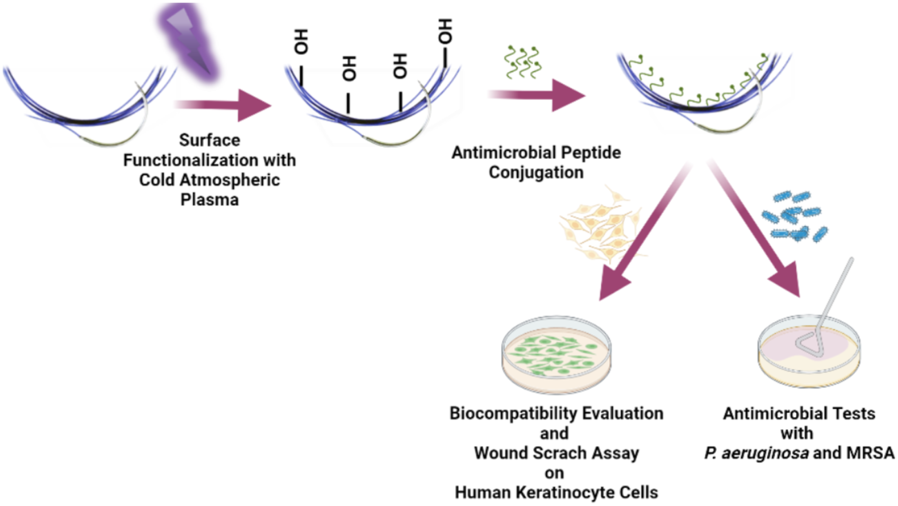
Surgical site infections are commonly encountered as a risk factor in clinics that increase the morbidity of a patient after a surgical operation. Surgical sutures are one of the leading factor for the formation of surgical site infections that induce bacterial colonization by their broad surface area. Current strategies to overcome with surgical site infections consist utilization of antibiotic agent coatings such as triclosan. However, the significant increase in antibiotic resistance majorly decreases their efficiency against recalcitrant pathogens such as; Pseudomonas aeruginosa and Staphylococcus aureus. Therefore, the development of a multi drug-resistant antimicrobial suture without any cytotoxic effect to combat surgical site infections is vital. Antimicrobial peptides are the first defense line which has a broad range of spectrum against Gram-positive, and Gram-negative bacteria and even viruses. In addition, antimicrobial peptides have a rapid killing mechanism which is enhanced by membrane disruption and inhibition of functional proteins in pathogens without the development of antimicrobial resistance. In the scope of the current study, the antimicrobial effect of antimicrobial peptide conjugated poly (glycolic acid-co-caprolactone) (PGCL) sutures were investigated against P. aeruginosa and methicillin-resistant S. aureus (MRSA) strains by using antimicrobial peptide sequences of KRFRIRVRV-NH2, RWRWRWRW-NH2 and their dual combination (1:1). In addition, in vitro wound scratch assays were performed to evaluate the effect of antimicrobial peptide conjugated sutures on keratinocyte cell lines. Our results indicated that antimicrobial peptide modified sutures could be a potential novel medical device to overcome surgical site infections by the superior acceleration of wound healing.
LankiewiczJDYokoeDSOlsenMA, et al.Beyond 30 days: does limiting the duration of surgical site infection follow-up limit detection?Infect Control Hosp Epidemiol2012; 33: 202–204. DOI: 10.1086/663715
2.
Surgical site infections after abdominal closure in colorectal surgery using triclosan-coated absorbable suture (PDS Plus) vs. uncoated sutures (PDS II): a randomized multicenter study. Surg Infect2011; 12: 483–489. DOI: 10.1089/sur.2011.001
3.
BanKAMineiJPLarongaC, et al.American College of Surgeons and Surgical Infection Society: surgical site infection guidelines, 2016 update. J Am Coll Surg2017; 224: 59–74. DOI: 10.1016/j.jamcollsurg.2016.10.029
4.
GarnerBHAndersonDJ. Surgical site infections: an update. Infect Dis Clin North Am2016; 30: 909–929. DOI: 10.1016/j.idc.2016.07.010
5.
LeaperDWilsonPAssadianO, et al.The role of antimicrobial sutures in preventing surgical site infection. Ann R Coll Surg Engl2017; 99: 439–443. DOI: 10.1308/rcsann.2017.0071
6.
PercivalSL. Importance of biofilm formation in surgical infection. Br J Surg2017; 104: e85–e94. DOI: 10.1002/bjs.10433
7.
WeigeltJALipskyBATabakYP, et al.Surgical site infections: causative pathogens and associated outcomes. Am J Infect Control2010; 38: 112–120. DOI: 10.1016/j.ajic.2009.06.010
8.
PangZRaudonisRGlickBR, et al.Antibiotic resistance in Pseudomonas aeruginosa: mechanisms and alternative therapeutic strategies. Biotechnol Adv2019; 37: 177–192. DOI: 10.1016/j.biotechadv.2018.11.013
9.
LydenJDellingerE. Surgical site infections. Hosp Med Clin2016; 5: 319–333. DOI: 10.1016/j.ehmc.2015.11.002
10.
OwensCDStoesselK. Surgical site infections: epidemiology, microbiology and prevention. J Hosp Infect2008; 70: 3–10. DOI: 10.1016/S0195-6701(08)60017-1
11.
SgangaGTasciniCSozioE, et al.Early recognition of methicillin-resistant Staphylococcus aureus surgical site infections using risk and protective factors identified by a group of Italian surgeons through Delphi method. World J Emerg Surg2017; 12: 25. DOI: 10.1186/s13017-017-0136-3
12.
ChuaRLimSKCheeCF, et al.Surgical site infection and development of antimicrobial sutures: a review. Eur Rev Med Pharmacol Sci2022; 26: 828–845. DOI: 10.26355/eurrev_202202_27991
13.
EdmistonCESeabrookGRGoheenMP, et al.Bacterial adherence to surgical sutures: can antibacterial-coated sutures reduce the risk of microbial contamination?J Am Coll Surg2006; 203: 481–489. DOI: 10.1016/j.jamcollsurg.2006.06.026
14.
KudurMHPaiSBSripathiH, et al.Sutures and suturing techniques in skin closure. Indian J Dermatol Venereol Leprol2009; 75: 425–434. DOI: 10.4103/0378-6323.53155
15.
DennisCSethuSNayakS, et al.Suture materials - current and emerging trends. J Biomed Mater Res A2016; 104: 1544–1559. DOI: 10.1002/jbm.a.35683
16.
KjaergardHK. Suture support: is it advantageous?Am J Surg2001; 182: 15s–20s. DOI: 10.1016/s0002-9610(01)00772-3
17.
MuganlıZOnakGErcanUK, et al.The effect of antimicrobial peptide conjugated PGCL sutures on in vitro wound healing. In: Medical Technologies Congress (TIPTEKNO), Izmir, Turkey, 3–5 October 2019: IEEE, pp. 1–4.
18.
Langley-HobbsSJ. Chapter 10 - sutures and general surgical implants. In: Langley-HobbsSJDemetriouJLLadlowJF (eds) Feline soft tissue and general surgery. Philadelphia, PA: W.B. Saunders, 2014, pp. 105–116.
19.
GavrilaDEVictorS. Degradation of polymer materials Biopropylene(BioPP) and Polyglycolide-co-caprolactone(PGCL) used in surgical sutures. In: e-Health and Bioengineering Conference, Iasi, Romania, 21–23 November 2019.
20.
DhomJBloesDAPeschelA, et al.Bacterial adhesion to suture material in a contaminated wound model: comparison of monofilament, braided, and barbed sutures. J Orthop Res, 2017; 35: 925–933. DOI: 10.1002/jor.23305
21.
ErcanUKIbisFDikyolC, et al.Prevention of bacterial colonization on non-thermal atmospheric plasma treated surgical sutures for control and prevention of surgical site infections. PLoS One2018; 13: e0202703. DOI: 10.1371/journal.pone.0202703
22.
SuzukiJBResnikRR. 11 - Wound dehiscence: incision line opening. In: ResnikRRMischCE (eds) Misch’s avoiding complications in oral implantology. Maryland Heights, MI. Mosby, 2018, pp. 402–439.
23.
TummalapalliMAnjumSKumariS, et al.Antimicrobial surgical sutures: recent developments and strategies. Polym Rev2016; 56: 607–630. DOI: 10.1080/15583724.2015.1119163
24.
MaheshLKumarVRJainA, et al.Bacterial adherence around sutures of different material at grafted site: a microbiological analysis. Materials2019; 12: 2848.
25.
YamanDPaksoyTUstaoğluG, et al.Evaluation of bacterial colonization and clinical properties of different suture materials in dentoalveoler surgery. J Oral Maxillofac Surg2022; 80: 313–326. DOI: 10.1016/j.joms.2021.09.014
26.
OnestiMGCarellaSScuderiN. Effectiveness of antimicrobial-coated sutures for the prevention of surgical site infection: a review of the literature. Eur Rev Med Pharmacol Sci2018; 22: 5729–5739. DOI: 10.26355/eurrev_201809_15841
27.
DaoudFCCoppryMMooreN, et al.Do triclosan sutures modify the microbial diversity of surgical site infections? A systematic review and meta-analysis. Microorganisms2022; 10: 927.
28.
ObermeierASchneiderJHarrasserN, et al.Viable adhered Staphylococcus aureus highly reduced on novel antimicrobial sutures using chlorhexidine and octenidine to avoid surgical site infection (SSI). PLoS One2018; 13: e0190912. DOI: 10.1371/journal.pone.0190912
29.
ObermeierASchneiderJWehnerS, et al.Novel high efficient coatings for anti-microbial surgical sutures using chlorhexidine in fatty acid slow-release carrier systems. PLoS One2014; 9: e101426. DOI: 10.1371/journal.pone.0101426
30.
ObermeierASchneiderJFöhrP, et al.In vitro evaluation of novel antimicrobial coatings for surgical sutures using octenidine. BMC Microbiol2015; 15: 186. DOI: 10.1186/s12866-015-0523-4
31.
BlinovAVNagdalianAAPovetkinSN, et al.Surface-oxidized polymer-stabilized silver nanoparticles as a covering component of suture materials. Micromachines2022; 13: 1105.
32.
JaswalTGuptaJ. A review on the toxicity of silver nanoparticles on human health. Mater Today Proc2021. DOI: 10.1016/j.matpr.2021.04.266. in press.
33.
SelvarajuGDUmapathyVRSumathiJonesC, et al.Fabrication and characterization of surgical sutures with propolis silver nano particles and analysis of its antimicrobial properties. J King Saud Univ - Sci2022; 34: 102082. DOI: 10.1016/j.jksus.2022.102082
34.
AdkinsJMAhmarRAYuHD, et al.Comparison of antimicrobial activity between bacitracin-soaked sutures and triclosan coated suture. J Surg Res2022; 270: 203–207. DOI: 10.1016/j.jss.2021.09.010
35.
PengXLiuGZhuL, et al.In vitro and in vivo study of novel antimicrobial gellan–polylysine polyion complex fibers as suture materials. Carbohydr Res2020; 496: 108115. DOI: 10.1016/j.carres.2020.108115
36.
FrancoARFernandesEMRodriguesMT, et al.Antimicrobial coating of spider silk to prevent bacterial attachment on silk surgical sutures. Acta Biomater2019; 99: 236–246. DOI: 10.1016/j.actbio.2019.09.004
37.
SwartjesJJSharmaPKvan KootenTG, et al.Current developments in antimicrobial surface coatings for biomedical applications. Curr Med Chem2015; 22: 2116–2129. DOI: 10.2174/0929867321666140916121355
LiangWDianaJ. The dual role of antimicrobial peptides in autoimmunity. Mini Rev2020; 11: 2077. DOI: 10.3389/fimmu.2020.02077
40.
MangoniMLMcDermottAMZasloffM. Antimicrobial peptides and wound healing: biological and therapeutic considerations. Exp Dermatol2016; 25: 167–173. DOI: 10.1111/exd.12929
41.
LimKLeongSSJ. 22 - antimicrobial coating development based on antimicrobial peptides. In: TiwariA (ed) Handbook of antimicrobial coatings. Amsterdam, Netherlands: Elsevier, 2018, pp. 509–532
42.
AndreaAMolchanovaNJenssenH. Antibiofilm peptides and peptidomimetics with focus on surface immobilization. Biomolecules2018; 8: 27. DOI: 10.3390/biom8020027
43.
KumarPKizhakkedathuJNStrausSK. Antimicrobial peptides: diversity, mechanism of action and strategies to improve the activity and biocompatibility In Vivo. Biomolecules2018; 8: 2018. DOI: 10.3390/biom8010004
44.
LeiJSunLHuangS, et al.The antimicrobial peptides and their potential clinical applications. Am J Transl Res2019; 11: 3919–3931.
45.
LinZWuTWangW, et al.Biofunctions of antimicrobial peptide-conjugated alginate/hyaluronic acid/collagen wound dressings promote wound healing of a mixed-bacteria-infected wound. Int J Biol Macromol2019; 140: 330–342. DOI: 10.1016/j.ijbiomac.2019.08.087
46.
LozeauLDGroshaJKoleD, et al.Collagen tethering of synthetic human antimicrobial peptides cathelicidin LL37 and its effects on antimicrobial activity and cytotoxicity. Acta Biomater2017; 52: 9–20. DOI: 10.1016/j.actbio.2016.12.047
47.
PirasAMaisettaGSandreschiS, et al.Chitosan nanoparticles loaded with the antimicrobial peptide temporin B exert a long-term antibacterial activity in vitro against clinical isolates of Staphylococcus epidermidis. Front Microbiol2015; 6: 372. DOI: 10.3389/fmicb.2015.00372
48.
ClarkSJowittTAHarrisLK, et al.The lexicon of antimicrobial peptides: a complete set of arginine and tryptophan sequences. Commun Biol2021; 4: 605. DOI: 10.1038/s42003-021-02137-7
49.
HouSLiuZYoungAW, et al.Effects of Trp- and Arg-containing antimicrobial-peptide structure on inhibition of Escherichia coli planktonic growth and biofilm formation. Appl Environ Microbiol2010; 76: 1967–1974. DOI: 10.1128/AEM.02321-09
50.
de la Fuente-NunezCKorolikVBainsM, et al.Inhibition of bacterial biofilm formation and swarming motility by a small synthetic cationic peptide. Antimicrob Agents Chemother2012; 56: 2696–2704. DOI: 10.1128/aac.00064-12
KaramanOKumarAMoeinzadehS, et al.Effect of surface modification of nanofibres with glutamic acid peptide on calcium phosphate nucleation and osteogenic differentiation of marrow stromal cells. J Tissue Eng Regen Med2016; 10: E132–E146. DOI: 10.1002/term.1775
53.
HeXMaJJabbariE. Effect of grafting RGD and BMP-2 protein-derived peptides to a hydrogel substrate on osteogenic differentiation of marrow stromal cells. Langmuir2008; 24: 12508–12516. DOI: 10.1021/la802447v
54.
SeleciMAg SeleciDYalçınkayaE, et al.Amine-intercalated montmorillonite matrices for enzyme immobilization and biosensing applications. RSC Adv2012; 2: 2112–2118. DOI: 10.1039/C2RA01225A
55.
TimurSOdaciDDincerA, et al.Biosensing approach for glutathione detection using glutathione reductase and sulfhydryl oxidase bienzymatic system. Talanta2008; 74: 1492–1497. DOI: 10.1016/j.talanta.2007.09.026
56.
PamulaEDryzekE. Structural changes in surface-modified polymers for medical applications. Acta Physica Pol A2008; 113: 1485. DOI: 10.12693/APhysPolA.113.1485
57.
LeaperDAssadianOHubnerNO, et al.Antimicrobial sutures and prevention of surgical site infection: assessment of the safety of the antiseptic triclosan. Int Wound J2011; 8: 556–566. DOI: 10.1111/j.1742-481X.2011.00841.x
58.
MahlapuuMHåkanssonJRingstadL, et al.Antimicrobial peptides: an emerging category of therapeutic agents. Front Cell Infect Microbiol2016; 6: 194. DOI: 10.3389/fcimb.2016.00194
59.
SpohnRDarukaLLázárV, et al.Integrated evolutionary analysis reveals antimicrobial peptides with limited resistance. Nat Commun2019; 10: 4538. DOI: 10.1038/s41467-019-12364-6
60.
MojsoskaBZuckermannRNJenssenH. Structure-activity relationship study of novel peptoids that mimic the structure of antimicrobial peptides. Antimicrob Agents Chemother2015; 59: 4112–4120. DOI: 10.1128/AAC.00237-15
61.
RathinakumarRWalkenhorstWFWimleyWC. Broad-Spectrum antimicrobial peptides by rational combinatorial design and high-throughput screening: the importance of interfacial activity. J Am Chem Soc2009; 131: 7609–7617. DOI: 10.1021/ja8093247
62.
LiJKohJ-JLiuS, et al.Membrane active antimicrobial peptides: translating mechanistic insights to design. Front Neurosci2017; 11: 73. DOI: 10.3389/fnins.2017.00073
63.
PiotrowskaUSobczakMOledzkaE. Current state of a dual behaviour of antimicrobial peptides-Therapeutic agents and promising delivery vectors. Chem Biol Drug Des2017; 90: 1079–1093. DOI: 10.1111/cbdd.13031
64.
YinLMEdwardsMALiJ, et al.Roles of hydrophobicity and charge distribution of cationic antimicrobial peptides in peptide-membrane interactions. J Biol Chem2012; 287: 7738–7745. DOI: 10.1074/jbc.M111.303602
65.
ThamriALétourneauMDjoboulianA, et al.Peptide modification results in the formation of a dimer with a 60-fold enhanced antimicrobial activity. PLoS One2017; 12: e0173783–e0173783. DOI: 10.1371/journal.pone.0173783
66.
YasirMWillcoxMDPDuttaD. Action of antimicrobial peptides against bacterial biofilms. Materials2018; 11: 2468.
67.
PhambuNAlmarwaniBGarciaAM, et al.Chain length effect on the structure and stability of antimicrobial peptides of the (RW)n series. Biophys Chem2017; 227: 8–13. DOI: 10.1016/j.bpc.2017.05.009
68.
ChenXZhangMZhouC, et al.Control of bacterial persister cells by Trp/Arg-containing antimicrobial peptides. Appl Environ Microbiol2011; 77: 4878–4885. DOI: 10.1128/aem.02440-10
69.
StrømMBRekdalOSvendsenJS. Antimicrobial activity of short arginine- and tryptophan-rich peptides. J Peptide Sci2002; 8: 431–437. DOI: 10.1002/psc.398
70.
MohanKVKSainath RaoSGaoY, et al.Enhanced antimicrobial activity of peptide-cocktails against common bacterial contaminants of ex vivo stored platelets. Clin Microbiol Infect2014; 20: O39–O46. DOI: 10.1111/1469-0691.12326
71.
AltVBechertTSteinrückeP, et al.An in vitro assessment of the antibacterial properties and cytotoxicity of nanoparticulate silver bone cement. Biomaterials2004; 25: 4383–4391.
72.
WiegandCHiplerUC. Evaluation of biocompatibility and cytotoxicity using keratinocyte and fibroblast cultures. Skin Pharmacol Physiol2009; 22: 74–82. DOI: 10.1159/000178866
73.
ChenYGuarnieri MichaelTVasil AdrianaI, et al.Role of peptide hydrophobicity in the mechanism of action of α-helical antimicrobial peptides. Antimicrob Agents Chemother2007; 51: 1398–1406. DOI: 10.1128/AAC.00925-06
74.
BacalumMRaduM. Cationic antimicrobial peptides cytotoxicity on mammalian cells: an analysis using therapeutic index integrative concept. Int J Pept Res Ther2015; 21: 47–55. DOI: 10.1007/s10989-014-9430-z
75.
KaltaliogluKCoskun-CevherS. A bioactive molecule in a complex wound healing process: platelet-derived growth factor. Int J Dermatol2015; 54: 972–977. DOI: 10.1111/ijd.12731
76.
WenXZhuMLiZ, et al.Dual effects of bisphenol A on wound healing, involvement of estrogen receptor β. Ecotoxicol Environ Saf2022; 231: 113207. DOI: 10.1016/j.ecoenv.2022.113207
77.
SchneiderOMoruzziAFuchsS, et al.Fusing spheroids to aligned μ-tissues in a heart-on-chip featuring oxygen sensing and electrical pacing capabilities. Mater Today Bio2022; 15: 100280. DOI: 10.1016/j.mtbio.2022.100280
78.
GuoZZhangZZhangN, et al.A Mg2+/polydopamine composite hydrogel for the acceleration of infected wound healing. Bioactive Mater2021; 15: 203–213. DOI: 10.1016/j.bioactmat.2021.11.036
79.
ZhengY-bMaL-dWuJ-l, et al.Design and fabrication of an integrated 3D dynamic multicellular liver-on-a-chip and its application in hepatotoxicity screening. Talanta2022; 241: 123262. DOI: 10.1016/j.talanta.2022.123262
80.
HasaninMSwielamEMAtwaNA, et al.Novel design of bandages using cotton pads, doped with chitosan, glycogen and ZnO nanoparticles, having enhanced antimicrobial and wounds healing effects. Int J Biol Macromol2022; 197: 121–130. DOI: 10.1016/j.ijbiomac.2021.12.106
81.
NasseriSSharifiM. Therapeutic potential of antimicrobial peptides for wound healing. Int J Pept Res Ther2022; 28: 38. DOI: 10.1007/s10989-021-10350-5
82.
de SouzaGSde Jesus SonegoLSantos MundimAC, et al.Antimicrobial-wound healing peptides: Dual-function molecules for the treatment of skin injuries. Peptides2022; 148: 170707. DOI: 10.1016/j.peptides.2021.170707
83.
TakahashiMUmeharaYYueH, et al.The antimicrobial peptide human β-defensin-3 accelerates wound healing by promoting angiogenesis, cell migration, and proliferation through the FGFR/JAK2/STAT3 signaling pathway. Front Immunol2021; 12: 712781. DOI: 10.3389/fimmu.2021.712781
84.
ZhouYLiuGHuangH, et al.Advances and impact of arginine-based materials in wound healing. J Mater Chem B2021; 9: 6738–6750. DOI: 10.1039/D1TB00958C10.1039/D1TB00958C