
Abstract
Objectives
Crocin, the principal water-soluble active constituent of saffron, possesses numerous pharmacological activities. The present investigation examined the potential antidiabetic and antioxidant characteristics of Crocin in rats with type-2 diabetes by administering it orally and intraperitoneally (i.p.).
Methods
After 2 weeks of a high-fat diet, streptozotocin (STZ) (i.p., 40 mg/kg) was administered to male adult rats to induce type-2 diabetes mellitus. Body weight and fasting blood glucose (FBG) were measured on days zero, weeks 1, and 2. At the end of 2 weeks of drug administration in their respective groups, fasting insulin and glucose levels were estimated, and insulin resistance (HOMA-IR) was determined. Intraperitoneal glucose (IPGTT) and insulin tolerance tests (ITT) were carried out. Histopathological investigation and biochemical parameters were estimated in pancreatic tissues.
Results
The Crocin (100 mg/kg) treatment has significantly improved body weight, abatement of FBG, fasting insulin, and HOMA-IR. Likewise, Crocin treatment significantly improved the glucose and insulin challenges. We observed a significantly marked elevation in endogenous antioxidant enzymes in Crocin-treated groups. Similarly, Crocin treatment reversed the histopathological changes and restored the normal integrity and function of the pancreas.
Conclusion
The overall finding indicates that intraperitoneal administration of Crocin demonstrated better control of glycemic level and body weight. Further, it has improved insulin levels in the serum and potentiated antioxidant properties.
Introduction
Diabetes mellitus (DM) is a global health issue growing more rapidly than expected. As per the latest International Diabetes Federation (IDF) Diabetes Atlas, the worldwide occurrence rate of diabetes has reached 10.5%, with nearly half of the adults (44.7%) undiagnosed. According to IDF forecasts, 783 million adults will have diabetes by 2045, accounting for one in every eight adults. This would be a 46% rise over the same period, more than double the predicted population growth of 20%.1,2 Diabetes statistics in Gulf Cooperation Council (GCC) countries have significantly increased over the last two decades. Among the MENA (Middle Eastern and North African) countries, Saudi Arabia is placed fourth in the diabetic population (20–79 years). By 2045, the IDF estimates that one-quarter of Saudi adults will develop diabetes.1,3
Diabetes mellitus is a complex disease caused by a genetic predisposition triggered by environmental circumstances.4,5 The common risk factors associated with the patient's lifestyle are food, physical activity, smoking, and pancreatic infections.6–8 Additionally, each region’s socioeconomic situation impacts patients’ ability to receive appropriate therapeutic methods. 9 Even though the government of Saudi Arabia has made significant efforts to control diseases and minimize rising prevalence rates, it appears that management will be complex due to the increasingly high prevalence of unhealthy lifestyles and the overall increase in population, especially the high percentage of geriatric people due to high life expectancy. Insulin resistance is the most common cause of chronic hyperglycemia in T2DM. It activates numerous factors and pathways linked to decreased insulin sensitivity and dysfunction of β-cells. 10 Healthcare practitioners frequently lack the skills to begin, intensify, or de-escalate therapy when required. 11 This perplexity frequently results in underachievement of the anticipated goal and the induction of life-threatening hypoglycemia. As a result, there is a need to investigate treatment drugs that are both efficacious and have a high safety profile. Herbal medications are widely employed in most countries' health care. 12 As per the World Health Organization (WHO), in underdeveloped countries such as Africa and Asia, over 80% of the population uses this form of medicine for primary care. 13 Herbal medications have been in high demand in developed countries for the last 20 years. 14 Approximately 75% of the French population and 80% of the US population take herbal medication at least once a year.15,16 Egypt (86%) and Saudi Arabia (88%) have witnessed similar figures. Consequently, there is a renewed focus on investigating the potential medicinal uses of this group of substances.17,18
Gardenia jasminoids fruit extract and Crocus sativus stigmas extract both include Crocin, which is one of the most biologically active chemicals.19,20 Crocin has effects that are anticancer, antiulcer, and antioxidant.21,22 Crocin treatment has reduced nephropathies, including gentamicin, carbon tetrachloride, doxorubicin, and methotrexate-induced nephropathies.23,24 Crocin has been demonstrated to prevent and treat atherosclerosis, myocardial ischemia, hemorrhagic shock, cancer, and cerebral disorders. 25 Crocin protects neuronal mitochondria against the development of Alzheimer’s disease. 26 Crocin decreased the inflammatory response and lessened anxiety-like behaviors in rats exposed to amyloid-beta-induced neurotoxicity. 27 Crocin showed protective effects against malathion-induced cardiotoxicity in rats. 28 It has also been demonstrated to provide protection against acrolein-induced lung toxicity in rats. 29 Despite the lack of data on Crocin’s effect on T2DM, specific experimental trials have revealed its glucose-lowering effect, in addition to its antioxidant and anti-hyperlipidemic properties, in STZ diabetic models.30–33 Crocin also reduced the severity of diabetic rats’ kidney damage (nephropathy). 34 Crocin is unique in that it is a water-soluble protein. They are mostly hydrolyzed after oral delivery and are not absorbed. 35 Previous in vitro research has shown that upon oral administration, saffron’s Crocin is most likely not available in the systemic compartment. Crocin is converted into trans-crocetin in the colon, which will be taken up to the systemic circulation by passive trans-cellular diffusion. This trans-crocetin exhibits protective properties against cerebral damage, 36 and it is this trans-crocetin that is responsible for the pharmacological effects of Crocin when it is taken orally. 37 Consequently, it was in our best interest to find out how Crocin would demonstrate its pharmacological potential when given via the intraperitoneal route of administration. This route is recognized for rapid drug absorption and diffusion into the surrounding tissues. The drugs that have been absorbed can either be attached to tissue proteins, degraded by tissue enzymes, or removed by lymph or capillary blood. However, it is unknown whether Crocin is converted to crocetin via this route.
Continuing our efforts to establish the therapeutic effects of Crocin,38,39 we conducted this investigation to assess the antidiabetic potential of two dosages of Crocin, both orally and intraperitoneally, in high-fat diet and low-dose streptozotocin-induced diabetic rats.
Materials and methods
Experimental animals
Adult male Wistar rats, 150–200 g in body weight, were purchased from a local animal supplier. The animals were accommodated for 1 week in a regular laboratory setting (temperature: 25 ± 2°C, 12-h dark/light cycle). Free access to a standard diet and water ad libitum was allowed for all animals. The ethical board of AlMaarefa University approved this study with number 09-20062021, and it was carried out in compliance with the National Institute of Health's (NIH) guidelines for the care and use of laboratory animals.
Materials
Crocin in powder form was procured from the Tokyo Chemical Industry, Toshima, Tokyo, Japan (C1527), with a percentage purity of 99.9% and lot number QE4EB-LL. The powder forms of metformin hydrochloride (1396309) and streptozotocin (S0130) were obtained from Sigma Aldrich Co., located in Darmstadt, Germany. Other chemicals of analytical quality were purchased from Sattec, Saudi Trading & Technology Co. Ltd. in Riyad, Saudi Arabia.
Experimental design and induction of type-2 diabetes
After 1 week of habituation, the animals were separated into eight groups (n = 6 in each). Group 1 (normal control; 10 mL/kg normal saline); Group 2 (diabetic control; DC); Group 3 (standard drug; metformin); Group 4 (negative control; 100 mg/kg Crocin i.p.); Group 5 (DC + Crocin 50 mg/kg i.p.); Group 6 (DC + Crocin 100 mg/kg i.p.); Group 7 (DC + Crocin 50 mg/kg p.o.), and Group 8 (DC + Crocin 100 mg/kg p.o.). For 2 weeks, the animals in groups 1 and 2 were fed a conventional, normal pellet diet. Whereas animals in groups 3 to 8 were initially fed a high-fat diet (HFD: 66.5% standard pellet feed, 13.5% lard, and 20% sugar) for 2 weeks, 40 40 mg/kg streptozotocin (STZ) made from 0.5% normal saline was injected intraperitoneally (i.p.) to induce type-2 diabetes. To avoid hypoglycemia, the animals were given a 5% glucose solution for the first 12 h. As previously stated, the HFD diet was prepared. 41 Fasting blood glucose (FBG) levels were assessed 3 days after STZ injection using a digital glucometer with test strips (Contour, Ascensia Diabetes Care Holdings AG, Switzerland), and animals were considered diabetic if FBG levels were >250 mg/dl, and those animals were included in the study. All animals were given free access to a typical standard diet and water from this day forward (day zero) until the end of the investigation. Using an intraperitoneal (i.p.) method, the normal and diabetes control groups were given 10 mL/kg of normal saline for up to 2 weeks. However, as specified in the research study design, the other groups were administered their respective pharmacological treatments daily for a duration of 2 weeks. All powder medications, such as STZ, metformin, and Crocin, were produced at the necessary concentrations in 0.5% normal saline.
Body weight, blood glucose, and insulin measurement
The animals were weighed on days 0, 7, and 14. Likewise, the blood samples collected on days zero, 7, and 14 were withdrawn by tail clipping in overnight-fasted rats to measure the fasting blood glucose levels. Fasting Serum insulin was determined on day 14 by the radioimmunoassay method using an insulin assay kit from Cisbio International, France. Insulin resistance was calculated (HOMA-IR) using the following formula: HOMA-IR = serum insulin (mmol/L) *(blood glucose (mmol/L)/22.5. 42
Intraperitoneal glucose and insulin tolerance test
IPGTT was done on overnight starved animals at the end of the treatment (day 14) by injection of glucose (2 g/kg, i.p.), and blood glucose levels were measured at 0-, 30-, 60-, and 120-min time intervals. Insulin (2 U/kg, i.v.) was given to overnight-fasted rats 48 h after the IPGTT to assess their insulin tolerance (ITT). Following insulin injection, blood samples were obtained at 0-, 10-, 20-, and 30-min intervals to evaluate blood glucose levels. The area under the curve (AUC) was calculated using the trapezoid rule to demonstrate the significant change in tolerance. 43
Determination of biochemical parameters
On day 16 of the investigation, the 12-h-starved animals were given an intraperitoneal injection of ketamine hydrochloride (90 mg/kg) and xylene hydrochloride (10 mg/kg) to produce anesthesia. 44 The pancreas was carefully taken from ethically sacrificed rats and placed in Petri dishes with an ice-cold buffer. Small tissue parts were stored at −800°C in liquid nitrogen for biochemical estimations. The other parts of the pancreas were fixed in formalin for histopathological observations. Superoxide dismutase (SOD), catalase (CAT), and glutathione (GSH) levels were measured in pancreatic tissue homogenate. CAT activity was determined using the Sinha technique. 45 Similarly, the protocol outlined by Markland 46 was used to estimate SOD, while Kelly and Beutler 47 provided the method for measuring glutathione activity.
Morphological and histological evaluation
Images of each isolated pancreas were captured to observe morphological changes. The extracted pancreatic tissues were embedded in paraffin after being treated with 10% formalin. They are cut into a thick section of 5 µm and examined under a fluorescence microscope using the hematoxylin and eosin staining procedure, Olympus, Tokyo, Japan, under 40x resolution.
Statistical analysis
GraphPad Prism version 6 (San Diego, USA) was used to analyze the data. The study results are presented as mean± SD. Tukey’s test was used after a one-way analysis of variance to calculate the differences between the different study groups.
Results
Effect of Crocin on body weight
Effect of Crocin on alteration in body weight of rats.

Data are demonstrated as mean ± SD (n = 6) at p < .05. One-way ANOVA followed by Tukey’s t-test to compare means. *p < .05,
Effect of Crocin on glycemic control in HFD-STZ-induced diabetic rats
Effect of Crocin on FBG levels in HFD-STZ-induced diabetic rats.

Values are expressed as Mean ± SEM. The values of percentage reduction in glycemia are shown in brackets. *p < .05,
Effect of Crocin on serum metabolic indexes in HFD-STZ diabetic rats
Effect of Crocin on serum metabolic indexes in HFD-STZ diabetic rats.

#p < .001 compared to normal control group; *p < .05, **p < .01, ***p < .001 compared to diabetic control group; NC: normal control.
Impact of Crocin intervention on IPGTT and ITT in rats
Effect of Crocin on intraperitoneal glucose tolerance test in HFD-STZ diabetic rats.

#p < .001; compared to normal control group; *p < .05, **p < .01, ***p < .001 compared to diabetic control group. The letters (a, b, c), represent the significant difference among the groups ap < .05, bp < .01, cp < .001.
Effect of Crocin on insulin tolerance test in HFD-STZ diabetic rats.
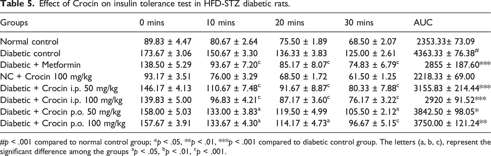
#p < .001 compared to normal control group; *p < .05, **p < .01, ***p < .001 compared to diabetic control group. The letters (a, b, c), represent the significant difference among the groups ap < .05, bp < .01, cp < .001.
Impact of Crocin on the endogenous in-vivo antioxidant levels
Impact of Crocin on the endogenous in-vivo antioxidant levels in HFD-STZ diabetic rats.

Data are indicated as mean ± SD (n = 6) at p < .05. One-way ANOVA thereafter by Tukey’s t-test is performed to compare means. #p < .001, compared to normal control group; *p < .05, **p < .01, ***p < .001 compared to diabetic control group.
Macroscopic and microscopic examination of rat pancreas
The morphological observation reveals normal appearance and texture in normal and negative control groups. The diabetic control group showed modest shrinkage compared to drug-treated groups (Figure 1). Histopathological examination of the normal and negative control (Crocin 100 mg/kg) pancreas revealed normal pancreatic acini and islets of Langerhans with normal cellularity and exocrine portion (Figure 2(a) and (d)). The pancreas of diabetic control rats indicates the shrinkage, reduced size, and number of pancreatic islets with necrosis and degranulation of cells as evident from karyopyknosis, vacuolation, and hydropic connective tissue invasion (Figure 2(b)). The pancreas of diabetic rats treated with Crocin showed the presence of normal islets of Langerhans, cellularity, and an exocrine portion (Figure 2(e)–(h)). Rats treated with metformin revealed intact and normal-sized islets of Langerhans cells (Figure 2(c)). Morphological alterations in the pancreas of various animal groups. Histopathological changes in the pancreas of different groups of animals. (a) Normal control; (b) Diabetic control; (c) Standard drug: Metformin); (d) Negative control: 100 mg/kg crocin i.p. in normal; (e) DC + Crocin 50 mg/kg i.p; (f) DC + Crocin 100 mg/kg i.p; (g) DC + Crocin 50 mg/kg p.o; (h) DC + Crocin 100 mg/kg p.o.

Discussion
The failure of beta-cells is the key factor that leads to the progression of type-2 diabetes. The prevalence of type-2 diabetes is a critical public health concern that has a considerable impact on people’s lives as well as the costs of medical care. Despite significant efforts in research, public health measures, and clinical care, the disease is spreading rapidly because of rapid economic development and urbanization.48,49 Metformin, glitazones, linagliptin, empagliflozin, and insulin are some of the drugs often used to treat type-2 diabetes. However, questions remain about whether these drugs can help with β cell dysfunction. Furthermore, they have been associated with a variety of negative outcomes such as hypoglycemia, GI distress, weight gain, stroke, and lactic acidosis. Thus, in the quest for complementary and alternative medicines, the focus is now on phytochemicals obtained from natural sources. Medicinal plants with bioactive chemicals and biological selectivity are important sources of alternative treatment strategies for a wide range of serious diseases. The unique properties of nutraceuticals have piqued the curiosity of researchers in the plant kingdom for promising possibilities in treating a variety of ailments, including diabetes. Crocin, an active component of saffron, has sparked substantial interest in scientific research during the last three decades due to its significant biological potential and antioxidant characteristics. 50 Crocin has been extensively studied in mice for its antidiabetic potential; prior studies have revealed that Crocin is not absorbed into the bloodstream after oral administration and is primarily eliminated in the feces. 51 As a result, this study looked into the antidiabetic potential of two Crocin dosages (50 and 100 mg/kg) given via two different routes (oral and intraperitoneal) in adult male Wistar rats. The HFD/STZ animal model was used to induce type-2 diabetes. Several methodologies were used to study the impact on insulin resistance and beta-cell dysfunction, as well as morphological and histological changes in pancreatic tissues. In addition, to determine Crocin’s antioxidant potential, we examined antioxidants such as SOD, CAT, and GSH.
The results of the study indicate that following a week of therapy, the animal’s body weight was unaffected by the administration of Crocin. On the other hand, on day 14, there was a considerable increase in body weight among Crocin-treated groups compared to diabetic rats, except for the Crocin 50 mg/kg oral therapy, which had no effect on body weight. An earlier study, which found identical results, revealed that a lower dose of oral Crocin had no meaningful effect on body weight. Furthermore, when compared to the normal control group, all doses of Crocin were able to considerably (p < .01) lower blood glucose levels. A previous study 52 discovered a non-statistically significant decrease in blood glucose levels after 2 weeks of administration of 150 mg/kg oral Crocin to type-2 diabetic rats. Another study 53 showed comparable results in STZ-induced diabetic rats with 60 mg/kg i.p. Crocin. When the results were compared to animals given the normal dose of metformin, mice given 50 mg/kg of Crocin intraperitoneally showed a more significant effect. Fasting insulin levels were significantly lower (p < .01; p < .001) in mice treated with intraperitoneal Crocin 50 and 100 mg/kg, respectively, in our investigation. Only a higher dose (100 mg/kg) of oral Crocin showed a significant (p < .05) reduction in fasting glucose level. Similar outcomes were seen in previous research, where oral Crocin at a dose of 150 mg/kg markedly reduced fasting insulin. 51
HOMA- β and HOMA-IR values were also computed. Insulin resistance, as indicated by HOMA-IR, is common in obesity, metabolic syndrome, and type-2 diabetes. 53 In our investigation, animals treated with intraperitoneal Crocin and a higher dose of oral Crocin (100 mg/kg) had a significant (p < .001) drop in HOMA-IR. The effect of 50 mg/kg oral Crocin on HOMA-IR, on the other hand, was less significant (p < .01). HOMA-β also assesses the function of the beta-cell in T1DM animals. We found no significant effect of Crocin on HOAM-β since our model is based on type-2 diabetes. However, oral Crocin therapy had no statistically significant effect on the HOAM-β score. Similarly, an earlier 41 study found a significant (p < .05) rise in HOAM-β score in animals given 150 mg/kg oral Crocin for 6 weeks. This suggests that long-term oral Crocin treatment would considerably increase cell mass, which could be beneficial in type-1 diabetes. Crocin-treated animals tolerated intraperitoneal glucose challenges significantly better than diabetic animals. Interestingly, a greater dose of Crocin (100 mg/kg) delivered orally and intraperitoneally demonstrated considerable (p < .001) tolerance to exogenously provided glucose after 120 min.
The insulin tolerance test assesses the degree of peripheral glucose utilization. 54 Crocin treatment increased insulin tolerance in HFD-STZ diabetic rats, as shown by significantly lower plasma glucose levels and AUC. The intraperitoneal and oral administration of 100 mg/kg oral Crocin demonstrated the best results significantly (p < .001). In the current study, 2 weeks of HFD intervention were followed by a single low-dose (40 mg/kg) administration of STZ intraperitoneally induced type-2 diabetes. HFD causes obesity and hyperinsulinemia, and low-dose STZ causes partial degeneration of β-cells by generating free radicals, thus reducing insulin production. Hence, any drug that can reduce oxidative stress can be beneficial in protecting the β-cells’ function. In the current study, both doses of Crocin decreased pancreatic oxidation by escalating the levels of endogenous antioxidant enzymes. Significant improvement (p < .001) among all three antioxidant parameters was found in animals treated with Crocin 100 mg/kg i.p. an effect comparable with standard metformin. Unfortunately, low-dose oral Crocin (50 mg/kg) demonstrated the least significant (p < .05) antioxidant potential. Thus, it is evident from the above results that Crocin administered by the intraperitoneal route has better therapeutic potential compared to the oral route.
Further, the morphological observation of the isolated pancreas revealed slight atrophy among the diabetic control group, indicating cell destruction. Histopathological evaluation was done to evaluate the cellular damage. Animals treated with Crocin demonstrated low karyopyknotic, vacuolation, hydropic, and necrotic cells, cell degranulation, and connective tissue invasion, unlike the diabetic group.
It is well known that combining a high-fat diet with STZ induces the generation of oxidative free radicals, which leads to catalase and glutathione inactivation. Catalase and glutathione are responsible for catalyzing the breakdown of hydrogen peroxide. In our research, we observed that animals given HFD-STZ had significantly lower levels of SOD, CAT, and GSH activity. Crocin treatment (both oral and intraperitoneal) protected pancreatic cells from free radical damage caused by hydrogen peroxide, thereby reducing oxidative stress. Our findings are consistent with our previous research and other published literature.38,55,56 The intraperitoneal route was superior to the oral route, probably due to Crocin's capacity to permeate the blood-brain barrier and localize inside the brain with increased bioavailability when injected intraperitoneally. 57 To summarize, Crocin’s antihyperglycemic, antioxidant, and β cell-protecting properties may help develop alternate therapy regimens for T2DM. However, more study is required to determine the mechanistic pathways and molecular mechanisms of Crocin and identify it as an antidiabetic medication.
The limitations of the study include short-term treatment with Crocin for 2 weeks. It is recommended to expand the course of treatment to find out the potential long-term benefits of Crocin among diabetic rats. Further, only six animals were used in each of the groups included in this study. It is recommended to increase the sample size of the animals in each group to improve the reliability of the findings. The outcome of this study was drawn based on pharmacodynamic and histopathology data. Therefore, future research should consider the pharmacokinetic analysis of Crocin, which would provide help in the extrapolation of the information on dosage and route of administration of Crocin from animals to humans.
Conclusions
Overall results show that intraperitoneal administration of Crocin displayed superior glycemic management, body weight change, enhanced serum insulin levels, and antioxidant effects compared to oral treatment. Improvements in fasting glucose levels, body weight, and insulin and HOMA-IR levels were seen with the higher dose of Crocin (100 mg/kg administered i.p.), like those seen with conventional metformin. Crocin therapy increased insulin tolerance and peripheral glucose utilization comparable to metformin. Crocin intervention restored antioxidant enzyme levels (SOD, CAT, and GSH) to normal levels, suggesting its antioxidant potential. Histopathology of the pancreas supports the protective action of Crocin, as the intervention of Crocin restored the normal integrity and function of the pancreas. Most of these effects are attributed to its antioxidant potential. In relation to the parameters examined in our procedure, the precise mode of action of Crocin is not fully understood. Our findings, especially in comparing the routes of administration, are encouraging, but more work needs to be done to investigate each individual process. Furthermore, future research should include a thorough molecular and cellular analysis to find out exactly how Crocin-mediated antidiabetic potential works, both when given by mouth and intraperitoneally.
Footnotes
Acknowledgments
The authors would like to acknowledge the financial support provided by the Researchers Supporting Project number (RSP2024R431) at King Saud University, Riyadh, Saudi Arabia.
Authors’ contributions
S.M.B.A and B.A.M. conceived the research idea and designed the work. R.O and I.A.S wrote the first draft of the manuscript. A.H., A.A., F.M.A.A, and M.M.A. carried out the experiments and interpretation of results and M.E.A. performed the statistical analysis. S.M.B.A supervised the research project and updated the manuscript. All authors contributed to the editing of the revised manuscript and approved the final manuscript.
Declaration of conflicting interests
The author(s) declared no potential conflicts of interest with respect to the research, authorship, and/or publication of this article.
Funding
The author(s) disclosed receipt of the following financial support for the research, authorship, and/or publication of this article: The authors are thankful for the financial support offered by Researchers Supporting Project number (RSP2024R431) at King Saud University, Riyadh, Saudi Arabia. The authors also would like to thank AlMaarefa University, Riyadh, Saudi Arabia for supporting this research.
Ethical statement
Animal welfare
Animals were handled according to the International Guidelines for the Care and Handling of Experimental Animals. The present study followed international, national, and/or institutional guidelines for humane animal treatment and complied with relevant legislation.
Data availability statement
All data and materials are available and can be submitted when needed.