
Abstract
Objective
Multiple myeloma (MM) is a plasma cell malignancy characterized by abnormal plasma cell proliferation in the bone marrow. Circulating exosomal miRNA-451 is associated with the progression of many tumors, but the relationship between its expression and MM has not been reported. In this study, we aimed to investigate the clinical value of miRNA-451 as a biomarker for diagnosis and prognosis of multiple myeloma.
Methods
A total of 120 patients with multiple myeloma and 120 healthy control people were recruited in this study. The miRNA-451 expression in serum exosomes of participants was measured by quantitative real-time polymerase chain reaction, and the diagnostic value of miRNA-451 for multiple myeloma was assessed by receiver operating characteristic (ROC) curve. The correlation between miRNA-451 expression and plasma cells ratio and M protein content was analyzed by Pearson correlation coefficient. The prognosis of different miRNA-451 expression was evaluated by survival curves.
Results
Results suggested that serum exosomal miRNA-451 expression was significantly decreased in patients with multiple myeloma rather than in the healthy controls. The ROC curve showed that area under the curve value of miRNA-451 was 0.888, suggesting that miRNA-451 had diagnostic value to multiple myeloma. Moreover, there was a negative correlation between miRNA-451 expression and plasma cells ratio or M protein content. Survival curves showed that patients with high miRNA-451 expression had a longer survival time, suggesting the value of miRNA-451 as a prognostic indicator of multiple myeloma.
Conclusion
We demonstrated the relationship between miRNA-451 expression and multiple myeloma, indicating that miRNA-451 in circulating exosomes may be an effective diagnostic biomarker and prognostic indicator for multiple myeloma.
Introduction
Multiple myeloma (MM) is a hematological malignancy characterized by abnormal proliferation of plasma cells in the bone marrow and increased levels of M protein, accounting for approximately 13% of all hematological malignancies.1,2 Although much progress has been made in understanding the pathogenesis of MM and in the development of potentially effective treatments, MM remains incurable. Because of the different clinical manifestations and different diagnostic periods of MM, patients have different therapeutic effects and prognostic outcomes, and only 10–15% of patients achieve expected survival. 3 Therefore, diagnosis of MM at its early stage allows for timely intervention, which can improve the survival rate of patients. In addition, early diagnosis is also helpful for risk stratification of patients with different stages of MM, and for formulating appropriate treatment strategies to improve the treatment effect and prognosis of patients.
To date, many diagnostic and prognostic biomarkers have been used for risk stratification of MM patients. Among these biomarkers, the most widely used are the International Staging System (ISS), a simple risk stratification algorithm based on the levels of Sβ2 M and serum albumin, which consists of three stages: stage I, Sβ2 M less than 3.5 mg/L plus serum albumin ≥ 3.5 g/dL (median survival, 62 months); stage II, neither stage I nor III (median survival, 44 months); and stage III, Sβ2 M ≥ 5.5 mg/L (median survival, 29 months). 4 Revised ISS (R-ISS) is another effective prognostic stratification approach that combines ISS with chromosomal abnormalities (mainly t(4; 14) translocation and 17p deletion) and lactate dehydrogenase to provide more accurate prognostic stratification in patients with MM, which is more accurate than the prediction of ISS of median survival. 5 However, survival times of patients with similar prognosis remain different. Therefore, it is necessary to investigate new MM biomarkers to help predict prognosis and accurately stratify risk management in patients with MM.
Exosome is a type of extracellular vesicle with a diameter of 50–140 nm that is widely distributed in body fluids such as blood and urine. 6 Exosome biogenesis consists of cargo sorting, multivesicular body (MVB) formation and maturation, transport of MVBs, and MVB fusion with the plasma membrane, resulting in the high heterogeneity of exosomes. 7 Exosomes are involved in the mediation of multiple physiological processes including immune response and tissue repair. 8 Moreover, exosomes carry a variety of biomolecules, and have become a new and important medium for signaling between cells. 8 Exosomes have also been reported to promote tumorigenesis in a variety of cancers, and one of the major ways is through microRNA (miRNA) delivery.9,10 miRNA is a small, endogenous non-coding RNA with an approximately length of 22 nt. miRNAs are evolutionarily conserved and regulate gene expression at the post-transcriptional level by targeting messenger RNAs, which play important roles in cell survival, cell proliferation and differentiation biological activities.11,12 miRNA is one of the most abundant RNAs in exosomes and is involved in exosome-mediated cell communication. 13 Numerous studies have reported that miRNA levels are dysregulated in cancers, and tumor-derived exosomes reflect miRNA expression in tumor cells.14,15 Some studies have reported that circulating exosomal miRNAs are used as biomarkers for early cancer diagnosis, suggesting that circulating exosomal miRNAs may be novel biomarkers to be applied in clinical diagnosis.16,17
In recent years, some studies reported the relationship between miRNAs and the development of MM. For example, high levels of miRNA-181a and miRNA-8074 are both correlated with high risk of death and poor prognosis.18,19 At present, the research on miRNA-451 is not sufficient. Previously studies reported that miRNA-451 has low expression in lung cancer and glioma cells, which is closely related to the malignant progression of tumors.20,21 miR-451 not only directly affects the biological function of tumor cells, but also indirectly affects the invasion and metastasis of tumor cells by secreting exosomes into the tumor microenvironment. 22 Lu et al. 23 reported that miRNA-451 inhibits malignant progression of multiple myeloma RPMI-8226 cells by targeting c-Myc. However, the potential of miRNA-451 in the diagnosis and prognosis of MM has not been reported yet.
In this study, we measured the expression of miRNA-451 in serum exosomes of patients with MM, and investigated its clinical value of MM. The results may provide a novel diagnosis and prognosis biomarker of MM.
Material and methods
Ethical statement
This study was performed in line with the principles of the Declaration of Helsinki. Approval was granted by the Ethics Committee of the Second Affiliated Hospital of Guizhou Medical University. Written informed consent was obtained from each participant. All methods were carried out in accordance with relevant guidelines and regulations.
Clinical data
From July 2021 to June 2022, 120 patients with MM who were firstly diagnosed in the hospital (MM group), and 120 age- and sex-matched healthy controls (HC group) were recruited for this study. The inclusion criteria were: (a) age ≥ 18 years old; (b) patients with MM who were first diagnosed according to the International Myeloma Task Force guidelines; and (c) those who had not received any treatment before blood sample collection. Exclusion criteria were: (a) those with a history of malignancy or a combination of other malignancies; (b) people with other organ dysfunctions such as heart, liver, kidney, etc.; and (c) patients with infectious diseases. After hospital admission, the clinical data (including age, gender, MM classification and staging, etc.) and laboratory indicators were collected from each group. After treatment, patients were followed up for 2 years to record disease progression, recurrence, and death. The clinical characteristics are shown in Table 1.
Clinical characteristics of the participants.
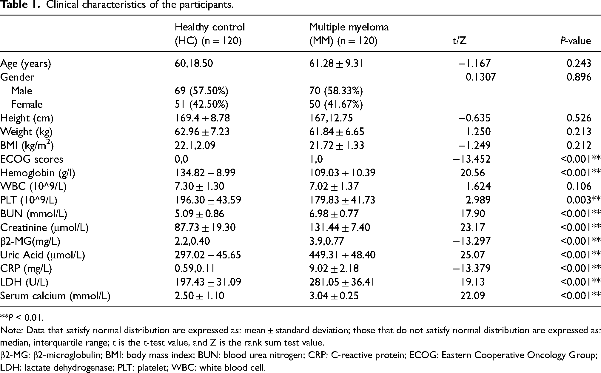
**P < 0.01.
Note: Data that satisfy normal distribution are expressed as: mean ± standard deviation; those that do not satisfy normal distribution are expressed as: median, interquartile range; t is the t-test value, and Z is the rank sum test value.
β2-MG: β2-microglobulin; BMI: body mass index; BUN: blood urea nitrogen; CRP: C-reactive protein; ECOG: Eastern Cooperative Oncology Group; LDH: lactate dehydrogenase; PLT: platelet; WBC: white blood cell.
Serum collection, exosomes extraction, purification, and identification
Blood was collected on an empty stomach in the morning and then centrifuged at 300×g for 10 min. Then, the serums in the upper layer were collected, and centrifuged at 4°C at 17,000×g for 10 min to completely remove the residual blood cells. The top serum was carefully aspirated, transferred to a new centrifuge tube and stored in a −80°C refrigerator. Next, the exosomes were extracted using an ExoQuickTM kit (System Biosciences, Palo Alto, CA, USA). The serums were mixed with the reagent and incubated for 30 min at 4°C, and then centrifuged for 2 min at 15,000×g. The supernatant was then discarded and the precipitate was re-suspended in 50 μL phosphate-buffered saline to obtain exosomes. The obtained exosomes were transferred to the upper tube of the exosome purification column (Yeasen, Shanghai, China), and then centrifuged at 3000×g for 10 min at 4°C. The liquid at the bottom of the tube was the purified exosomes. The exosomes were loaded on a copper mesh, stained with uranyl acetate, and observed using a transmission electron microscope (TEM). The size of exosomes was detected by nanoparticle tracking analysis (NTA) using a NanoSight NS300 instrument (Malvern, Worcestershire, UK).
Western blot
Total protein extraction was conducted using RIPA lysis (Thermo Scientific, Waltham, MA, USA) and quantified using a BCA kit (Thermo Scientific). Samples were loaded on 10% SDS-PAGE for electrophoretic separation, transferred to a PVDF membrane and incubated with anti-CD63 (1: 1000, ab134045, Abcam, Cambridge, MA, USA) and anti-TSG101 (1: 1000, ab125011, Abcam) overnight at 4°C. After incubation, the membrane was washed with 1×TBST and incubated with secondary antibodies (1: 1000, ab205718, Abcam) for 2 h. Finally, the membrane was washed with 1×TBST for three times and visualized using the ECL reagent (Thermo Scientific).
Quantitative real-time polymerase chain reaction
Total RNA of the exosomes was isolated by Trizol reagent (Invitrogen, Carlsbad, CA, USA) and quantified using a Qubit RNA HS assay kit (Invitrogen). The cDNA was synthesized using the miRcute miRNA First-Strand cDNA synthesis kit (TIANGEN, Beijing, China) and analyzed by quantitative real-time PCR (qPCR) using the SYBR qPCR master mix kit (Invitrogen). Data were analyzed by the 2−ΔΔCT method, and the miRNA expression was normalized to U6. The primers of miRNA-451 for qPCR were as follows: 5ʹ-AAACCGTTACCATTACTGAG-3ʹ (sense) and 5ʹ-CTCAACTGGTGTCGTGGAGTCGG-3ʹ (antisense).
Statistical analysis
Data analysis was processed using the SPSS 22.0 software, and the results are shown as mean ± SD. In the comparison of clinical characteristics between the HC group and the MM group, the student's t-test was used to analyze the data that met the normal distribution; and the rank sum test was used to analyze the data that did not meet the normal distribution. Gender in Table 1 and the number of clinical classifications was analyzed by chi-square test. The expression of miRNA-451 in the two groups was analyzed by rank sum test. P < 0.05 was considered as statistically significant. The receiver operating characteristic (ROC) curve was used to assess the diagnostic value of miRNA-451 for MM, the area under the curve (AUC) > 0.5 was considered to have a diagnostic value. The difference of serum exosome miRNA-451 expression between patients with MM and HC was compared by the Mann–Whitney U test, and the correlation between miRNA-451 expression and plasma cell ratio or M protein content was analyzed by the Pearson correlation coefficient.
After miRNA-451 expression was measured, the patients were ranked according to the level of expression from low to high, with the first 60 considered to have a low level of expression and the next 60 a high level of expression. The survival curves of high and low miRNA-451 expression patients were plotted by the Kaplan–Meier method and compared using the Log-Rank method.
Results
Clinical characteristics of the subjects
Before the start of the study, we first compared the clinical characteristics of patients with MM with the HC group. As shown in Table 1, we found that hemoglobin and platelets in patients with MM was significantly decreased. However, Eastern Cooperative Oncology Group scores, blood urea nitrogen, creatinine, β2-microglobulin, uric acid, C-reactive protein, lactated hydrogenase, and serum calcium were all increased in patients with MM. These results indicated that patients with MM exhibited impaired mobility and a higher disease severity, accompanied by compromised renal function.
Identification of exosomes
Exosomes were identified by TEM. Results showed that the exosomes exhibited a spherical characterized by bilayer vesicles (Figure 1(a)). NTA analysis demonstrated that the diameter distribution of exosomes was predominantly concentrated at 100–200 nm (Figure 1(b)). Western blot suggested that the protein levels of CD63 and TSG101 were upregulated in exosomes (Figure 1(c)). In conclusion, these results demonstrated that the particles were exosomes.
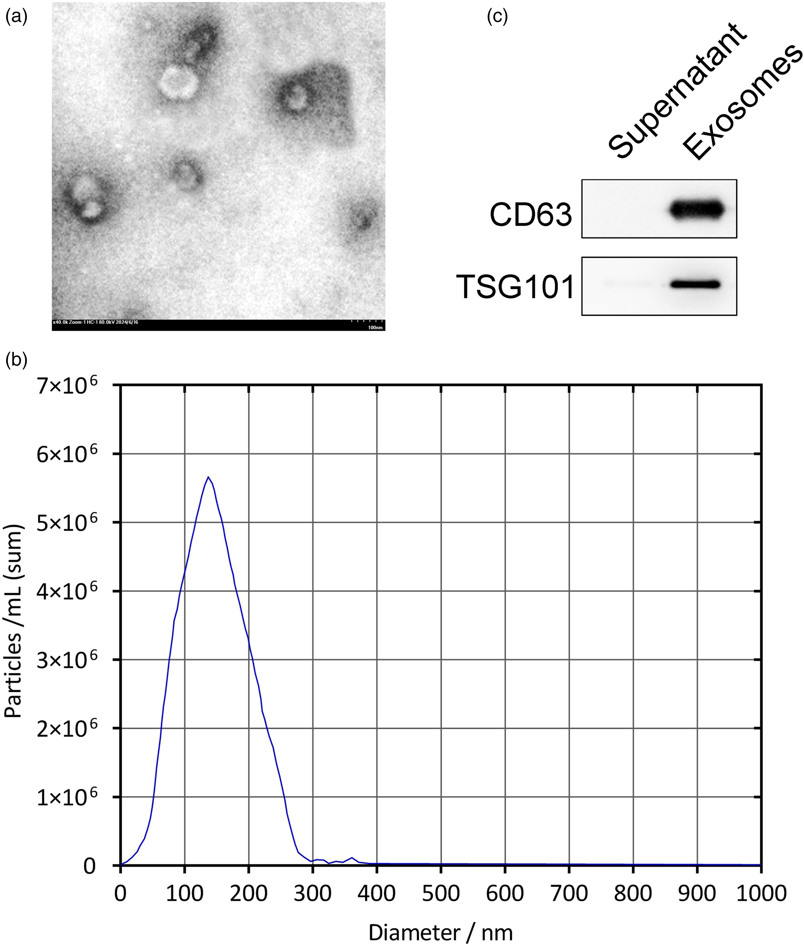
Identification of exosomes. (a) TEM was performed to evaluate the characteristic of exosomes. (b) The size of exosomes was measured by NTA analysis. (c) Western blot was performed to detected the protein levels of exosome labeled proteins.
miRNA-451 may be an effective biomarker for the diagnosis of MM
To investigate the value of miRNA-451 as a biomarker of MM, we first detected the expression of miRNA-451 in the serum exosomes of 120 patients with MM. Compared with the HC group, miRNA-451 expression was significantly decreased in patients with MM (Figure 2(a)). Next, we drew a ROC curve to identify the diagnostic value of miRNA-451 for MM. The results showed that the AUC value was 0.888 (P < 0.001; 95% confidence interval: 0.846, 0.930), suggesting the high diagnostic accuracy of miRNA-451 for MM and indicating that serum exosomal miRNA-451 may be an effective biomarker for the diagnosis of MM (Figure 2(b)). Taken together, these results suggested the potential of serum exosomal miRNA-451 as a diagnostic biomarker of MM.
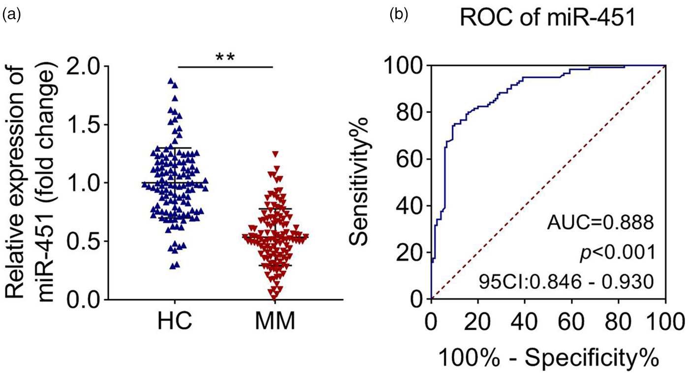
miRNA-451 may be an effective biomarker for the diagnosis of MM. (a) The serum exosomal miRNA-451 expression of the HC group and patients with MM was measured by qPCR. (b) The diagnostic value of miRNA-451 for MM was evaluated by the ROC curve. **P < 0.01.
miRNA-451 expression is negatively correlated with plasma cells and M protein content
The abnormal proliferation of plasma cells and the increase of M protein are the markers of MM. To further confirm the relationship between miRNA-451 and MM, we then analyzed the correlation between miRNA-451 expression and plasma cells or M protein content. As shown in Figure 3(a), there was a negative correlation between miRNA-451 expression and plasma cells ratio (r = −0.624, P < 0.001). There is also a negative correlation between miRNA-451 expression and M protein content (Figure 3(b); r = −0.547, P < 0.001). These results suggested a negative correlation between miRNA-451 expression and other MM biomarkers, further demonstrating the potential of miRNA-451 as a diagnostic biomarker of MM.
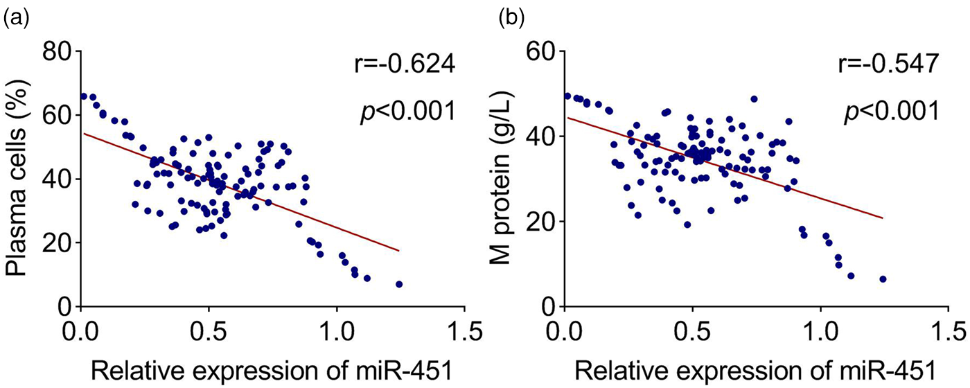
Serum exosomal miRNA-451 expression is negatively correlated with plasma cells ratio and M protein content. (a) and (b) The correlation between miRNA-451 and plasma cells ratio and M protein content was analyzed by the Pearson correlation coefficient.
Relationship between mir-451 expression level and clinical classification
To better understand the clinical value of miRNA-451, we divided the patients into high and low miRNA-451 expression groups, with 60 patients in each group, and measured the correlation between miRNA-451 expression and clinical classification. In Table 2, the results showed that among patients with high and low miRNA-451 expression, there was a significant difference ISS and R-ISS staging. There were more patients in ISS/R-ISS stages I and II than in stage III, suggesting that patients with high miRNA-451 expression had a better prognosis.
Relationship between miR-451 expression level and clinical classification.
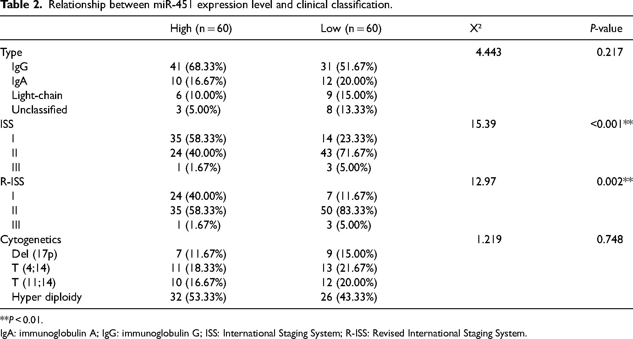
**P < 0.01.
IgA: immunoglobulin A; IgG: immunoglobulin G; ISS: International Staging System; R-ISS: Revised International Staging System.
miRNA-451 could be a prognostic indicator for MM
Next, we plotted survival curves to further investigate the correlation between miRNA-451 expression and prognosis in patients with MM. Results showed that the survival time was significantly shortened in patients with low miRNA-451 expression, suggesting that those with high miRNA-451 expression had a higher survival rate (Figure 4(a)). These results suggested that miRNA-451 could be a prognostic indicator of MM.
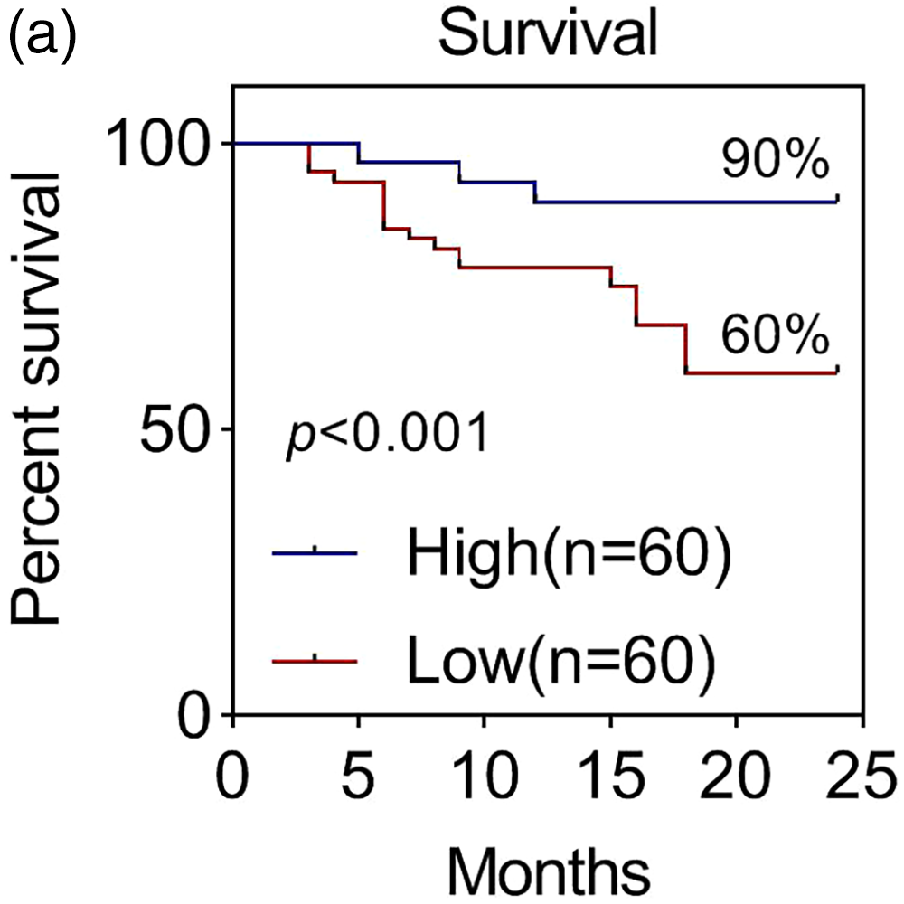
miRNA-451 can be a prognostic indicator for MM. (a) Survival curves were performed to exhibit the effect of high and low expression levels of miRNA-451 on the prognosis of patients with MM.
Discussion
MM is a plasma cell malignancy characterized by abnormal plasma cell proliferation in the bone marrow. Patients with MM have a poor prognosis and a low survival rate. Therefore, the development of effective diagnostic biomarkers of MM is of great importance to improve the prognosis and quality of life of patients. 24 In addition to the two commonly used prognostic staging systems, ISS and R-ISS, several diagnostic and prognostic biomarkers for MM have been developed. For example, Edwards et al. 25 demonstrated that the peripheral blood monocyte count is a dynamic prognostic biomarker in MM, and an abnormal absolute monocyte count is associated with inferior overall survival. Moreover, Li et al. 26 confirmed that the increase of circulating plasma cells has a negative impact on the overall survival and progression-free survival of patients with MM, which is a promising prognostic biomarker for risk stratification and disease surveillance. Collectively, the development of more sensitive and accurate biomarkers is necessary to improve patient prognosis.
miRNA is a non-coding RNA of approximately 21 nucleotides in length that binds to the untranslated region of the 3ʹ end of the target mRNA to cause mRNA degradation or translation inhibition, thereby regulating multiple biological processes. miRNAs are widely used as biomarkers in the diagnosis and prognosis of multiple cancers. For example, Zhang et al. 27 demonstrated that seven miRNAs, including miR-103a-3p, miR-127–3p, miR-151a-5p and miR-17-5p, etc., were significantly overexpressed in colorectal cancer, suggesting that these miRNAs can be used as potential biomarkers for colorectal cancer diagnosis. In recent years, some studies investigated the relationship between miRNAs and MM. Leotta et al. 28 demonstrated that miR-125a-5p antagonism results in the activation of p53 pathway in MM cells. Leone et al. 29 first found that miR-21 is highly expressed in MM and plays its role as an oncogene, suggesting the function of miRNA in the diagnosis of MM.
Currently, circulating miRNAs in body fluids have become more clinically attractive biomarkers due to their non-invasive methods of acquisition, and have been reported as reliable biomarkers for diagnosis and prognosis for many types of cancer, including MM. For instance, Zhou et al. 17 demonstrated that exosomal miR-15a-5p is a promising and effective diagnostic biomarker for the early detection of endometrial cancer. miR-21-5p in urinary extracellular vesicles has also shown to be a novel biomarker of urothelial carcinoma with a specificity of 95.8%. 30 Szudy-Szczyrek et al. 18 found that high expression of miRNA-8074 in patients with MM is significantly associated with a higher risk of death, suggesting the potential of circulating serum miRNA-8074 as a prognostic biomarker for MM. In recent years, several exosomal miRNAs have also emerged as effective biomarkers for MM. Lee et al. 31 revealed that secretion of miR-1305 mediated by exosome in MM cells promotes tumor aggressiveness under hypoxia, suggesting that it may be a biomarker for predicting prognosis of MM. Manier et al. 32 investigated the relationship between miRNA levels and patient outcomes in 156 patients with MM, and found that miRNA let-7b and miR-18a were significantly correlated with progression-free survival and overall survival, indicating that miRNA let-7b and miR-18a can be used as prognosis biomarkers of MM. Zhang et al. 33 explored the relationship between serum exosomal miRNAs and clinical significance in patients with MM, and found that the expression of let-7c-5p, let-7d-5p, miR-140-3p, miR-185-5p, and miR-425-5p in patients with MM is significantly lower than those of HCs. This evidence suggests the potential of exosomal miRNAs as the diagnostic and prognostic biomarker of MM.
However, clinical studies on the role of circulating exosomal miRNA in MM are lacking; especially, the effects of circulating exosomal miRNA-451 on MM have not been reported. miRNA-451 is differentially expressed in different diseases, which mediates multiple disease processes, especially cancers. Wang et al. 34 revealed that miRNA-451 is involved in tumor suppression of malignant proliferation of head and neck squamous cell carcinomas by regulating c-Myc expression. A meta-analysis studied the clinical utility of miRNA-451 for the diagnosis of human cancers, and revealed that miRNA-451 has moderate diagnostic capacity for cancers and can be a potential biomarker for early screening, and its diagnostic utility is independent of population and cancer type. 35 Few studies have explored the relationship between miRNA-451 and MM; only Du et al. 36 have demonstrated that miRNA-451 regulates the stemness of side population cells through PI3 K/Akt/mTOR signaling pathway in MM. Whether miRNA-451 could be used as a biomarker of MM remains unclear. The ideal biomarkers should be easily accessible, easy to detect, and have high sensitivity in disease diagnosis. In our study, we demonstrated that circulating exosomal miRNA-451 expression of patients with MM was significantly lower than that in the HC group, and had a high diagnostic value for MM (AUC = 0.888). Moreover, we demonstrated that patients with MM with high miRNA-451 expression had a better prognosis. These results indicating that miRNA-451 is a reliable diagnostic and prognostic biomarker of MM.
Notably, Mimmi et al. 37 identified miRNAs in different exosome subtypes, angiotensin-converting enzyme 2 expressing circulating exosomes (ExoACE2) and non-ExoACE2, in the plasma of patients with COVID-19, and found that let-7g-5p and hsa-miR-4454 + miR-7975 were upregulated, while hsa-miR-208a-3p and has-miR-323-3p were downregulated in ExoACE2 vs. non-ExoACE2, which further detailed the characterization of potential biomarkers in patients with COVID-19. This study demonstrates differences in miRNA expression across exosomal subtypes. Therefore, isolation of different subtypes of exosomes may be potentially relevant for the development of more accurate MM biomarkers, which may be investigated in our future work.
In conclusion, we have demonstrated that miRNA-451 expression is correlated with the diagnosis and prognosis of MM, and has a high diagnostic accuracy of MM with AUC equaling 0.888, suggesting that miRNA-451 can be used as a diagnostic and prognostic biomarker of MM.
Footnotes
Acknowledgements
Not applicable.
Authors’ contributions
JZ conceived the study, conducted the experiments, and was a major contributor in writing the manuscript; CL, HL, BZ, HS, and BH analyzed the data. All authors read and approved the final manuscript.
Availability of data and materials
The datasets used and/or analyzed during the current study are available from the corresponding author on reasonable request.
Consent for publication
All authors approved the final manuscript and the submission to this journal.
Declaration of conflicting interests
The authors declared no potential conflicts of interest with respect to the research, authorship, and/or publication of this article.
Funding
The authors disclosed receipt of the following financial support for the research, authorship, and/or publication of this article: This study was supported by 2021 Guizhou Provincial Health Commission Science and Technology Fund Project (gzwkj2021-159).