
Abstract
Aim
To compare the relationship between white matter hyperintensities (WMH) on brain magnetic resonance imaging and retinal nerve fiber layer (RNFL), choroid, and ganglion cell layer (GCL) thicknesses in migraine patients and healthy subjects. We also assessed the role of cerebral hypoperfusion in the formation of these WMH lesions.
Methods
We enrolled 35 migraine patients without WMH, 37 migraine patients with WMH, and 37 healthy control subjects examined in the Neurology outpatient clinic of our tertiary center from May to December 2015. RFNL, choroid, and GCL thicknesses were measured by optic coherence tomography.
Results
There were no differences in the RFNL, choroid, or GCL thicknesses between migraine patients with and without WMH (p > 0.05). Choroid layer thicknesses were significantly lower in migraine patients compared to control subjects (p < 0.05), while there were no differences in RFNL and GCL thicknesses (p > 0.05).
Conclusions
The ‘only cerebral hypoperfusion’ theory was insufficient to explain the pathophysiology of WMH lesions in migraine patients. In addition, the thinning of the choroid thicknesses in migraine patients suggests a potential causative role for cerebral hypoperfusion and decreased perfusion pressure of the choroid layer.
Introduction
Migraine is a primary headache disorder characterized by recurrent headache attacks, and has a one-year prevalence of 16.4% in the Turkish population (1,2). Migraine is associated with a variety of structural brain lesions, including clinically silent infarct-like lesions in the posterior circulation territory, white matter hyperintensities (WMH), and grey and white matter volume changes (3–5). WMH are the most common lesions (6), and are characterized by multiple small punctate discrete lesions without mass effect in fluid attenuated inversion recovery (FLAIR) and T2 brain magnetic resonance imaging (MRI) scans, predominantly located in deep white matter in the anterior circulation territory and periventricular areas (7,8). Migraine patients have a four-fold greater risk of WMH (9), which is higher in female subjects and in patients with higher attack frequency and longer disease duration (10).
There are several hypotheses regarding the underlying mechanisms of WMH (6,11–14), although the pathogenesis and clinical significance of these lesions remains to be fully determined. One of these proposed mechanisms involves attack-related oligemia and focal hypoperfusion (15). Oligemia induced by repeated migraine attacks, especially with aura and high frequency, was reported to affect the small deep penetrating arteries, and this local hypoperfusion may cause minor brain lesions (6). The associations between migraine, WMH, and ischemic stroke are complex (16), and these lesions also have a predictive value for further stroke (17,18).
Hypoperfusion associated with migraine attacks can also involve areas other than the brain, including the eyes (19). As a vascular ocular structure, the choroid is directly affected by perfusion pressure (20), and although vasospasm of the cerebral and retinal blood vessels is a transient phenomenon, the chronic nature of migraine can result in structural abnormalities of the retina and choroid layer (19,21). A reduction in blood supply to the retina microcirculation may also cause ganglion cell death in some migraine patients (22).
Optic coherence tomography (OCT) is becoming an increasingly used imaging method for evaluating the thickness of the optic nerve head, retinal nerve fiber layer (RNFL), ganglion cell layer (GCL), and the choroid layer in various neuro-ophthalmologic diseases (21). Thus, the overall aim of this study was to use OCT to compare the relationship between WMH and RNFL, choroid, and GCL thicknesses in migraine patients with healthy individuals, and to determine the role of hypoperfusion in the pathogenesis of the structural lesions.
Methods
Study population
This study was approved by the local ethical committee (Baskent University, Human Ethics Committee, Study No: KA 15/92 Ankara, Turkey), and written informed consent was obtained from all patients before recruitment into the study. We enrolled patients admitted to the Neurology outpatient clinics of Baskent University Hospital (from May to December 2015) with a complaint of headache and a diagnosis of migraine according to The International Classification of Headache Disorders, 3rd edition (beta version) (1). Patients were recruited according to the following inclusion criteria: 18–55 years of age (to exclude RFNL, GCL, and choroid layer thickness differences and development of WMH due to aging); without systemic comorbidities such as diabetes mellitus, hypertension, or hyperlipidemia, or neurological diseases such as cerebrovascular diseases or demyelinating diseases (to exclude other possible influences on WMH); without history of ocular surface disorder, previous ocular surgery, ocular injury, any congenital or acquired retinal disorder, or high myopic and hyperopic refractive errors (to exclude potential effects on RFNL, GCL, and choroid layer thicknesses); and who had had brain MRI scans as a part of their evaluation. Randomly assigned healthy subjects of similar age distribution, without any headache complaint, systemic comorbidity, neurological or ocular disease, were used as control group.
Overall, 121 patients were eligible for the study, but 12 patients were excluded because of incomplete layer thickness measurements. In total, 109 patients (35 migraine patients without WMH, 37 migraine patients with WMH, and 37 healthy control subjects) were evaluated. Detailed histories, demographic characteristics, information on the frequency of migraine attacks, attack duration, age at onset, migraine treatments, the presence of aura, WMH in brain MRI, and familial migraine history were recorded for each patient. A pain score was determined for each migraine patient based on the Visual Analogue Scale (VAS) (23).
Brain MRI scans and WMH evaluation
Brain MRI examinations were performed with a 1.5 T MRI scanner (Siemens, Avanto, Germany) with a 16-channel phased array head coil. The scanning protocol included fluid attenuated inversion recovery (FLAIR) axial (repetition time/echo time [TR/TE]: 8000/84 ms, slice thickness: 5.5 mm, field of view [FOV]: 22 cm, matrix: 256 × 157, bandwidth: 190, flip angle: 150°, number of slices: 20); axial T1-weighted images (T1W) (TR/TE: 410/9.2 ms, slice thickness: 5.5 mm, FOV: 22 cm, matrix: 448 × 186, bandwidth: 130, flip angle: 90°, number of slices: 20); axial T2-weighted (T2W) images (TR/TE: 3630/103 ms, slice thickness: 5.5 mm, FOV: 22 cm, matrix: 512 × 325, bandwidth: 191, flip angle: 150°, number of slices: 20); coronal T2W images (TR/TE: 3630/103 ms, slice thickness: 5.5 mm, FOV: 22 cm, matrix: 512 × 302, bandwidth: 191, flip angle: 150°, number of slices: 20); sagittal T2W images (TR/TE: 3630/103 ms, slice thickness: 5.5 mm, FOV: 22 cm, matrix: 512 × 358, bandwidth: 191, flip angle: 150°, number of slices: 20).
WMH were evaluated by an investigator (FK) blinded to migraine diagnosis and clinical data. The WMH were counted on FLAIR images, and patients were grouped into four subgroups according to the number of lesions: 0, 1–3, 4–8, ≥9. The location and distribution of the lesions were described according to the methodology reported by Barkhof et al. in multiple sclerosis patients (24). Patients were further divided into juxtacortical, subcortical/deep white matter, callosal/subcallosal, and periventricular subgroups. The locations of WMH were classified as frontal, temporal, parietal, occipital, and infratentorial.
OCT imaging
Both eyes of the participants were evaluated, and the right eye was included for analysis. All patients had undergone a detailed ophthalmic examination, including visual acuity testing, biomicroscopy, intraocular pressure measurement with non-contact tonometry, fundus examination, and RFNL, GCL, and choroid layer thickness measurements. An OCT device (Spectralis; Heidelberg Engineering, Heidelberg, Germany) was used to assess tissue thicknesses. Scans for all participants were performed with pupillary dilatation under the same intensity of dim room lighting by the same experienced technician with the same OCT device. An internal fixation target was also used in all scans, with the real-time eye tracking system to adjust for eye motion.
The macular thickness (µm) was determined automatically and analyzed by the OCT software. The fast macular thickness map included a 25-line raster volume scan, 20 × 20°, and was centered on the fovea. In the raster scans used for the macular measurements, the scans were obtained in high-speed mode with the automated real-time feature enabled and set at nine frames. The infrared scanning laser ophthalmoscope scan angle was set at 30° for all acquired scans. The results obtained from the macular scan were classified by segments. The mean thickness values of the RNFL and GCL were assessed for all the subfields in the Early Treatment Diabetic Retinopathy Study (ETDRS) map (25). The concentric circle diameters were 1 mL in the fovea, 3 mL in the parafoveal region, and 6 mL in the perifoveal region. The Heidelberg built-in software was used to assess RNFL and GCL thicknesses, and measurements were recorded by sector. The map was divided into nine ETDRS macular fields: central sector, superior internal sector, temporal internal sector, inferior internal sector, nasal internal sector, superior external sector, temporal external sector, inferior external sector, and nasal external sector.
The choroid layer thickness was measured by enhanced depth imaging using the spectral-domain OCT. Each section, consisting of 30 average scans, was obtained in a 15 × 30° rectangle centered at the macula. Choroid layer thickness was determined as the distance from the outer surface of the hyper-reflective line, referred to as the ‘retinal pigment epithelium’ layer, to the hyper-reflective line of the inner scleral border. Choroid layer thickness was measured at the fovea, 500 µm nasal, and 500 µm temporal to the fovea in a horizontal scan section (26). Manual measurements were performed by two physicians blinded to the groups (IA, LA), and the average of the two measurements was taken to avoid potential bias.
Statistical analyses
Numerical data were presented as mean ± standard deviation (SD), median, minimum, and maximum values. Categorical data were described as percentages. Normality of data distribution was checked using the Shapiro–Wilk test. Data with a non-normal distribution were tested with the Mann–Whitney U test between two groups or the Kruskal–Wallis test for more than two groups. Categorical variables were analyzed using the chi-square test, Fisher Exact test, or the Fisher–Freeman–Halton test. Numerical data were analyzed by Spearman correlation analyses. For all analyses, a two-tailed p value < 0.05 was considered statistically significant. Statistical analyses were performed using statistical software (IBM-SPSS 21.0 for Windows, Chicago, Illinois, USA).
Results
Demographic data of patients and controls.
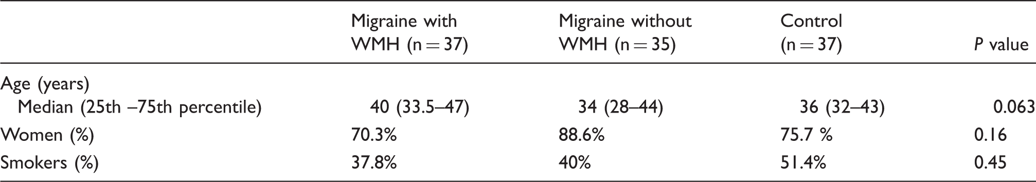
WMH = white matter hyperintensity.
There were no differences in the presence of aura between migraine patients with and without WMH (p = 0.246). Among the 72 migraine patients, four patients (11.4%) had aura in the group with WMH, and eight patients (21.6%) had aura in the group without WMH. Among patients with aura, 10 patients reported visual symptoms such as light flashes, light refraction, and black dots in the visual area.
Baseline characteristics of migraine patients with/without WMH.

VAS = Visual Analogue Scale.
In the group of migraine with WMH, 25 patients (67.6%) had 1–3 lesions, seven patients (18.9%) had 4–8 lesions, and five patients (13.5%) had more than nine lesions. With respect to the localization of WMH, 34 patients had supratentorial deep, 10 patients had periventricular, two patients had infratentorial, and one patient had callosal WMH.
Optic coherence tomography (OCT) analysis of the retinal nerve fiber layer (RNFL) and ganglion cell layer GCL) thicknesses.

Data are median (25th–75th percentile). c = central sector; int sup = superior internal sector; int inf = inferior internal sector; int temp = temporal internal sector; int nasal = nasal internal sector; ext sup = superior external sector; ext inf = inferior external sector; ext temp = temporal external sector; ext nasal = nasal external sector.
OCT analysis results of the choroid layer thicknesses.

Data are median (25th – 75th percentile).
When migraine patients were grouped according to the presence of aura, there were no differences in the demographic characteristics, baseline characteristics, or the median (25th–75th percentile) choroid layer thicknesses between migraine patients with and without aura. However, there was a difference in the median (25th–75th percentile) RNFL thickness in the internal inferior region between the migraine patients with (28.00 µm (25.25–30.00)) and without aura (25.50 µm (24.00–28.00)) (p = 0.015). Furthermore, there was a significant difference in the median GCL thickness in the external inferior region between the migraine patients with (32.50 µm [32.00–36.00]) and without aura (36.00 µm (34.00–38.00]) (p = 0.014).
In migraine patients there were negative, weak correlations between the median GCL thicknesses in the external temporal (r = −0.279, p = 0.017), external nasal (r = −0.234, p = 0,048), external superior (r = −0.236, p = 0.046), and internal nasal (r = −0.245, p = 0.035) regions and migraine disease duration. There were no correlations between the median RFNL, GCL, or choroid layer thicknesses and attack duration, frequency of attacks, or VAS scores in migraine patients. When migraine patients with WMH were evaluated according to the number of lesions on MRI scans, there were no correlations between the number of lesions and the median RFNL, GCL, or choroid layer thicknesses.
Discussion
There are numerous reports investigating RFNL, GCL, and choroid layer thicknesses separately in migraine patients. However, in the present study, we provide novel data on the relationship between the presence of WMH and RFNL, GCL, and choroid layer thicknesses in migraine patients, and evaluated the role of cerebral hypoperfusion in the pathogenesis of these lesions. Furthermore, we compared changes in RFNL, GCL, and choroid layer thicknesses between migraine patients and healthy individuals.
The pathogenesis of WMH is still unclear and several hypotheses have been suggested (6). One of these hypotheses includes oligemia and focal hypoperfusion induced by repeated migraine attacks. Another proposed mechanism for the formation of WMH is endothelial dysfunction. It was reported that proinflammatory cytokines elevated during the migraine attacks and interictal periods, and caused endothelial dysfunction and increased aggregation of platelets (11–13). Moreover, genetic risk factors (methylene tetrahydrofolate reductase gene polymorphisms, insertion/deletion polymorphism of the angiotensin converting enzyme), mitochondrial dysfunction and vasoconstrictor agents used for migraine treatment may cause development of WMH (6,10,14,16).
The main finding of our study was that there were no differences in the RFNL, GCL, and choroid layer thicknesses between the migraine patients with WMH and without WMH. This suggests that hypoperfusion theory alone is insufficient to account for the pathophysiology of WMH, and other factors than hypoperfusion or multiple factors may be included in the formation of WMH, which requires further studies.
We also found no differences in the RFNL and GCL thicknesses of migraine patients compared with healthy individuals. In support, Tan et al. reported that RNFL thickness was unaffected in migraine patients as measured by scanning laser polarimetry (27). By contrast, several studies have suggested higher resistance in the retinal central artery during migraine periods, confirming the presence of vascular alterations in the ocular system due to headache pathogenesis (28). Martinez et al. also reported retinal abnormalities in migraine patients using Stratus OCT, with a significant reduction in RFNL thickness in the temporal quadrants (22). Gipponi et al. showed a reduction in RNFL thickness in the superior retinal quadrant in migraine patients compared with normal subjects using spectral domain OCT (19). Furthermore, Sorkhabi et al. found a significant reduction in nasal quadrant RNFL thickness in migraine patients compared with age-matched healthy subjects using Stratus OCT (29). The discrepancy between our findings and these studies may reflect differences in the measurement techniques, the number or the characteristics of participants, and the ratios of women and men studied.
A number of studies have suggested a casual effect of reduced blood flow or hypoxia on ocular injury. For example, Bourke et al. reported a correlation between untreated systemic hypertension and choroidopathy (30), while Regatieri et al. reported an association of retinal tissue hypoxia with choroid layer thinning in diabetic retinopathy (31). Anemia also causes chronic tissue hypoxia depending on its severity, and factors such as hypoxia, venous stasis, vasospasm or increased vascular permeability have been proposed to change choroid layer thicknesses (32). In a study, significant reduction in choroid layer thickness was found in patients with iron deficiency anemia as a result of inadequate oxygen transportation to tissues, and a significant correlation between hemoglobin levels and choroid layer thickness was observed (32). Also, Sımsek et al. found that choroid layer thickness was significantly thinner in all quadrants in children with beta thalassemia major, and reported positive correlation between choroid layer thickness and hemoglobin levels and negative correlation between choroid layer thickness and ferritin levels (33). Moreover, womens' menstrual phase may cause a difference in the choroid layer thickness. Hormonal changes in menstruation associated with altered sympathetic outflow may affect the choroidal vascularity and the choroid layer thickness. In a study, it is reported that choroid layer thickness is reduced by 6.47% between the early follicular and midluteal phase (34).
Choroid layer thinning is also common in migraine patients, as migraine is a neurovascular disease and causes a reduction in blood flow in the central retinal artery and posterior ciliary artery (21). In the present study, there was a significant reduction in the median choroid layer thicknesses measured at the fovea, 500 µm temporal to the fovea, and 500 µm nasal to the fovea in migraine patients compared with controls (p = 0.01). In support, choroid layer thicknesses were reported to be reduced at 500 µm nasal, 1000 µm nasal, 500 µm temporal, 1000 µm temporal, and 1500 µm temporal to the fovea in migraine patients compared with controls (20). Furthermore, Karalezli et al. assessed choroid layer thicknesses of 20 newly diagnosed migraine patients, and observed a significant reduction in median choroid layer thickness in migraine patients compared with controls (35). We also observed a longer mean attack duration in migraine patients with WMH than in patients without WMH. These differences in the length of attack duration may be caused by longer periods of intracerebral hypoperfusion or local inflammatory changes and excitotoxicity-induced neuronal damage, as previously reported (6,10,16).
This study had several limitations. First, the choroid layer thickness measurements were performed manually, which remains a potential cause of interobserver bias. To overcome this limitation, the thickness of the choroid was measured by two independent blinded investigators. A further limitation was that measurements were collected at different hours of the day. It has been shown in a study that the diurnal rhythm causes a change of approximately 20–30 µm in choroid layer thickness (36). However, Pollithy et al. reported no evidence of a diurnal rhythm in choroid layer thickness (37). It is also possible that the medication used for migraine attacks or for prophylactic treatment may have affected ocular measurements. For example, Kara et al. investigated mean peripapillary RNFL thickness before and after topiramate treatment. Significant thinning in the temporal quadrant (p = 0.008) and a trend towards decreased subfoveal choroid layer thickness was reported after two weeks of topiramate use (38).
Conclusions
Our data suggest that the cerebral hypoperfusion theory is insufficient to explain the pathophysiology of WMH lesions in migraine patients. Given the association of increased risk of stroke in patients with WMH, future studies in larger series of patients are required to further understand the pathogenesis of WMH. In contrast, the thinning of the choroid thicknesses observed in migraine patients compared with healthy controls supports a causative role for cerebral hypoperfusion and decreased perfusion of the choroid layer in migraine patients. Thus, OCT is a useful technique for evaluating the retinal and choroid pathologies in migraine, and to examine the efficacy of treatments for retinal abnormalities.
Footnotes
Clinical implications
Migraine patients have a higher risk for developing white matter hyperintensities than the healthy population.
One of the proposed mechanisms for pathogenesis of white matter hyperintensities involves attack-related oligemia and focal hypoperfusion, but this theory alone is insufficient to explain the pathophysiology, and suggests that the causes of lesion formation are likely to be multifactorial.
Hypoperfusion-associated migraine attacks can also affect the eye, and vasospasm of the cerebral and retinal blood vessels can result in structural abnormalities of the retina and choroid layer.
Our study supports the notion that choroid layer thickness is reduced in migraine patients compared with healthy individuals.
Acknowledgements
The authors thank the staff all participants of the study for their important contributions.
Declaration of conflicting interests
The authors declared no potential conflicts of interest with respect to the research, authorship, and/or publication of this article.
Funding
The authors disclosed receipt of the following financial support for the research, authorship, and/or publication of this article: This work was supported by the Baskent University Medical Research Funding [project number KA 15/92].