
Abstract
Introduction
Interictal deficits of elementary visuo-cognitive functions are well documented in patients with migraine and are mostly explained in terms of neocortical hyperexcitability. It has been suggested that the basal ganglia and the hippocampi might also be affected in migraine. If so, a deterioration of learning and memory processes related to these structures is expected.
Methods
A visual learning paradigm thought to be capable of dissociating learning/memory processes mediated by the basal ganglia from processes mediated by the hippocampus (the Rutgers Acquired Equivalence Test) was applied to a group of patients with migraine without aura and to age- and sex-matched controls.
Results
Patients with migraine showed a significantly poorer performance in both main phases of the test and the deficit in the phase considered to be dependent on the hippocampi was especially marked.
Conclusions
These results can be interpreted as behavioural support for findings that have suggested the involvement of the basal ganglia and the hippocampi in migraine, but further research is needed to clarify these findings.
Introduction
It has been known for some time that certain elements of visuo-cognitive processing function suboptimally in patients with migraine, both with and without aura (1–9). The functions studied previously have been predominantly elementary functions, such as contrast sensitivity and local contour integration, and the observed deficits have mostly been explained in terms of neocortical hyperexcitability and the subsequent increased internal noise due, in particular, to deficient top-down inhibition (4,7,10–12).
A recent imaging study, however, showed that the basal ganglia can also be affected by migraine without aura (13) and there is evidence from rodents to suggest that cortical spreading depression – a hallmark feature of migraine with aura – can spread on to the hippocampus through the entorhinal cortex and alter signal processing there (14,15). The roles of the basal ganglia and the hippocampi have been shown in a wide range of higher cognitive functions, the most important of which are the various types of learning and memory (16,17). Therefore, if these structures are affected in migraine, deficits would be expected in related learning and memory functions.
In 2003, Myers et al. (18) found that a cognitive task called the acquired equivalence paradigm may be the optimum tool with which to analyse separately the functioning of the hippocampi and the basal ganglia, as these structures support performance in different phases of the paradigm, as evidenced by the performance of patients with Parkinson’s disease and hippocampal atrophy on a computerized version of the task.
In an acquired equivalence task, the participant learns that two or more stimuli are equivalent in terms of being mapped onto the same outcomes or responses. This is referred to as functional equivalence because the stimuli are grouped, not according to their inherent characteristics, but on a functional feature (i.e. the outcome associated with them) (19). The computerized version of the task Myers et al. (18) used, the Rutgers Acquired Equivalence Task (RAET), consists of two main phases. In the acquisition phase, the participants have to learn to associate pairs of visual stimuli (antecedents and consequents) and the equivalence testing or transfer phase includes new pairings of familiar antecedents and consequents. If the functional equivalence has been successfully established, the participants will have no difficulty with these new pairings. Myers et al. (18) found that patients with Parkinson’s disease performed significantly worse in the acquisition phase (but transferred well), whereas patients with mild to moderate hippocampal atrophy showed the opposite pattern.
Considering the latest findings in connection with the involvement of these brain structures in patients with migraine, we decided to test a group of adult migraineurs with RAET to see if we could find evidence for involvement at the behavioural level. We acquired written permission from Catherine Myers and her colleagues at Rutgers University to use RAET and then prepared a Hungarian version of the test and administered it to a group of patients with migraine without aura.
Methods
The 22 migraineurs (two men, 20 women, age range (median) 20–52 (42.5) years were patients of the Neurology and Stroke Department of the Hospital of Kecskemét, Hungary. The inclusion criterion was a diagnosis of migraine without aura as determined by the same neurologist according to the ICHD-2. Only patients with no other neurological, psychiatric or ophthalmological disorder were eligible for this study. A negative history was verified from the patients’ files. A further exclusion criterion was colour vision deficiency, which was tested using Ishihara plates before testing. For all patients, at least five days had passed since the last attack at the time of testing. The study size was determined by the timeframe (six months) and the rigorous application of the diagnostic and inclusion/exclusion criteria. In total, 37 patients with migraine were approached in the study period. Ten were excluded because they had migraine with aura (or attacks both with and without aura); other neurological condition(s) were present in three patients; in one patient psychiatric comorbidity led to exclusion; and one patient dropped out because of a computer failure at the end of the acquisition phase. This left us with the final sample of 22 patients.
Ten of the 22 patients were receiving flunarizine as interval treatment at a dose of 10 mg/day. Other prophylactic drugs included topiramate (25 mg/day; one patient) and mitrazapine (15 mg/day; one patient). Ten patients received no interval treatment. Migraine abortive drugs were used by all patients. These were dominantly sumatriptane and non-steroidal anti-inflammatory drugs (ibuprofen, naproxen, diclofenac sodium, metamizole sodium, indomethacin). Two patients also used ergotamine tartarate for migraine abortive purposes.
As the study was carried out according to a one case–one control design, the control group consisted of 22 healthy volunteers matched to the migraineur group in sex, age and level of education (n
Demographic and migraine characteristics of the participants.
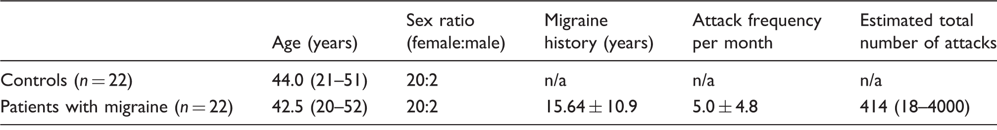
Data are presented as median (range) or mean ± SD values.
The testing software was written in Assembly for Windows. The software was a modified form of RAET (18), used and modified with the written permission of Myers et al. (18). The tests were run on a Lenovo ThinkPad T430 laptop computer. The testing sessions took place in a quiet room with the participants sitting at a comfortable distance from the computer screen. One participant was tested at a time and no time limit was set so that the participants could concentrate on the task.
The testing was carried out according to the method of Myers et al. (18). The acquired equivalence paradigm was structured as follows (see also Table 2). On each trial of the task, the participants saw a face and a pair of fish and had to learn through trial and error which of the fish went with which face (Figure 1). There were four faces (A1, A2, B1, B2) and four possible fish (X1, X2, Y1, Y2), referred to as antecedents and consequents, respectively. In the initial training stages the participants were expected to learn that when A1 or A2 appeared, the correct answer was to choose fish X1 over fish Y1; given face B1 or B2, the correct answer was to choose fish Y1 over fish X1. If the associations were successfully learned, the participants also learnt that face A1 and A2 were equivalent with respect to the associated fish (faces B1 and B2 likewise). Next, the participants learned a new set of pairs. Given face A1, they had to choose fish X2 over Y2 and given face B1 they had to choose fish Y2 over X2. The participants were then given a transfer test to see if they would choose fish X2 or Y2 given face A2 or B2. Having learned that faces A1 and A2 were equivalent, the participants may generalize from learning that if A1 goes with Y1, then A2 also goes with X2; the same holds for B2 (equivalent to B1) and Y2 (associated with B1). Although the formal description may give the impression that the task is difficult, healthy children (20) and participants with mental retardation (21,22) have been shown to reliably make this kind of generalization.
Example screen events during one trial. (a) Stimuli appear. (b) Participant responds and corrective feedback is given. Translation of Hungarian: (a) ‘Which fish belongs to her? The LEFT or the RIGHT one?’; (b) ‘Right!’. Summary of the acquired equivalence paradigm (18).

The participants’ task throughout the acquisition and testing phases was to indicate their choice in each trial by pressing one of two keyboard buttons labelled LEFT and RIGHT. Visual feedback on the correctness of choice was provided in the acquisition phases, but not in the transfer phase. The original version of the test uses audio-visual feedback, but as our aim was to test visual learning, the audio responses were omitted. New associations were introduced one by one during the acquisition stages. New associations were presented mixed with trials of previously learned associations. The participants had to achieve a certain number of consecutive correct answers after the presentation of each new association (four after the presentation of the first association, and 4, 6, 8, 10 and 12 with the introduction of each new association) to be allowed to proceed. This meant an increased number of the required consecutive correct trials compared with the original paradigm, which made getting through the acquisition phase by mere guessing less probable. Similarly, there were 48 trials in the transfer phase (12 trials of new and 36 trials of previously learned associations), as opposed to the 16 trials of the original paradigm.
The results were analysed in three groups: the results from the acquisition phases; the results from the ‘old associations’ part of the test phase (i.e. when the participant was presented an already learned association); and the results from the transfer part of the test phase (i.e. hitherto not learned associations). The number of correct and wrong answers were recorded in all phases, as well as the ratio of these numbers to the total number of trials during the respective phase. The number of trials necessary for the completion of the acquisition phase was also recorded.
Statistical analyses were conducted in SPSS 21.0 (IBM, USA) to compare the migraineurs and controls in terms of error ratios and the number of trials necessary for the completion of the acquisition phase. As the Shapiro–Wilk test indicated a normal distribution for all studied variables, one-way ANOVA was used for the comparisons. Additional linear regression analyses were performed to determine whether any of the examined migraine characteristics (e.g. migraine history in years, attack frequency per month) had an effect on the target variables in the migraine group (error rates and the number of acquisition trials). The effect of interval treatment as a long-term influence (and thus a potential confounder) was also tested.
Results
All the migraineurs and controls were able to complete both phases of the task. The mean error ratio during the acquisition phase was significantly higher in the migraine group than in the control group (0.16 vs. 0.078 mean error ratios for migraineurs and controls, respectively; F = 9.078; df = 1; p = 0.011, two-tailed; η2 = 0.144). This is also reflected in the fact that the migraine group needed significantly more trials than the controls for the completion of the acquisition phase (mean number of trials n
As the migraine group needed significantly more trials to reach criterion, they were over-trained. To check if this over-training could have led to the significantly poorer performance of the migraineurs on the generalization part of the task, we conducted ANCOVA with the number of teaching trials as a covariate. The significance decreased, but the effect remained highly significant (F = 11.364; df = 1; p = 0.002) (Figure 2). The most marked difference between migraineurs and controls was therefore observed in the transfer phase, which is well illustrated by the distribution of the participants’ performance expressed as their error ratios. In the control group, 19 of the 22 participants (86.36%) stayed below an error ratio of 0.01. Twelve of these participants (54.54% of the control sample) made no error at all. The maximum error ratio in the control group was 0.5, reached by only one participant. In contrast, in the migraineur group, 14 (63%) participants were characterized by error ratios > 0.5, including four participants (18% of the migraineur sample) whose error ratio was 1.0. Only five (22.7%) participants made no error in this group.
Error ratios in the three main phases of the paradigm. Learning error ratio and known pairs error ratio characterize learning efficiency in the acquisition phase, whereas the transfer error ratio characterizes the ability of the participant to generalize the acquired rule of equivalence to novel recombinations of previously trained stimuli. Black: migraineurs; grey: controls; columns: means; error bars: standard deviation.
Interval treatment (flunarizine) had no effect on performance in any of the test stages (patients receiving interval treatment vs. patients not receiving interval treatment; p = 0.46, 0.98 and 0.30 for acquisition, testing for acquired pairs and transfer, respectively). According to the regression analyses, age did not have a significant influence on any of the test variables in either group and, in the migraine group, neither migraine duration in years, attack frequency per month nor the estimated total number of attacks during the participant’s lifetime made a significant difference in any of the test variables. In other words, the test variables were independent of these factors.
Discussion
These results indicate that migraineurs acquired visual stimulus pairings and the pairing rule with significantly greater difficulty than matched controls, but, having acquired them, their recall performance for already seen stimulus pairs was on par with that of controls. However, when it came to generalizing the pairing rule to hitherto not seen stimuli, they performed predominantly at chance level or worse, whereas most controls applied the rule correctly with an error ratio < 1%.
The acquisition phase of the paradigm applied in this study involves two different kinds of learning: associative learning of the individual stimulus pairs and the implicit learning of stimulus categories. Trial and error associative learning of the individual stimulus pairs with immediate feedback after each trial assumes the intact functioning of the dorsal striatum (16) and is highly dependent on the dopaminergic input (23).
Although the neural basis of the implicit learning of categories (i.e. the rule of which stimuli belong together as functional equivalents in our paradigm) is not well understood, it is widely accepted that the basal ganglia play a key part in this form of learning (24). In their elegant series of experiments, Meeter et al. (19) showed that the stimuli in RAET become functional equivalents through an alteration of their representation in the participants’ memory. Implicit category learning is hypothesized to depend on posterior cortical areas supported by cortico-striatal circuits, connecting these to the basal ganglia, specifically the caudate (25,26). Considering what we know about associative learning and the findings of Meeter et al. (19), the role of these areas in this paradigm is to associate the individual stimulus pairs and to alter stimulus representations in such a way that functionally equivalent stimuli belong to the same category. It comes as no surprise that patients with Parkinson’s disease showed a significant deficit in this phase (18).
In view of the assumed role of the basal ganglia in the memory processes of the acquisition phase, our results seem to be a behavioural corroboration of the findings of Maleki et al. (13) based on functional magnetic resonance imaging. They concluded that ‘migraine attacks the basal ganglia’ (13). We also took into consideration the fact that the calcium channel antagonist flunarizine – the prophylactic drug used by half of our patients – is also a dopamine receptor antagonist (27,28) that has been shown to cause learning and memory deficits in animal models (29,30). As trial and error learning is dopamine-mediated, we expected that flunarizine could interfere with it. The results did not show this, however: flunarizine did not have any influence on the performance of patients in the acquisition phase. Given the small sample size, it cannot be generally concluded that the drug has no influence on learning of this type, but, in this study, deficient performance in the acquisition phase (i.e. trial and error learning and implicit rule acquisition/functional equivalence) appeared to be related to migraine, which supports the hypothesized involvement of the basal ganglia in migraine. It should not be forgotten, however, that feedback-guided associative learning is not pure implicit learning (31). Participants make conscious efforts to memorize the associations and they might apply various strategies to reach this end. The wrong strategy, consciously applied, can have profound effects on the outcome. As we did not examine this aspect in this study (i.e. conscious rule/strategy application), this possibility cannot be ruled out.
As suggested by Myers et al. (18), the hippocampi are structures of key importance in the transfer phase, when the rule of functional equivalence has to be applied to new stimuli: patients with hippocampal atrophy cannot make this kind of generalization. Our results suggested that migraineurs have similar difficulties. More than 60% of the studied migraine group performed below chance level and nearly 20% (four participants) appeared to have generalized reliably according to a rule other than that defined by the task (no correct response). On one hand, this could indicate hippocampal dysfunction in migraine. Based on the results of this study, we cannot argue for or against this possibility and published data on hippocampal involvement in migraine is almost non-existent. In rodent models, cortical spreading depression has been described to spread on to the hippocampi and cause functional alterations (14,15), but, as we studied patients with migraine without aura, the effect of cortical spreading depression does not seem to be a plausible explanation – and it is not known whether the same effect is seen in humans. An alternative explanation is that this is a secondary deficit that follows logically from the suboptimum functioning of the basal ganglia. If the modification of stimulus representations cannot happen and thus functional equivalence between the antecendents and consequents is not formed, there is no rule that the hippocampi could apply to new stimuli.
Four participants in the migraine group failed to give correct responses in the transfer phase. Could it be that they applied a wrong rule/strategy in the acquisition phase and they carried this over to the transfer phase? If so, it should be reflected in their performance in the acquisition phase. To examine this possibility, we compared the acquisition phase indices of 100% incorrect responders with those of 100% correct responders. The only difference we found was that the 100% incorrect responders needed almost twice as many trials to accomplish the acquisition phase (130 vs. 73), but they finally acquired the rule at the same level as the 100% correct responders. The same is true for those who generalized exactly at chance level (144 teaching trials). This suggests that the generalization error is not secondary to failure in the acquisition phase.
But does this task really depend on the basal ganglia and the hippocampi, or is there something else that is common to the various disorders that have been described to lead to a deficient performance? Such disorders include Parkinson’s disease and hippocampal atrophy (18), mild Alzheimer's disease (31) and long-term cocaine use (32).
Any psychophysical task that requires the participants to maintain attention for a longer period raises the question of attention deficit. Although we did not monitor attention in any direct way, we observed no deterioration in performance as a function of time (which would indicate difficulty in maintaining attention) and the (limited amount of) published data on visual attention in migraine is definitely against the idea that migraine is characterized by attention deficit by default (33–35), which is not to be confused with an inability to filter visual noise (36). Therefore we do not consider the observed alterations to be attention-related. This led us to conclude that, even if any or all of the other disorders are characterized by a general attention deficit or difficulty in maintaining attention, migraine does not share this feature, so in migraine it is not a likely explanation for the deficient performance.
How do migraine patients perform compared with patients with other disorders in which a deficient performance on RAET was found? In patients with Parkinson’s disease (18) and those with long-term cocaine use (32), the marked deficit characterizes the acquisition phase, but if the participants reach criterion, the transfer is almost intact. Both phases are affected in patients with migraine, but the deficit of the transfer phase is much more obvious. The availability of fronto-striatal dopamine is limited in patients with Parkinson’s disease and the availability of D2 dopamine receptors is limited in those with long-term cocaine use (37). We might therefore conclude – without limiting the problem to the basal ganglia – that the acquisition phase of the task is dependent on the intact functioning of the fronto-striatal dopamine system. Given the known association of migraine with extrapyramidal disorders (38–40), impaired dopamine-mediated learning would come as no surprise, but the role of flunarizine has to be clarified before any conclusion can be drawn.
Our findings show the closest similarity to the results found in patients with mild Alzheimer’s disease (31) – that is, mild impairment in the acquisition phase with profound impairment in the transfer phase. This finding provides further support to the assumption that the transfer phase is dependent on the hippocampi (and their connections) because hippocampal atrophy is a key feature of the early stages of Alzheimer’s disease (41). If we accept this assumption as a working hypothesis, our results might be interpreted as behavioural support for the involvement of the hippocampi in migraine – without aura in this specific example.
These are only simplifying hypotheses, however. The only strong conclusion our data allow is that certain learning and memory processes linked in previously published work to the basal ganglia and the hippocampi are deficient in patients with migraine, which, in our opinion, deserves further exploration, especially because important questions remain unanswered.
One of these questions is the issue of sex differences. As a result of the uneven distribution of the sexes in our sample (even if reasonable given the population), sex-related conclusions cannot be drawn from the results. This uneven distribution followed from the rigorous application of the diagnostic criteria and, in this sense, it might be regarded as the cost of having a study group free of false positives; however, a more balanced sample would be desirable. Another important question is how flunarizine influences the performance on this task. Although we found no statistically significant difference between patients in this respect, a 10 vs. 10 comparison is far too weak to allow any conclusions to be drawn. As data are available on the learning impairments caused by other calcium channel blockers (e.g. verapamil) in humans, it is difficult to accept that flunarizine has no such effect, especially because it is also a dopamine receptor antagonist. The application of the same paradigm to a population of patients with migraine with aura would help to clarify these findings.
Conclusions
We sought to provide behavioural evidence for the involvement of the basal ganglia and the hippocampi in migraine. We applied a learning and generalization paradigm that has been suggested to rely on these structures. We found a mild (but significant) impairment in associative learning with profoundly impaired generalization. Our results support the involvement of the basal ganglia and the hippocampi (more precisely, their functional networks) in migraine without aura. Although we cannot provide an exact explanation for our findings, these results emphasize the importance of further research into how migraine (with and without aura) affects subcortical structures and non-neocortical areas.
Article highlights
Adult migraineurs and controls were tested with a computerized acquired equivalence paradigm assumed to rely on the basal ganglia and the hippocampi. The paradigm involved associative learning, implicit rule extraction and rule generalization. The migraineurs showed significantly poorer performance than the controls in the phases related to the basal ganglia (associative learning and implicit rule extraction). Most of the migraineurs performed below chance level in the phase related to the hippocampus (rule generalization) and some of them could not make the generalization at all. The results seem to support the hypothesized involvement of the basal ganglia and the hippocampi in migraine without aura. The raw data of the study are available from the corresponding author by email.
Footnotes
Acknowledgements
Attila Öze and Attila Nagy contributed equally to the study. The authors express their gratitude to Catherine E Myers at Rutgers University for her generously allowing us to re-code and use the fish test in Hungarian and to Dr László Berente, leader of the Department of Neurology at the István Bugyi Hospital, Szentes, Hungary, for his generous professional support. We are especially grateful to Anna Pihokker, Petra Rózsa and Dr Gabriella Eördegh for the recruitment of healthy control participants.
Declaration of conflicting interests
The authors declared no potential conflicts of interest with respect to the research, authorship, and/or publication of this article.
Funding
The authors disclosed receipt of the following financial support for the research, authorship, and/or publication of this article: This work was supported by Hungarian Brain Research Program Grant KTIA_13_NAP-A-I/15 and by OTKA Hungary Grant No. K83810.