
Abstract
Background
Fixed-functional appliances (FFAs) are commonly used to correct skeletal Class II malocclusions in adolescents, enhancing mandibular growth and facial esthetics. Dolphin Imaging’s Visual Treatment Objective (VTO) module simulates soft tissue changes, but its predictive accuracy after FFA therapy remains uncertain, especially across sexes.
Aim
To evaluate and compare Dolphin VTO soft-tissue prediction accuracy between males and females following FFA treatment.
Materials and Methods
Lateral cephalograms from 40 Class II adolescents (20 males, 20 females) were analyzed. Predicted and actual post-treatment soft-tissue values were compared using paired and independent t-tests.
Results
Dolphin VTO predictions for soft-tissue changes post fixed-functional therapy had moderate accuracy, with the highest errors in angular parameters and areas involving vertical projection. Males had the greatest error in the H-angle, while females showed the largest discrepancy in the soft tissue facial angle (S.T. angle). A statistically significant gender difference was seen only in the S.T. angle (P = .04), with females showing greater deviation. Other parameters showed no significant gender-based differences, though variability was slightly higher in females.
Conclusion
Dolphin VTO is generally accurate, but gender-specific discrepancies should be considered in treatment planning and monitoring soft tissue changes during and after FFA treatment to ensure that patients achieve their desired facial esthetics.
Introduction
Accurate prediction of soft-tissue changes following orthodontic intervention is essential for effective treatment planning and patient communication. Fixed-functional appliances (FFAs) are widely used to correct skeletal Class II malocclusions by inducing both skeletal and dentoalveolar changes. As these changes are often reflected in the soft-tissue profile, clinicians increasingly rely on virtual treatment objective (VTO) tools, such as Dolphin Imaging software, to simulate post-treatment outcomes. However, soft-tissue responses to therapy may vary with gender, necessitating a more nuanced understanding of prediction accuracy. 1
Several studies have evaluated the accuracy of Dolphin VTO in predicting soft-tissue changes. In 2004, Hajeer et al. demonstrated that Dolphin Imaging software could reasonably predict post-treatment soft-tissue profiles in Class II cases, though accuracy varied among regions of the face. 2 In 2024, Maniyar et al. found a moderate correlation between predicted and actual soft-tissue changes, highlighting discrepancies in the perioral region. 3 Furthermore, research by Alhumadi et al. in 2022 emphasized the need for individualized predictions, noting gender-based differences in soft-tissue thickness and morphology. 4
Prior investigations into Dolphin VTO have largely focused on orthognathic surgery or extraction-based orthodontics, reporting mixed results,5, 6 while others reveal clinically significant discrepancies across multiple landmarks.7–9 Holdaway’s soft tissue analysis remains widely used for quantitative comparison. Comparative gender studies on the effects of FFAs in orthodontics often reveal that girls and boys respond differently to the treatment, especially in terms of mandibular growth and development.10, 11 Given the paucity of data on FFA-specific outcomes, this study aims to rigorously compare Dolphin VTO predictions against actual post-FFA soft tissue changes in a cohort of Class II adolescent boys and girls.
Materials and Methods
Study Design
A comparative retrospective study at the Department of Orthodontics and Dentofacial Orthopedics was conducted, and institutional ethical committee approval was obtained for the conduct of the study. This study was a record-based descriptive study using lateral cephalograms.
Sample Size
Based on Zhang et al.’s variance estimates (SD1 = 0.47, SD2 = 0.39, ∆ = 0.25 mm), α = 0.05 and power = 0.80; the sample size calculated was n ≈ 40. 1
Data Acquisition
Lateral cephalograms from 40 Class II adolescents (20 males, 20 females) were analyzed. Pre- and post-treatment lateral cephalograms were imported into Dolphin Imaging software 11.95 (USA). Dolphin’s VTO module generated predicted post-therapy tracings using default soft tissue ratios.
Soft Tissue Parameters
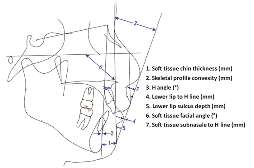
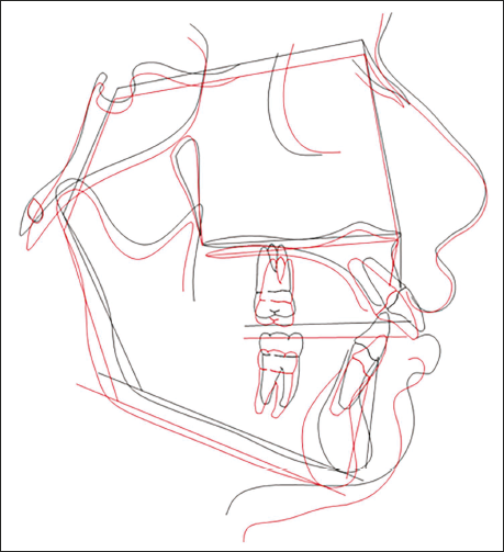
Seven landmarks from Holdaway’s soft tissue analysis were measured on both predicted and actual tracings (Figure 1): convexity (A–N′), lower-lip to H-line, soft tissue facial angle (S.T. angle) (FH–N′Pg), subnasale to H-line, H-angle, inferior sulcus to H-line and chin thickness (Pg–Pg′) (Figure 1). The lateral cephalograms were superimposed to determine the prediction errors (Figure 2).
Parameters of Holdaway Soft Tissue Analysis.
Cephalometric Superimposition of the Actual Changes After Treatment (Red Line) and the Visual Treatment Objective (VTO) Predicted Changes (Black Line) for Calculating the Predication Errors.
Inclusion and Exclusion Criteria
Inclusion Criteria
Age 12-25 years.
Skeletal maturity index ≥8.
Class II molar relationship.
Clear pre- and post-FFA cephalograms suitable for landmark identification.
Exclusion Criteria
History of facial trauma, congenital syndromes, or craniofacial deformities.
Prior orthognathic or maxillofacial surgery.
Temporomandibular joint disorders.
Poor radiographic quality or incomplete records.
Statistical Analysis
Data were analyzed using Statistical Package for the Social Sciences (SPSS) statistical software (IBM Corp) (v.26.0). A paired t-test was used to compare the statistical differences between the predicted and actual treatment outcomes of the parameters used in the Holdaway soft tissue analysis across both genders. P values of <.05 were considered statistically significant.
Results
Descriptive Statistics
Mean predicted versus actual values showed close agreement (all differences ≤2 mm). S.T. angle had the most variation between the sexes.
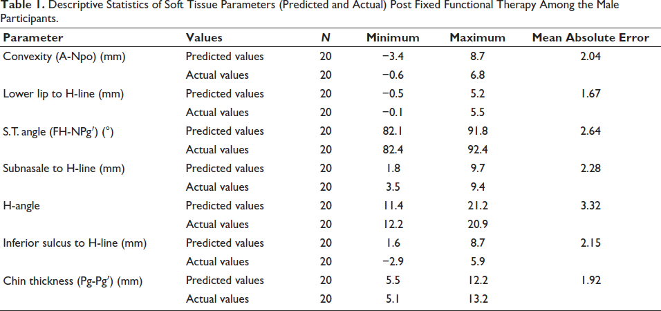
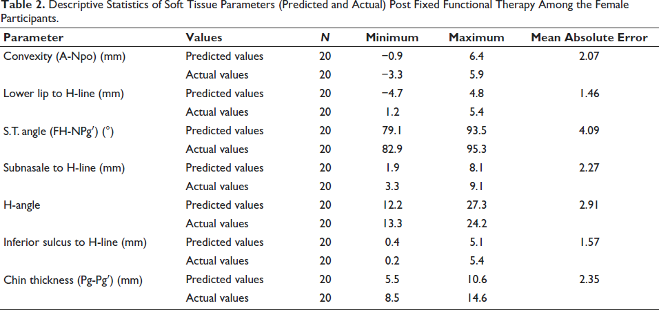
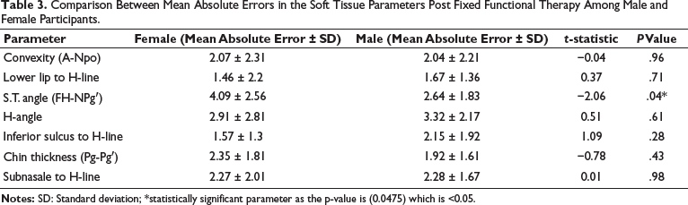
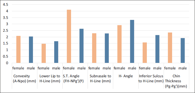
Tables 1 and 2 present predicted and actual soft-tissue measurements for male and female patients post FFA therapy. In both groups, differences between predicted and actual values were evident across several parameters, indicating varying degrees of prediction error. For males, the highest mean absolute error (MAE) was noted in the H-angle (3.32°), followed by the S.T. angle (2.64°) and the subnasale to H-line (2.28 mm). For females, the largest discrepancy was found in the S.T. angle (4.09°), followed by the chin thickness (2.35 mm) and the H-angle (2.91°).
Descriptive Statistics of Soft Tissue Parameters (Predicted and Actual) Post Fixed Functional Therapy Among the Male Participants.
Descriptive Statistics of Soft Tissue Parameters (Predicted and Actual) Post Fixed Functional Therapy Among the Female Participants.
These results suggest that Dolphin VTO predictions were moderately accurate but tended to under or overestimate certain facial regions, especially angular parameters and areas involving vertical projection (e.g., lips, chin).
Table 3 and Figure 3 highlight key differences in prediction error between male and female groups. The S.T. angle showed a statistically significant difference (P = .04), with females demonstrating greater deviation between predicted and actual values than males. This may reflect the complex soft-tissue response and variability in mandibular morphology in females post-treatment. For all other parameters, there were no statistically significant differences in MAE between genders (P > .05), although trends suggest slightly higher variability in females in some measurements (e.g., chin thickness, H-angle).
Comparison Between Mean Absolute Errors in the Soft Tissue Parameters Post Fixed Functional Therapy Among Male and Female Participants.
Comparison Between Mean Absolute Errors in the Soft Tissue Parameters Post Fixed Functional Therapy Among Male and Female Participants.
Discussion
This study assessed Dolphin VTO’s predictive precision for soft tissue changes after FFA therapy between male and female participants. While certain parameters (subnasale to H-line, H-angle) showed no significant differences, three parameters were predicted more accurately in females than in males.
Parameters that were more accurately predicted in females included lower lip to H-line, S.T. angle, and inferior sulcus to H-line. Lower lip and inferior sulcus predictions are better for females, which may be due to sex-based differences in soft tissue morphology.
Parameters that were more accurately predicted in males included convexity (A-Npo), which was notably better predicted, and chin thickness was also more accurately predicted in males.
Of the seven soft-tissue variables analyzed, only the S.T. angle showed a statistically significant difference in prediction accuracy between males and females (P = .0475), with females exhibiting higher MAE than males. This suggests a potential limitation in the VTO system’s ability to accurately model post-treatment changes in this specific anatomical region among female patients.
All other variables—including convexity, lower lip prominence, H-angle, inferior sulcus depth, chin thickness, and subnasale position—did not show statistically significant differences between sexes. This implies that the Dolphin VTO system maintains a relatively consistent level of prediction accuracy for most soft-tissue features, regardless of patient sex.
Our findings align partially with previous comparative studies that suggest gender-related variability in orthodontic outcomes. Certain studies have documented differential soft-tissue adaptation to FFAs between gender.12, 13 However, few studies have quantitatively evaluated these differences in the context of automated VTO predictions, making our results a novel contribution in this area.
Clinical Significance
The observed disparity in S.T. angle predictions may reflect underlying biological differences in the way male and female soft tissues adapt to skeletal changes during fixed functional therapy. For clinicians, these findings highlight the importance of considering gender-specific prediction variances when using VTO tools to plan or communicate expected treatment outcomes. In particular, extra caution may be warranted when interpreting soft-tissue angular predictions for female patients.
This study has several limitations. First, the relatively small sample sizes in both male and female groups may limit the statistical power and generalizability of the findings. Second, the exclusive use of a single VTO prediction system—Dolphin Imaging—means that the results may not be applicable to other prediction platforms with different algorithms or modeling approaches. Lastly, the accuracy of soft-tissue predictions can be influenced by additional variables such as patient age, individual growth patterns, and the timing of treatment, none of which were controlled for in this analysis.
Future Directions
Three-dimensional imaging [cone beam computed tomography (CBCT), stereophotogrammetry] and patient-specific soft tissue modeling (finite element analysis) could enhance predictive accuracy. Incorporating dynamic factors like muscle activity and tissue biomechanics into VTO algorithms warrants further research. Longitudinal follow-ups are needed to correlate prediction accuracy with actual treatment satisfaction and esthetic outcomes.
Conclusion
Comparative studies investigating the effects of FFAs on soft tissue changes after treatment, considering gender differences, show that while both genders experience some soft tissue changes, the magnitude and specific characteristics of these changes can vary. It is crucial to consider gender-specific differences in treatment planning and monitoring soft tissue changes during and after FFA treatment. This can help optimize treatment outcomes and ensure that patients achieve their desired facial esthetics.
Footnotes
Declaration of Conflicting Interests
The authors declared no potential conflicts of interest with respect to the research, authorship, and/or publication of this article.
Ethical Approval
The study was conducted in accordance with the Declaration of Helsinki and was approved by the Ethics Committee of KLE, VK Institute of Dental Sciences (no. 218) on November 16, 2023, with the need for written informed consent waived.
Funding
The authors received no financial support for the research, authorship, and/or publication of this article.
Informed Consent
The study being lateral-cephalogram-based, retrospective, and descriptive in nature. No patient photographs or personal descriptions were involved in the study.