
Abstract
Absolute anchorage systems like mini-implants, bone screws, and mini-plates have been a landmark evolution in the modern orthodontic world providing an added armamentarium in the hands of orthodontists to treat their patients with utmost efficiency. Unlike other systems, the extra radicular placement and limited surgical exposure make the bone screws such as infrazygomatic crest screws (IZC screws) superior, and to capitalize on these benefits, stability of the implant is vital that relies on their precise placement and angulation. Hence, this article presents a simple implant guide with elastomeric impression material for the precise positioning of IZC screws with utmost ease in fabrication.
Introduction
The introduction of absolute anchorage systems has offered significant benefits in anchorage preservation and has opened up a new envelope in treatment planning. Systematic reviews of the literature document that from both biomechanical and surgical perspectives, bone screws like infrazygomatic crest screws (IZC) in the maxilla and buccal shelf screws in the mandible are the silver lining because they are less invasive, extra radicular in location, close to the center of resistance, and do not interfere with tooth movement, thereby increasing the envelope of discrepancy. 1
The infrazygomatic crest is a bony ridge running along the curvature between the alveolar and zygomatic processes of the maxilla which is clinically palpable above the roots of maxillary first and second molars in adults. Literature evidence emphasizes that the accurate placement of extra radicular implant per se is of paramount importance for its stability with due consideration to the proximity of anatomically significant structures like maxillary sinus and tooth roots.2, 3 Lin et al 2 preferred IZCs to be placed between the first and second molar regions as the alveolar bone is thicker on the buccal surface of the second maxillary molar whereas Liou et al 3 suggested a more anterior placement, closer to the mesiobuccal root of the first molar, orienting screws at 14–16 mm above the maxillary occlusal plane with an angle of insertion between 55 and 70 degrees to the maxillary occlusal plane to achieve maximal buccal bone engagement.
Direct insertion of orthodontic implants without guidance resulted in an additional 20% injury rate during screw positioning 4 which led to the development of various implant guides 5 such as Chen’s double film method, Pinhead soft tissue penetration method, and 3D CAD/CAM surgical splints. All these methods had the added disadvantage of being cumbersome in fabrication and increased radiation exposure. The aim of this article is to provide an alternate simple and effective implant guide using elastomeric impression material for precision in IZC placement.
Clinical Procedure
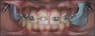
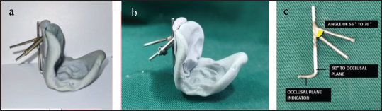
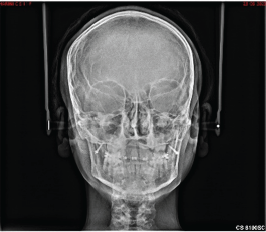
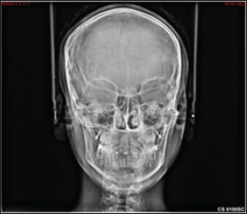
The patient was described about the procedure and informed consent was obtained in written form. Since this was a regular clinical case, ethical approval was not applicable. A Monophase impression was made involving the entire occlusal surface of the second premolars, molars, and the full sulcus in the buccal aspect of the maxillary arch using mucocompressive elastomeric impression material. Considering Lin et al’s 2 or Liou et al’s suggestions for IZC placement as a reference, either the site between the first and second molar region or the mesiobuccal cusp of the upper first molar was delineated in the set impression material respectively (Figures 1 and 2). Vertically, a heavy 14-gauge stainless steel wire approximating the diameter of bone screws was inserted into the elastomeric impression material at about 14–16 mm above the maxillary occlusal plane with an angle of insertion between 55 and 70 degrees to the maxillary occlusal plane verified with the help of a customized angle indicator made of 19-gauge stainless steel wire with a set angle of 60 degrees. Once the pilot hole is made in the impression material, the bone screw of comparable diameter was inserted into the hole such that it had a snug fit (Figures 3a–c). If the implant is driven directly into the impression material, the chances of material contamination between the threads might be present and this predrilling will prevent such incorporation in implant threads. With the impression and the angulated implant in place, a posteroanterior frontal radiograph was taken to assess the position of IZC before the final placement (Figure 4). If the implant position was satisfactory, the entire assembly was autoclaved and the bone screw was inserted into the infrazygomatic crest through the void created by the 14-gauge wire which will ensure precise placement of the implant. An additional posteroanterior cephalogram after placement can also be done to ensure the placement of IZC screw, which is not always necessary to limit patient exposure (Figure 5).
Frontal View
Buccal View.

Elastomeric Impression Material with Customized Angle Indicator
Posteroanterior Radiograph with Implant Guide.
Posteroanterior Radiograph After Implant Placement
Conclusion
This simple and effective implant guide made of elastomeric impression with a customized angle indicator provides insight into the precise placement of IZC implants. It differs from other implant guides mentioned in the literature with ease in fabrication, economical, time-saving, reliable, and less cumbersome to patients with limited radiation exposure.
Footnotes
Declaration of Conflicting Interests
The authors declared no potential conflicts of interest with respect to the research, authorship and/or publication of this article.
Funding
The authors received no financial support for the research, authorship and/or publication of this article.