
Abstract
Maxillary canine impaction is a malocclusion feature of very low prevalence (0.9%-2.2%) with a preponderance to females over males in the ratio of 2:1. However, when it presents, it is a complex orthodontic challenge. Treatment choices vary from no treatment to surgical exposure followed by orthodontic traction. Surgical exposure and orthodontic traction is an option that requires careful consideration of factors such as labiolingual position of the crown, vertical position of the tooth relative to mucogingival junction, amount of attached gingiva, and mesiodistal position of the crown. Age is an important factor in the prognosis of impacted maxillary canine treatment. Prognosis is lower in an adult compared to a younger patient and worsens with age. We report this case of impacted left maxillary canine (23) with palatal displacement in a young adult. A modified cantilever system was used for an early movement of canine from the lateral incisor which showed a grade II mobility. This along with controlled mechanics resulted in bringing the impacted 23 into class I position in the arch, thereby achieving a stable canine-guided occlusion within a period of 16 months.
Introduction
Impaction of maxillary canine is an anomaly that is low in prevalence1, 2 but is a complex orthodontic problem to manage. Two-thirds of maxillary canine impactions are palatal. The sequelae of untreated impacted maxillary canine teeth include loss of arch length, internal and external resorption of the canine as well as the adjacent teeth, dentigerous cyst formation, and referred pain. 3 Management of impacted maxillary canine involves options ranging from no treatment to surgical exposure followed by orthodontic traction. The latter option requires careful consideration of factors including labiolingual position of the crown, vertical position of the tooth relative to mucogingival junction, amount of attached gingiva, and mesiodistal position of the crown. Age is a detrimental factor in the prognosis. Success rate among adults is 69.5% compared to 100% among younger age group with similar presentation, the prognosis worsening with age. 4 Mechanics involved in the orthodontic traction need to be precise to make the path that the canine takes as close to the normal eruptive path as possible. 5 Proximity and involvement of the lateral incisors require adjuvant interventions. This report highlights a case of impacted left maxillary canine in a 20-year-old male seeking treatment for spacing and mobility of left lateral incisor.
Case Report
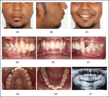
A 20-year-old male patient reported to the department of Orthodontics and Dentofacial Orthopedics with complaints of spacing and mobility of teeth in the upper front teeth region (Figure 1A-H). Clinical evaluation showed the molar relationship to be class I. He presented with an overjet of 3 mm, 80% overbite, retained remnant of 63 and 22 with grade II mobility. His maxillary dental midline had shifted to the left by 2 mm. A palatal bulge was evident in the region of 23. Orthopantomography (OPG) revealed an impacted 23(1I). The canine was diagnosed to be palatally impacted with the OPG using the method advocated by Chaushu et al. 6 The tooth exhibited 40° angulation to the midline, sector II vertical height, sector III anteroposterior position of the root, sector IV overlap over adjacent incisor, and no evidence of resorption of adjacent incisor. 7 The prognosis was average to poor.
(A-C) Pretreatment Extraoral View (Frontal, Profile, Frontal Smile), (D-H) Intraoral View (Right Lateral, Frontal, Left Lateral, Upper Occlusal [Circle Shows Palatal Bulge], Lower Occlusal), (I) OPG.
Alternative options of extraction/surgical relocation were given to the patient along with information about risks and benefits of all the options. The patient gave an informed consent for treatment to bring the impacted 23 into the arch.
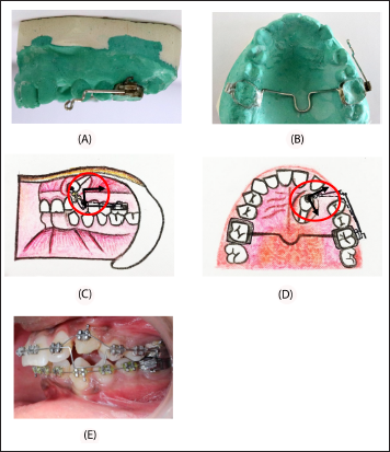
A modified cantilever system (MCS) (Figure 2A, B) consisting of a soldered extension (19 gauge SS) to headgear slot of first molar tube with transpalatal arch was fabricated and cemented. The impacted canine was surgically exposed using closed flap technique. Eruption chain was bonded and traction force applied by tying it to the anterior arm of MCS (2C, D). A distally applied force vector resulted in distal tipping of 23 crown away from the root of 22, since the point of application of force is restricted to the crown of 23 no reactionary forces were involved affecting other teeth particularly the mobile 22. This would not have been the case if conventional method of aligning the arch with continuous arch wires and retraction of canine was done simultaneously. Alignment and levelling of upper and lower arches was done with 0.022 × 0.028 dimension MBT 3M metal brackets. Upon completion of alignment and levelling with 0.019 × 0.025 SS wire, traction force was applied using 0.016 NiTi piggybacked on the main arch wire (Figure 2E). The arches were coordinated and finishing done using 0.019 × 0.025 HANT followed by 0.022 × 0.028 NiTi. At the end of 16 months, the treatment goals were met with (Figure 3A-H). Periodontal support to 22 was restored primarily by the use of MCS that led to early distal movement of canine without further compromising the periodontal health of 22.
(B-D) Modified Cantilever Spring, (E) Box Loop with Piggy Back Wire.
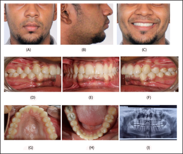
(A-C) Posttreatment Extraoral View (Frontal, Profile, Frontal Smile), (D-H) Intraoral View (Right Lateral, Frontal, Left Lateral, Upper Occlusal, Lower Occlusal), OPG.
Discussion
A tortuous course of eruption and long period of development makes permanent canines most prone for impaction. 8 During this course, the canines closely approximate the roots of lateral incisors. An estimated 0.71% of 10 to 13-year-old children have been shown to have resorbed permanent incisors due to ectopic eruption of canines. 9 Root integrity and periodontal support to the lateral incisors are important factors to be considered in gauging the prognosis of surgical exposure and traction of palatally impacted canines into the arch. Distalizing the canine away from the incisor root at the earliest is recommended. 10 Age is another determinant for the prognosis of treatment involving the traction of impacted canine into the arch. This case is being highlighted owing to its extremely good outcome despite the presence of 2 factors that could have undermined its prognosis viz: age and a periodontally compromised lateral incisor. However, these factors are not deterrents if one uses early distalization and proper directional forces of traction. 11 Application of MCS effectively distalized the canine which resulted in the improvement of periodontal status of the lateral incisor in a short period of time. This also resulted in ease of applying proper vector of forces later during the comprehensive phase of aligning and levelling of the arch. At the end treatment, we were able to achieve all the treatment goals including a class I positioning of the impacted canine along with improvement of periodontal support of the lateral incisor root.
Conclusion
Treatment of impacted canines in adult patients is a challenge wherein the risk-benefit assessment has to be made by the operator and conveyed to the patient unequivocally. In patients such as ours where the lateral showed grade II mobility, careful consideration should be given to the factor of proximity of canine to the lateral incisor root. Consolidation of arch with a rigid wire is needed to apply traction from the arch wire which consumes considerable period of treatment time. MCS is an effective way for early distalization of impacted canine prior to the beginning of traction. This prevents further compromise to the periodontal health of the lateral incisor. By initially distalizing the canine and applying precise mechanics, we were able to bring the impacted canine into the arch through the attached gingiva in a period of 16 months. One year post treatment, the patient showed a stable occlusion with good canine guidance, 22 with improved periodontal health, attached gingiva with adequate width, and aesthetics which immensely enhanced the confidence of the patient.
Footnotes
Declaration of Conflicting Interests
The authors declared no potential conflicts of interest with respect to the research, authorship, and/or publication of this article.
Funding
The authors received no financial support for the research, authorship, and/or publication of this article.
Statement of Informed Consent and Ethical Approval
Necessary ethical clearances and informed consent was received and obtained respectively before initiating the study from all participants.