
Abstract
Slaughterhouse inspections play a crucial role in the sanitary control of zoonoses and foodborne diseases. This study aimed to identify and analyze the frequencies of lymph node diseases in cattle slaughtered for human consumption, using the samples sent to the anatomic pathology service of the Federal Laboratory for Agricultural Defense (Laboratório Federal de Defesa Agropecuária), Minas Gerais, Brazil, from January 2015 to September 2022. In total, 2000 lymph node samples were analyzed, and additional information was individually retrieved. Lesions were most frequently identified in thoracic lymph nodes. Bacterial isolation and quantitative polymerase chain reaction (qPCR) were performed using samples suspected of tuberculosis. Tuberculosis cases accounted for 89.3% of the samples. Histopathology was more sensitive than other ancillary tests for diagnosing tuberculosis. Paraffin-embedded tissues from lymphoma cases were subjected to immunophenotyping using anti-CD3 and anti-CD79a immunohistochemistry. Frozen and/or paraffin-embedded tissues from lymphoma cases were used to identify the enzootic bovine leukosis (EBL) retrovirus through qPCR. Other diagnoses included primary (T- and B-cell lymphoma) and metastatic neoplasms (squamous cell carcinoma, pulmonary adenocarcinoma, undifferentiated carcinoma, undifferentiated adenocarcinoma, undifferentiated sarcoma, undifferentiated round cell tumor, mesothelioma, hepatic carcinoid, meningioma, and seminoma), actinogranulomas (pyogranulomatous lymphadenitis [actinobacillosis and actinomycosis]), idiopathic lymphadenitis (neutrophilic and/or histiocytic, granulomatous, and suppurative), and miscellaneous nonspecific lymphadenopathies (depletion/lymphoid atrophy, lymphangiectasia, erythrocyte drainage, parasitic eosinophilic lymphadenitis, follicular hyperplasia, and toxic granulomatous lymphadenitis). The combination of histopathology with complementary techniques is important for successful diagnosis, especially in complex cases of high epidemiological, economic, and zoosanitary importance, such as tuberculosis and EBL.
Brazil is home to one of the largest cattle herds, with >224 million cattle, as estimated by the Brazilian Institute of Geography and Statistics in 2021. 22 This massive production chain needs to be handled with great responsibility regarding planning, resource management, and ensuring health and disease control in all stages of animal production. In Brazil, veterinarians are responsible for meat inspection in slaughterhouses. 8 Slaughterhouse inspections are important for controlling zoonoses and foodborne diseases, including identifying exotic diseases and developing eradication programs,10,53 which can considerably reduce the economic losses resulting from the condemnation of carcasses.17,59
Although lymph nodes are consistently associated with various primary causes of condemnation in cattle slaughter facilities,35,47,48,54 their significance may be underestimated where the collection and examination of the organ is not mandatory. This oversight has led to notable epidemiological gaps in the understanding of the occurrence of certain diseases and their correlation with lymphatic distribution.
Therefore, this study aimed to evaluate the diagnostic approaches for identifying lymph node–associated diseases in cattle, as well as to identify and analyze the frequencies of various lymph node diseases in cattle slaughtered for human consumption, using the samples sent to the veterinary pathology diagnostic service of the Federal Laboratory for Agricultural Defense (Laboratório Federal de Defesa Agropecuária [LFDA]), Minas Gerais, Brazil, from January 2015 to September 2022.
Materials and Methods
Samples
This was a retrospective study that included cases diagnosed between January 2015 and September 2022. The cases included in this study were restricted to lymph nodes from cattle slaughtered for human consumption. All procedures were approved by the committee on ethics for the use of animals at the Universidade Federal de Minas Gerais (no. 222/2021). All 2000 samples were from the diagnostic service of the Veterinary Pathology Laboratory, from LFDA of Minas Gerais, Brazil, obtained by veterinarians from the Brazilian Federal Inspection Service (Serviço de Inspeção Federal) based on gross lesions observed on slaughter. Veterinarians enrolled in the Federal Inspection Service are expected to receive regular training in slaughter inspection and are required to follow the national guidelines for handling and collecting tissue samples for definitive diagnosis and corresponding ancillary tests.
Samples were placed in airtight containers containing 10% neutral-buffered formalin or were frozen in an airtight container for duplicated specimens (molecular identification). Samples were accompanied by official forms signed by the requesting veterinarian and were given a registration code from both the origin and LFDA. Thirty-six samples that were either confirmed or suspected of tuberculosis (TB) were retrieved from a previous validation study that aimed to assess the sensitivity and specificity of histopathology during an early phase of the period of study. These samples comprised a subset of cases (36/2000) and consisted of fixed and frozen lymph nodes sent for histopathology, and quantitative polymerase chain reaction (qPCR) and mycobacterial isolation, respectively.
Information regarding the time of entry into the Veterinary Pathology Laboratory, origin (federal unit and municipality of origin of the cattle and requested inspection service), sex, and approximate age (if available) were retrieved for each sample. In addition, information about the affected lymph nodes and simultaneously affected organs was listed individually whenever available.
Histopathology
Histological procedures were performed as previously described, 32 with minor modifications. Apart from hematoxylin and eosin, Goodpasture, Ziehl-Neelsen, periodic acid-Schiff, Grocott-Gomori’s methenamine silver, Masson’s trichrome, and Fontana-Masson histochemical stains were used on selected cases. Microscopic analysis established the types of pathological processes involved (such as infectious, neoplastic, or hyperplastic) and their respective subclassifications (such as bacterial, fungal, viral, protozoal, or toxic lymphadenitis; lymphomas; and hyperplasia). In cases where the definitive diagnosis of the main pathological process was not possible (such as etiology or neoplastic type), it was classified as “undetermined” or “undifferentiated.” Throughout the study period, definitive diagnoses were made individually for each sample, and the absolute and relative frequencies for each diagnosis were recorded. The cases were further subclassified into broad diagnostic categories according to their etiological proximity.
Immunophenotyping of Cases Suspected of Lymphoma
In total, 45 formalin-fixed paraffin-embedded samples were subjected to immunophenotypic analysis based on morphological and immunophenotypic features according to the Revised European American Lymphoma/World Health Organization classification adapted for animal use. 52 Immunohistochemical (IHC) analysis was performed using anticluster of differentiation (CD79a; ScyTek, West Logan, Utah; RA0071-C.5, clone JCB117 and HM47/A9; [1:300]) for B-lymphocytes and anti-CD3 (Sigma-Aldrich, Saint Louis, Missouri; C7930; [1:1000]) for T-lymphocytes as primary antibodies, which were incubated overnight at 4°C. A 10% hydrogen peroxidase solution was used to block the endogenous peroxidase activity. CD79a and CD3 were retrieved in a citrate buffer (pH 6.0) using a steamer cooker (96°C, 60 minutes). Following this, a protein block was applied for 30 minutes to prevent nonspecific binding. EnVision Flex/horseradish peroxidase (K800221-2, Dako, Carpinteria, California, USA) was used to amplify and detect the immunolabeling. The chromogen 3,3’-diaminobenzidine (ACB, ScyTek) was used for the visualization, before counterstaining with Harris hematoxylin solution. Tissue sections of lymph nodes from healthy slaughtered cattle were used as positive controls, and additional sections from the same samples incubated with a phosphate-buffered saline instead of primary antibodies were used as negative controls.
Samples tested for both markers were classified as reactive or nonreactive based on the presence or absence, respectively, of a strong/specific immunolabeling of either neoplastic or nonneoplastic lymphocytes (internal control). Samples classified as reactive were further subclassified as positive or negative, where positive samples corresponded to confirmed cases of lymphomas and negative samples corresponded to cases confirmed not to be lymphomas.
Classification of Bovine Lymphomas
Thirty cases of lymphoma previously subjected to histological and IHC analyses were originated from the 45 cases initially described and were classified based on topography, cellular distribution, morphology, and mitotic index, following the criteria established by the World Health Organization for the use of animal species, 52 and a previous study, 26 with minimum variations.
To establish a proportional nuclear size grading, based on the standard for dogs, an adjusted nuclear size was determined by multiplying the relative diameter of red blood cells (RBCs) by 1.4 for each of the categories and used as follows: small (<2 RBCs), intermediate (2–3 RBCs), and large (>3 RBCs), as bovine RBCs correspond to approximately 0.7 times the size of the cells found in dogs.2,50
The mitotic count was assessed as the sum number of mitoses in 10 high-power fields (×400). The field diameter was recorded as 0.55 mm and the field of view was 0.237 mm2 at a magnification of ×400. The mitotic index was recorded separately and graded as follows: low (0–5 mitoses/0.237 mm2), intermediate (6–10 mitoses/0.237 mm2), and high (>10 mitoses/0.237 mm2), as established previously. 51
DNA Extraction and qPCR for Molecular Diagnosis
DNA was extracted from the frozen tissues in 36 cases either confirmed or suspected of granulomatous lymphadenitis due to TB and 7 cases of histologically confirmed lymphomas, using a Maxwell 16 Tissue DNA Purification Kit (Promega) according to the manufacturer’s instructions. The subsampling criterion included the availability of frozen tissues for DNA extraction. The formalin-fixed paraffin-embedded tissues of the 36 cases of granulomatous lymphadenitis were additionally subjected to DNA extraction, together with 30 samples of histologically confirmed lymphomas, according to the instructions provided by the QIAamp DNA FFPE Tissue kit (QIAGEN) manufacturer.
The qPCR test for the enzootic bovine leukosis retrovirus (EBLV) was performed as previously established, 16 using the Taqman Fast Advanced Master Mix kit (Thermo Fisher Scientific). Based on previous studies, qPCR was performed for Mycobacterium spp. 42 and Mycobacterium bovis. 34 The analyses were performed using the GoTaq Probe qPCR kit (Promega). Additional qPCR for beta-actin was performed to evaluate the integrity of the DNA and overall quality of the samples, as previously described. 7
Mycobacterial Isolation of Samples From Suspected TB Cases
The 36 cases of either confirmed or suspected granulomatous lymphadenitis due to TB that were subjected to histology and qPCR were also subjected to bacterial isolation of Mycobacterium spp. 24 for comparison.
Data Analyses
Data relevant to the identification of the samples (including identification number, date of delivery, municipality of origin of the cattle and the requesting inspection service, sex, age, affected lymph node, concomitantly affected organs, and gross diagnosis), etiological diagnosis, definitive diagnosis, and etiology were retrieved from case records and compiled in the Excel spreadsheets (Microsoft Office). Samples were subdivided into diagnostic groups based on the overall etiological affinity among the diagnoses as follows: actinogranuloma, idiopathic lymphadenitis, TB, B-cell lymphoma, neoplasia, and others.
Absolute and relative frequencies were obtained for the different diagnoses and percentages were determined accordingly for each diagnostic group. The most frequent causes of lymphatic involvement, the geographic location, and the main types of lymph nodes affected throughout the retrospective study were also determined, and a descriptive analysis of these data was conducted.
Thematic maps were generated for the geographic identification of the origin of cattle and inspection services, based on the diagnoses (Arcgis software 10.3). Multiple correspondence analyses (MCA) were conducted to correlate affected lymphatic sites, their diagnoses, and concomitantly affected organs or gross diagnoses, which were exclusively applied to the confirmed cases of TB and enzootic bovine leukosis (EBL), using biplot-type graphs (R software 4.3.0). Sensitivity and specificity tests were also performed to individually correlate the gross diagnosis with definitive diagnoses of TB and EBL to compare the results of complementary tests with bacterial isolation in cases suspected of TB. Cohen’s kappa coefficient (k) analysis was performed to assess the agreement between the techniques evaluated based on sensitivity and specificity values (Minitab software 21.3), considering a 95% confidence interval. The results were graded into the following 6 categories based on the kappa values: no agreement (k ≤ 0), slight agreement (0.01 < k ≤ 0.20), weak agreement (0.21 < k ≤ 0.40), moderate agreement (0.41 < k ≤ 0.60), strong agreement (0.61 < k ≤ 0.80), and very strong agreement (0.81 < k ≤ 1.00).
Results
A total of 2000 samples were obtained at the end of the study period (January 2015 to September 2022). In each year, July and November accounted for the months with the highest number of samples collected.
Lymph Node Diseases and Differential Diagnoses
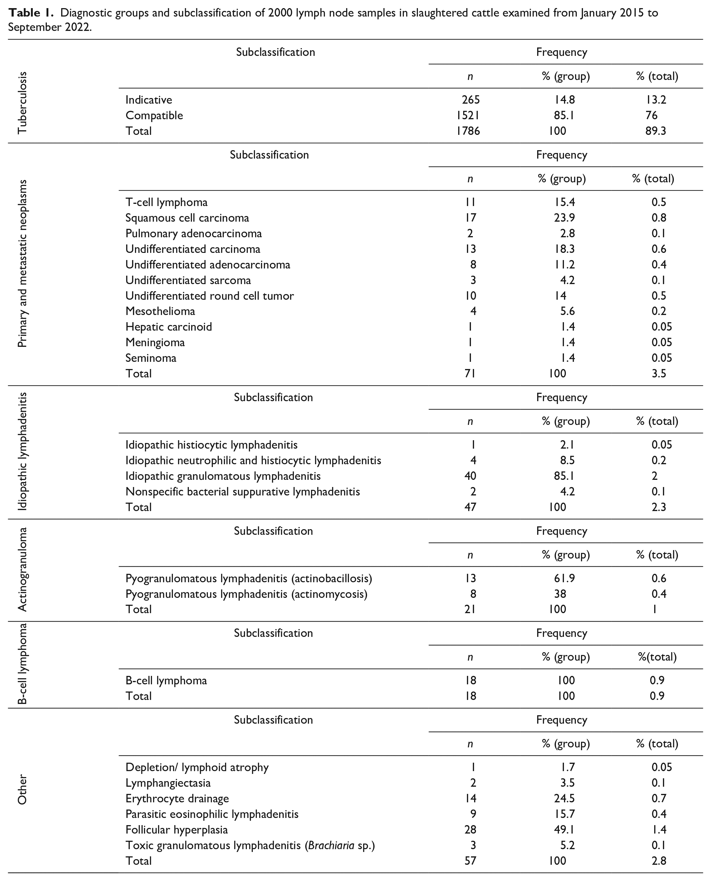
TB cases (Fig. 1a–c) represented 89.3% (1786/2000) of the total diagnoses, followed by neoplasms other than B-cell lymphomas (3.5%, 71/2000; Fig. 1d–f), idiopathic lymphadenitis (2.3%, 47/2000), actinogranulomas (1%, 21/2000; Fig. 1g–i), and B-cell lymphomas (0.9%, 18/2000; Fig. 1j–l), which were treated separately from the other neoplasms. Other causes of lymphadenopathy were recorded separately and represented 2.8% (57/2000) of the registered cases. The subclassification of each diagnostic group is presented in Table 1.
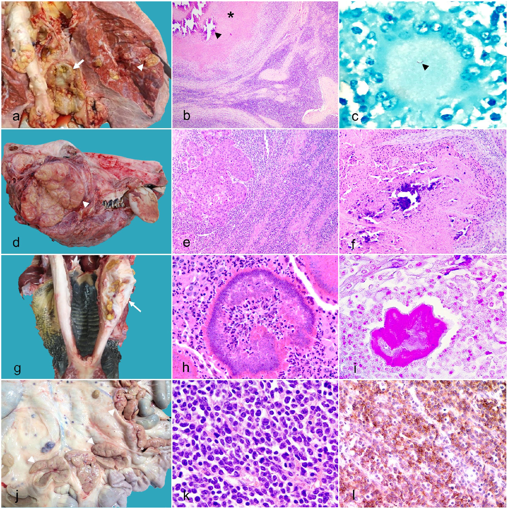
Gross and microscopic findings of lymph nodes in slaughtered cattle. (a–c) Granulomatous lymphadenitis due to tuberculosis. (a) Lung, dorsal view, and mediastinal lymph node, cut surface. Multifocal, granulomatous pneumonia (arrowhead) and lymphadenitis (arrow). (b) Granulomatous inflammation with extensive caseous necrosis (asterisk) and central mineralization (arrowhead). Hematoxylin and eosin (HE). (c) Acid-fast bacilli (arrowhead) within the cytoplasm of a multinucleated giant cell (Langhans-type). Ziehl-Neelsen. (d–f) Metastatic squamous cell carcinoma. (d) Head, lateral view. Infiltrative retrobulbar squamous cell carcinoma with involvement of parotid lymph node (arrowhead). (e) Infiltrative, nonencapsulated squamous cell carcinoma with coalescing islands of neoplastic cells. HE. (f) Extensive comedonecrosis, with central mineralization deposits. HE. (g–i) Granulomatous lymphadenitis due to actinomycosis. (g) Mandible, ventral view. Focally extensive, unilateral pyogranulomatous osteomyelitis (arrow). (h) Submandibular lymph node. Splendore-Hoeppli reaction (center) with interspersed degenerated neutrophils and epithelioid macrophages. HE. (i) Submandibular lymph node. Myriads of gram-negative bacterial bacilli. Goodpasture. (j–l) Enzootic bovine leukosis (B-cell lymphoma). (j) Mesenteric lymph nodes, cut surface (arrowheads). Note diffuse and marked enlargement of mesenteric lymph nodes, with homogeneous white appearance, and corticomedullary indistinction. (k) Mesenteric lymph node. Monomorphic population of neoplastic lymphocytes with centroblastic pattern (large nuclei, multiple mitotic figures, and infiltrative behavior). HE. (l) Mesenteric lymph node. Positive immunolabeling in neoplastic cells. Immunohistochemistry for CD79a.
Diagnostic groups and subclassification of 2000 lymph node samples in slaughtered cattle examined from January 2015 to September 2022.
Microscopic Analysis of Cases of TB
Cases of TB were often characterized by multiple mineralized granulomas (mature lesions; Fig. 1b) and poorly or nonmineralized granulomas (early mature lesions) with central to multicentric caseous necrosis and variable mineral deposits, externally surrounded by rims of fibrous connective tissue and lymphoplasmacytic infiltrates. TB cases were further subclassified as compatible (definitive diagnosis; 76%, 1521/2000) or indicative (presumptive diagnosis; 13.2%, 265/2000) upon the observation of mineralized caseonecrotic granulomatous lesions with or without intralesional acid-fast bacilli (Fig. 1c), respectively. In addition, the absence of typical caseonecrotic lesions in some lymph nodes allowed the differentiation of idiopathic granulomatous lymphadenitis from cases indicative of TB, where the microscopic feature of granulomatous inflammation with caseonecrotic lesions was identified and the visualization of acid-fast bacilli was not possible.
Microscopic, Molecular, and Microbiological Analysis of Suspected TB Cases
Suspected cases of TB-related lymphadenitis based on gross features during postmortem examination that were subjected to complementary testing comprised a subset of cases (36/2000; Supplemental Table S1). Among these, 31 (86%) were positive for intralesional acid-fast bacilli. In the qPCR analysis of paraffin-embedded and unfixed tissues, 12 (33%) and 22 (61%) cases, respectively, were positive for M. bovis. M. bovis was cultured and isolated in 28 (77%) cases. Of these, 23 (82%) cases were confirmed using qPCR. Quantification tests and DNA quality assessment for each sample through qPCR for the beta-actin gene showed relatively high values of postextraction total DNA concentration for all samples. Confirmation of TB diagnosis by any technique was not possible in 3 of the 36 cases tested. All cases included in this study were histologically classified as granulomatous lymphadenitis with typical caseonecrotic lesions.
Sensitivity and specificity tests comparing bacterial isolation with histopathology through the observation of acid-fast bacilli and qPCR assays of fixed and unfixed tissues indicated 96.4% sensitivity and 50% specificity for histopathology (Supplemental Table S2).
Histopathology, IHC, and qPCR of Suspected Lymphoma Cases
Data regarding histopathology, IHC, and qPCR analyses of confirmed cases of lymphoma are shown in Supplemental Table S3. A total of 18 cases of B-cell lymphoma were recorded of which diffuse large-cell lymphoma (monomorphic population of large B-lymphocytes with uniform, round, or cleft nuclei in diffuse growth; Figs. 1k, l) was the most common subtype. A total of 11 cases of T-cell lymphoma were recorded of which peripheral T-cell lymphoma (monomorphic population of uniform T-lymphocytes) was the most frequent subtype.
In addition, cases of anaplastic diffuse large B-cell lymphoma (pleomorphic population of large anaplastic B-cells with bizarre nuclei and abundant cytoplasm), lymphoplasmacytic B-cell lymphoma (monomorphic population of plasmacytoid B-lymphocytes with small nuclei), and small lymphocytic B-cell lymphoma (monomorphic population of small B-lymphocytes) were diagnosed as B-cell neoplasms. For T-cell lymphomas, subtypes of small lymphocytic T-cell lymphoma (monomorphic population of small T-lymphocytes) and anaplastic large T-cell lymphoma (monomorphic population of large T-lymphocytes with wide nuclear size and shape variation and multiple mitotic figures) were diagnosed.
Higher mitotic counts and mitotic index values were observed in animals diagnosed with B-cell lymphoma. Thoracic lymph nodes were frequently affected in T- and B-cell lymphomas but were particularly frequent in cases of B-cell lymphoma.
Of the 30 confirmed cases of lymphoma, unfixed tissues of 7 cases were tested by qPCR for EBLV of which 3 cases were positive for the tested virus. These 3 cases were also reactive for CD79a, confirming a B-cell origin. Five of the 30 cases, for which paraffin-embedded tissues were tested by qPCR, were positive for EBLV. Only 3 of these 5 cases were CD79a reactive, confirming a B-cell origin, whereas the remaining were CD3 reactive. The most frequently affected organs in EBL cases were the liver, intestine, and lungs. In the cases histologically confirmed as lymphomas, EBL was considered as the most likely differential diagnosis based on the gross lesions in all but 3 cases in which the differential diagnosis of lymphadenitis was considered. Owing to the scarcity of data regarding the ages of the animals, it was not possible to determine an age pattern for diagnosis, although most individuals were aged ≥3 years.
Ten cases of round cell tumors with a lymphocytic appearance did not react positively for CD3 or CD79a by IHC. The lack of immunoreactivity in these cases might be due to overfixation of tissue samples. These cases were included in the undifferentiated round cell tumor group.
The beta-actin gene was detectable by qPCR in 77% (23/30) of the tested, paraffin-embedded tissues samples with relatively high postextraction total DNA concentration values. The 7 cases that tested negative for the gene were equally negative in the qPCR examinations of unfixed and paraffin-embedded tissues for EBLV, with only 2 of these cases being reactive for CD79a.
Microscopic Analysis of Nonlymphoid Neoplasms
Data related to other types of neoplasms diagnosed in this study are presented in Supplemental Table S4. Sixty neoplasms other than lymphoma were identified in lymph nodes, with a predominance of metastatic squamous cell carcinoma (17/2000; polygonal epithelial cells organized in solid islands with keratin lakes and central necrosis; Fig. 1e, f), undifferentiated carcinomas (13/2000; pleomorphic epithelial cells organized in solid arrangements), and undifferentiated round cell tumors (10/2000; pleomorphic round cells that did not react positively to any of the lymphocytic immunomarkers).
Other neoplasms that were identified in the lymph nodes at a lower frequency included undifferentiated adenocarcinoma (8/2000; pleomorphic epithelial cells organized in tubulo acinar arrangements without distinct features from primary site), metastatic mesothelioma (4/2000; fusiform cells organized in circumferential arrangements with peripheral cords of cuboidal cells), undifferentiated sarcomas (3/2000; pleomorphic fusiform mesenchymal cells associated with the amorphous matrix), hepatic carcinoid (1/2000; polygonal neuroendocrine cells arranged in dense packages surrounded by fibrovascular stroma), seminoma (1/2000; round to polygonal, individualized germ cells with a high nucleus-to-cytoplasm ratio), and meningioma (1/2000; fusiform meningothelial cells organized in whorls and radially around blood vessels).
Similar to the cases of lymphoma (Supplemental Table S3), the thoracic lymph nodes were the most frequently affected by nonlymphoid neoplasms. Among the simultaneously affected organs, lesions were most commonly identified in the liver, lungs, and serous membranes. Most animals diagnosed with nonlymphoid neoplasms were ≥3 years of age, with a single animal affected by undifferentiated sarcoma at 2 years of age. The age of 8 years was the highest among the studied animals. Among the differentials suspected based on the gross lesions, TB and EBL were the most commonly cited differential diagnoses, corresponding to almost all the samples analyzed.
Microscopic Analysis of Cases of Actinogranulomas
Cases of actinogranulomas were characterized by pyogranulomas with myriads of gram-positive filamentous bacteria (8/2000; actinomycosis; Fig. 1h, i) or gram-negative bacilli (13/2000; actinobacillosis), and were often associated with abundant, deeply eosinophilic (hyalinized) material, forming radiating, club-like projections (Splendore–Hoeppli reaction).
Microscopic Analysis of Cases of Other Lymphadenopathies
Cases included in this subsection consisted of a miscellaneous group of lymphadenopathies with various etiopathogeneses. Cases of toxic granulomatous lymphadenitis had the following 2 histological patterns: (1) foamy or epithelioid macrophages containing irregular or acicular intracytoplasmic crystals arranged in the sinuses and corticomedullary cords of the hepatic and mesenteric lymph nodes, and (2) epithelioid macrophages or multinucleated giant cells randomly arranged in the cortex and medulla of multiple lymph nodes. The toxic causes were related to Brachiaria spp. and granulomatous systemic diseases (Vicia spp. or citric pulp), respectively.
The main histological features observed in the confirmed cases of parasitic eosinophilic lymphadenitis consisted of expanded lymphoid capsules and repleted lymphatic sinuses by a large number of eosinophils. In contrast, RBC accumulation in the dilated corticomedullary sinuses was related to erythrocyte drainage.
The expansion of lymphoid follicles with enlargement of the germinal centers and corticomedullary cords was the main feature observed in cases of lymphoid follicular hyperplasia. The rarefaction of the lymphoid cortex (decreased number of follicle units) was related to lymphoid depletion/atrophy, and the expansion of the subcapsular and corticomedullary sinuses was interpreted as lymphangiectasia.
Anatomic Distribution of Affected Lymph Nodes
Information related to the affected lymph nodes in each diagnostic group is shown in Fig. 2. TB was primarily identified in the thoracic lymph nodes, particularly the tracheobronchial lymph nodes (929/2000; Supplemental Fig. S3). Abdominal lymph nodes were also affected by TB, with involvement of the hepatic (542/2000) and mesenteric (391/2000) lymph nodes. Confirmed cases of EBL (B-cell lymphoma) showed a wide range of affected lymph nodes, with a predominance of thoracic lymph nodes, such as the mediastinal (10/2000) and prepectoral (8/2000) lymph nodes (Supplemental Fig. S4). Other neoplasms followed a very similar pattern, with a predominant involvement of mediastinal (31/2000) and tracheobronchial (19/2000) lymph nodes. In the cases of actinogranulomas, the prescapular (8/2000) and retropharyngeal (7/2000) lymph nodes were most commonly affected, whereas idiopathic lymphadenitis cases mainly involved the tracheobronchial (19/2000) and mediastinal (17/2000) lymph nodes. Other lymphadenopathies were commonly identified in the mediastinal (25/2000) and prescapular (15/2000) lymph nodes.
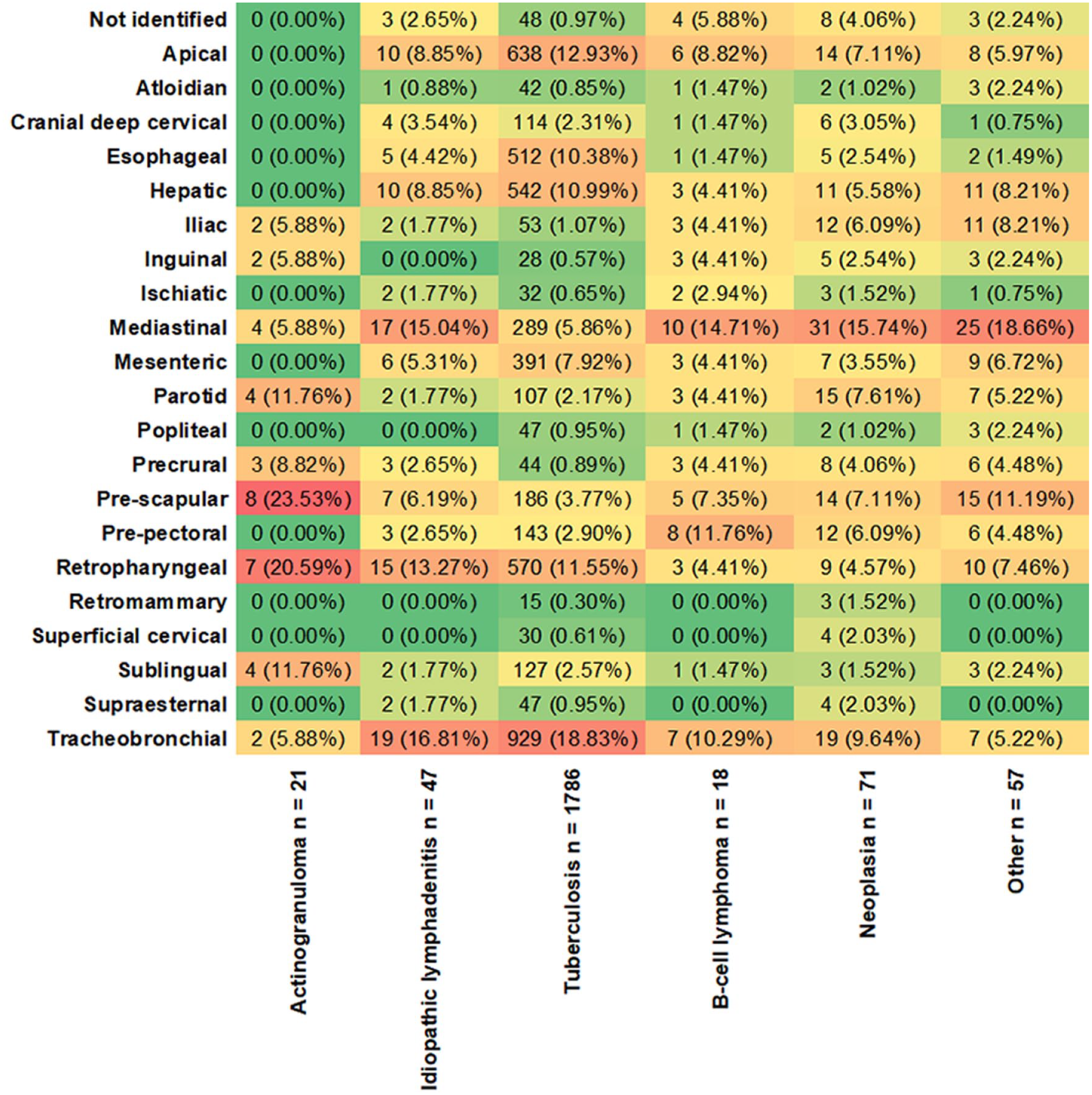
Correlation between diagnostic groups and affected lymphatic sites in slaughtered cattle. Numbers in the heatmap indicate absolute and relative values for each affected lymphatic site by diagnostic groups (columns). Values on the base of columns correspond to the absolute number of diagnoses for each listed group. Warmer colors represent higher relative values. Diagnostic groups must be analyzed separately.
MCA of Lymphatic Sites and Simultaneously Affected Organs in TB and EBL Cases
MCA for TB diagnosis (Supplemental Fig. S5) showed a greater correlation between the disease and thoracic or abdominal lymph nodes, such as hepatic and mesenteric lymph nodes, whereas lymph nodes in the inguinal and pelvic regions were minimally associated with the disease. Other lymph nodes were also affected in cases with TB, but they were mutually related to several other diseases analyzed together.
A complementary MCA was performed to correlate the TB diagnosis with simultaneously affected organs (Supplemental Fig. S6). In this analysis, the lungs were the concomitantly affected organ with the highest association in confirmed cases of TB, whereas the heart, peritoneum, and diaphragm had lower associations with the disease.
The same analysis for the diagnosis of EBL (B-cell lymphoma; Supplemental Fig. S7) showed a strong relationship between the disease and the following lymph nodes: atloidian, precrural, iliac, inguinal, ischiatic, and popliteal. Other lymph nodes related to the majority of EBL cases were also related to several other diseases, and it was not possible to establish a strong correlation between these other lymph nodes and confirmed EBL cases.
A complementary MCA was performed to correlate the EBL diagnosis with simultaneously affected organs (Supplemental Fig. S8). The analysis indicated that the abomasum, heart, diaphragm, intestine, peritoneum, and pleura were the organs that were most closely associated with the disease.
MCA also revealed that the diagnoses of TB and EBL did not correlate with any other gross differential diagnoses, which was different from each of these diagnoses alone. The sensitivity and specificity values for gross diagnoses were 95.8% and 44.8%, respectively, for TB, and 94.7% and 98.7%, respectively, for EBL.
The sex of the animals showed divergences in absolute terms, with considerably higher values for male (1310/2000) than for female animals (659/2000) or animals with unidentified sex (31/2000). However, the proportions were maintained within each diagnostic group, indicating no sex-based predisposition to any of the diagnoses.
Geographic Distribution of Origin of Samples and TB and EBL Cases
Data regarding the locations of inspection services are presented as thematic maps to determine the main Federal Inspection Services sent throughout Brazil (Supplemental Fig. S1). The inspection services located in 2 municipalities of the state of Minas Gerais (southeastern region of Brazil) were most notable in absolute terms, whereas a municipality in the state of Rio Grande do Sul (southern region of Brazil) was the second most representative for this study. In total, 37 Federal Inspection Services contributed to the caseload in this study. In 2.1% (43/2000) of the samples obtained from the inspection services, there was no information regarding the municipality of origin.
An additional thematic map was generated to visualize the locations of the cattle (Supplemental Fig. S2), proving the importance of the southern and southeastern regions for the number of cases included in our study. The 2000 samples used in this study were obtained from 12 states and 428 municipalities. The state of Minas Gerais was responsible for 68.5% of sample shipments, followed by the states of Rio Grande do Sul (8.6%), Paraná (7.4%; southern region of Brazil), Maranhão (5%; northeastern region of Brazil), Pará (3.5%; northern region of Brazil), Mato Grosso (2.3%; central–west region of Brazil), Tocantins (2.2%; central region of Brazil), Rondônia (1.2%; central–west region of Brazil), Piauí (0.4%; northern region of Brazil), Goiás (0.2%; central–west region of Brazil), São Paulo (0.2%; southeastern region of Brazil), and Espírito Santo (0.05%; southeastern region of Brazil). Accordingly, absolute numbers of TB cases were most frequently found in the state of Minas Gerais, particularly in 2 municipalities (viz, Araguari and Estrela do Sul), whereas EBL cases were concentrated in the northern, northeastern, and central–west states.
Discussion
Studies on the differential diagnosis of lymph node diseases in slaughtered cattle, which include a large number of individual samples subjected to different diagnostic techniques, and the information on the location of affected lymph nodes are limited. The results of this study reinforce the importance of epidemiological studies and the association among different diagnostic techniques for studying the pathogenesis, etiology, and distribution dynamics of certain diseases that affect cattle. The importance of inspection services and cattle slaughter databases has been evidenced through programs for surveillance and control of diseases transmitted through these animals.9,25,27
Regarding the affected lymphatic sites in cases of TB, some cases had the involvement of only hepatic and mesenteric lymph nodes compared with that of the thoracic lymph nodes. In most of these cases, only the abdominal viscera (including the intestine, spleen, and liver) were simultaneously affected (alimentary route of infection), which is consistent with the findings of another study. 19 In calves, ingestion of contaminated milk has been proposed as an important source of infection, 15 although the aspiration of amniotic fluid (congenital route) is also considered in neonates with the respiratory form of the disease.15,36,58 For oral transmission to occur in the cases of TB, several factors are contributory, such as the maintenance of viable bacilli in the environment 6 and ingestion of numerous bacilli. 12
It is still possible that bacteria of the Mycobacterium avium subsp. paratuberculosis complex, associated with paratuberculosis (Johne’s disease), is also associated with alimentary lesions, considering that this agent is a frequent cause of intestinal lesions in cows.49,52 However, this etiological distinction was not possible in this study due to methodological limitations. In addition, the absence of typical caseonecrotic lesions in some lymph nodes allowed for the differentiation of idiopathic granulomatous lymphadenitis from cases indicative of TB in which the microscopic feature of granulomatous inflammation with caseonecrotic lesions was identified and the visualization of acid-fast bacilli was not possible. As paucibacillary lesions are frequently observed to indicate TB in cattle, the presence of acid-fast organisms is not always readily observed in histological sections. 39 Fresh samples for bacterial isolation were not provided in these cases.
Among the TB cases subjected to complementary diagnostic techniques (such as qPCR and bacterial isolation), histological examination associated with the acid-fast technique proved to be the most effective, as satisfactory results occurred in most of the cases, with sensitivity and specificity values of 96.4% and 50%, respectively, compared with those of the gold standard test (bacterial isolation). Although the histopathology specificity value was relatively low when compared with the results presented by other studies,13,28 the sensitivity and specificity values for most complementary techniques compared with those of bacterial isolation were similar to those observed in another study. 44 In this study, more cases were confirmed by histopathological analysis than by bacterial isolation. Thus, the presented results reinforce the paramount importance of histopathology in TB diagnosis in tissue samples.
The diagnosis of EBL-associated bovine lymphomas is quite challenging and requires the combined analysis of PCR with clinical and histopathological findings as the histological diagnosis of lymphoma alone does not confirm its relationship with the retrovirus. 3 For this study, qPCR assays identified the virus in the tested samples, including T-cell lymphoma cases that were positive for EBLV, although only a small proportion of the tested cases were confirmed as B-cell lymphoma. Furthermore, reactive cases for molecular tests aimed at retroviral identification do not necessarily correspond to the occurrence of B-cell lymphoma as EBLV can sustain long periods of latency and inactivity 55 and can co-occur with other lymphatic diseases.
Several factors can negatively affect PCR results when using genetic material extracted from formalin-fixed paraffin-embedded samples as a lower quality or absolute quantity of DNA and ribonucleic acid (RNA) is expected for these samples compared with the material extracted from unfixed tissues. 14 This is mainly due to the preanalytical processes that precede DNA extraction, such as sample conservation, formalin fixation time, paraffin quality, and time and storage conditions of the paraffin-embedded tissues. 4 In this study, the cycle threshold results within the positivity profile for the beta-actin gene suggest that the DNA quality of most samples was not necessarily impaired because of the extraction technique applied to the paraffin-embedded tissues. Consequently, higher cycle threshold values are expected for samples with genetic material extracted from paraffin-embedded tissue, which translates into a decrease in the sensitivity of the technique. 30
In contrast, immunophenotyping was efficient in the diagnosis of lymphoid neoplasms as the IHC technique allowed the differentiation of B and T-lymphocyte populations. Thus, a definitive diagnosis was made even in cases where molecular confirmation was not possible. However, the restricted profile of viral expression makes it impossible to apply this technique for the direct identification of the virus in cells. 18 In this regard, an in situ hybridization technique has been designed to identify viral RNA in cases of bovine lymphomas; 3 however, this was not performed in this study due to methodological limitations. In addition, the lack of reactivity in some of the tested cases for both immunomarkers possibly occurred because of the overall low quality of the samples, especially due to overfixation by formaldehyde. 40 The histological classification of EBL cases was similar to that in other studies in which diffuse large B-cell lymphoma was the most frequent subtype.37,55
The diagnosis of sporadic bovine leukosis in this study was supported by immunoreactivity for the specific antibody for T-lymphocytes and the absence of specific markers for B-lymphocytes. Some of these cases were older than the typical period of presentation of the disease; however, the same has been described for the manifestations of systemic 20 or localized 21 forms of the disease in older animals. Data regarding the frequency of sporadic bovine leukosis compared with bovine lymphomas caused by bovine leukemia virus, using tissue samples and immunophenotypic studies, were not found in the literature.
Squamous cell carcinoma was the most frequent type of cancer among the nonlymphoid neoplasms diagnosed in this study. However, in many of these cases, the gross diagnosis consisted of “TB.” Foci of intratumoral dystrophic mineralization are frequently observed in squamous cell carcinoma, particularly when associated with areas of comedonecrosis. In certain cases, this finding may lead to confusion on gross examination, resembling the mineralized granulomas typically found in TB cases. 29 Furthermore, the yellow and friable macroscopic appearance at the center of the proliferation areas may mimic zones of caseous necrosis, as observed in most chronic tuberculous granulomas, which have also been described in cases of pulmonary adenocarcinoma with metastases to the lymph nodes. 57 In the cases of pulmonary adenocarcinoma described here, the histological pattern was similar between the metastasis and the primary tumor and was morphologically indicative of a lower respiratory epithelial origin.
In some of the cases analyzed here, the histological classification was limited to terms, such as “undifferentiated carcinoma,” “adenocarcinoma,” or “sarcoma.” Histological subclassification of these neoplasms was not readily possible because of several factors, including marked cellular dedifferentiation of the lymphatic metastases, lack of clinical and epidemiological history or complementary information during the slaughter process, and the absence of complementary tissues (primarily affected organs).
Gross examination alone was effective for confirmed cases of TB and leukosis, with high rates of sensitivity (95.8% and 94.7%, respectively) and specificity (44.8% and 98.7%, respectively). Although the sensitivity and specificity values were not calculated individually for each of the other diagnostic categories, these results highlight the expertise of professionals from the official inspection service in Brazil.
Histopathology plays a crucial role in confirming the diagnosis as it enables the verification of pathological processes and confirmation of the etiology in most cases. Studies have described the importance of this diagnostic technique in the control and monitoring of bovine TB, emphasizing quick, economical, and reliable confirmation when compared with that of qPCR and bacteriology techniques. 28 Notably, the importance of histology lies not only in verifying the suspicions raised during the postmortem inspection but also in the diagnosis of different diseases that were previously listed in the list of differentials, 28 as seen repeatedly in this study.
Regarding the choice of lymph node trimming areas, the importance of the lesional microenvironment has been highlighted when evaluating the different stages of development of granulomatous lesions in cattle. 11 However, in this study, the staging of granulomatous lesions in cases of suspected TB was not possible because areas with earlier stage lesions were preferred for trimming over lesions with extensive foci of mineralization and necrosis to maximize the possibility of visualization of intralesional bacilli.
The shipment of samples to the federal laboratory occurred sporadically on a free-demand basis, with only a few contributing slaughter facilities. This factor also affected the geographic distribution data of the origin of the cattle, considering that the relative coverage of states and municipalities in the national territory was limited due to a small number of inspection services, with a notable contribution from the state of Minas Gerais, where the federal laboratory is located. Thus, the geographical distance to the LFDA located in Minas Gerais may have affected the sample submissions as samples sent from more distant locations took a much longer time between sending and diagnosis and had higher transport-related costs. Another consideration is the initiation of laboratory activities in animal pathology at this particular LFDA in 2015, which may explain the progressive increase in samples received throughout the study years. Considering the large representativeness of TB cases across all the evaluated samples (89.3%, 1786/2000), the geographical distribution data were quite similar among municipalities of origin of TB and municipalities of origin of cases in general submitted for analysis. Dairy farming is widely practiced in several Brazilian states, especially in Minas Gerais; 23 hence, numerous cattle are raised under intensive or semi-intensive management, which favors the transmission and propagation of the disease. 31
The nonmandatory submission of samples for histopathology negatively contributed to the number of samples received throughout the study. This apparent selection bias is partly justified by the efforts of the meat inspection services and LFDA to highlight histopathology as a diagnostic technique for TB, which was not evenly implemented for EBL or other diagnoses evaluated in this study. Nevertheless, these data prove that TB is quite recurrent in the national territory.5,35
EBL cases were dispersed across the states of Roraima (northwestern region of Brazil), Mato Grosso, Pará, and Maranhão. However, the representation of these cases may not be reliable compared with that of the national scenario, as confirmed EBL cases were only 0.9% of the total samples analyzed. Other studies have reported that EBL accounts for approximately 4% and 3% of the condemnation in slaughterhouses in Paraná 43 and Rio Grande do Sul, 47 respectively. However, these data reveal only a small fraction of the total cases of infection in cattle, considering the vast prevalence of infection in herds from all regions of Brazil, as shown by serological methods, in contrast to the low proportion of postinfection clinically ill cattle.38,46
Another study also indicated the possibility of a hidden spread of TB and EBL diseases in dairy herds, 33 in areas where animals are raised close to people with prolonged exposure, and explored the potential risks to public health, particularly those attributed to TB. Therefore, the clandestine trade of contaminated meat and milk may increase the risks of TB transmission. 1 However, the actual importance of TB transmission is poorly understood, although some studies have indicated a relatively low prevalence of M. bovis in human samples from TB-related illnesses.41,45
Data regarding the distribution of some of the aforementioned diseases in Brazil are limited, and their overall correlation with the affected lymph nodes/organs is still poorly understood. The results of this study, using correlation analyses between lymphatic sites and diagnostic groups, indicated a predilection for lymph nodes of the thoracic/respiratory chain in most of the diagnostic groups, especially in the MCA of cases indicative of or compatible with TB, as described in a bibliometric study. 35 Nonetheless, to the best of the authors’ knowledge, no other studies have performed MCA to correlate confirmed cases of TB and EBL with affected organs and lymph nodes in slaughtered cattle.
The results discussed here highlight the importance of pathological analysis for the differential diagnosis of lymph node diseases, characterizing and identifying affected lymph nodes, and generating valuable epidemiological information. Nevertheless, there is a lack of integration between the federal sphere and the inspection service as the current representativeness of this study did not include all the places with inspection services. To increase the relationship between inspectors and laboratory workers, the support network and continuing education program for meat inspection veterinarians “Catalan Slaughterhouse Support Network,” established in 2007 by the Catalan government, guarantees online support for veterinarians from the official inspection service. 56 The current relevance and success of such programs support the benefits of their implementation, particularly in countries with extensive agro-industrial activities and large territories, such as Brazil.
Conclusions
Lymph node examination should not be underestimated because it corresponds to an important draining lymphoid organ and is a required step in the inspection process. Histopathology has proven to be a fast, accessible, and reliable technique, although its association with complementary techniques is crucial for successful differential diagnosis, especially in complex cases of great epidemiological, economic, and zoosanitary importance, such as EBL and TB. This study also highlights the importance and overall expertise of the service provided by the Federal Laboratories for Agricultural Defense by emphasizing histopathology as a useful modality for TB surveillance under the National Program for the Control and Eradication of Brucellosis and TB. Thus, good communication among the property of origin, inspection sector, and support laboratory enables consistent gains in the diagnostic process and ultimately affects consumers, producers, and veterinarians of the inspection service. Therefore, continuing education programs need to be developed and constantly encouraged to guarantee the quality of the official inspection service and, consequently, the product at the table of the consumer.
Supplemental Material
sj-pdf-1-vet-10.1177_03009858241257908 – Supplemental material for Pathological findings and differential diagnoses of lymph node diseases in slaughtered cattle in Brazil: A study of 2000 samples
Supplemental material, sj-pdf-1-vet-10.1177_03009858241257908 for Pathological findings and differential diagnoses of lymph node diseases in slaughtered cattle in Brazil: A study of 2000 samples by Carlos E. B. Lopes, Fabiana G. Xavier, Rafael R. Nicolino, Luana F. M. Cordeiro, Leandro C. Rezende, Marcelo C. Lopes, Dayse H. L. Silva, Antônio A. Fonseca Júnior, Luciana R. Ferreira, Marcelo F. Camargos, Paulo M. Soares Filho, Ivy C. C. Souza and Roselene Ecco in Veterinary Pathology
Footnotes
Acknowledgements
The authors thank all the personnel from the Laboratories of Veterinary Pathology, Diagnosis of Viral Diseases, and Diagnosis of Bacterial Diseases of the Federal Laboratory for Agricultural Defense (Laboratório Federal de Defesa Agropecuária) for their technical assistance. The authors thank all the veterinarians from the official inspection service who were directly or indirectly involved in this study, with special mention to Dr Marlise Neves Milhomem and Dr Arlene dos Santos da Silva. The authors thank the Sector of Veterinary Pathology of UFRGS for providing representative images for some of the lesions described.
Author Contributions
RE, FGX, and CEBL designed and conducted the analyses. ICCS, MFC, and PMSF contributed to the experimental design. FGX, LCR, and CEBL performed histological evaluations. CEBL, LFMC, and MCL performed immunohistochemical (IHC) analysis. LRF, AAFJ, DHLS, and CEBL performed molecular analysis. RRN performed statistical analysis. CEBL wrote the manuscript draft. RE, with contributions from other authors, revised the manuscript.
Declaration of Conflicting Interests
The author(s) declared no potential conflicts of interest with respect to the research, authorship, and/or publication of this article.
Funding
The author(s) disclosed receipt of the following financial support for the research, authorship, and/or publication of this article: RE was supported by a fellowship from Conselho Nacional de Desenvolvimento Científico e Tecnológico (National Council for Scientific and Technological Development), Brazil. CEBL, DHLS, and MCL were supported by the Coordenação de Aperfeiçoamento de Pessoal de Nível Superior (Coordination of Improvement of Higher Education Personnel), Brazil, Finance Code 001.
Supplemental Material for this article is available online.
References
Supplementary Material
Please find the following supplemental material available below.
For Open Access articles published under a Creative Commons License, all supplemental material carries the same license as the article it is associated with.
For non-Open Access articles published, all supplemental material carries a non-exclusive license, and permission requests for re-use of supplemental material or any part of supplemental material shall be sent directly to the copyright owner as specified in the copyright notice associated with the article.