
Abstract
Bats have a fundamental ecological role, and no wildlife disease has decimated more individuals than white-nose syndrome (WNS). This impactful mycosis has raised the importance of monitoring disease threats to bat populations. In this study, we aimed to investigate gross skin lesions in neotropical bats by histopathology to survey the occurrence of dermatitis that could resemble WNS cases in Brazil. Eleven species of free-ranging bats were sampled from the rabies surveillance program in 9 municipalities of Northern Paraná. Members of the Molossidae family were the most frequent ones among the 126 analyzed individuals, and 4 cases of dermatitis in 2 black mastiff bats (Molossus rufus), 1 great fruit-eating bat (Artibeus lituratus), and a big free-tailed bat (Nyctinomops macrotis) were detected. Gross lesions included alopecia, macules, discoloration, and hyperkeratosis. Among the bats with gross lesions, dermal thickening and mild inflammation were observed histologically. Two M. rufus bats had dermal fungal invasion; however, none resembled WNS.
With over 180 species identified in Brazil, bats represent one of the most diverse mammals in the country. They are known for their ecological role in seed dispersion, insect control, and pollination of a great range of plants. Despite their wide distribution and important environmental service, little is known about disease threats to free-ranging bats, contrary to several published studies of their role as human and livestock disease reservoirs. 11
The vulnerability of wild bat populations has gained attention, especially after millions of deaths of Northern American bats from white-nose syndrome (WNS). The disease is caused by the psychrophilic fungus Pseudogymnoascus destructans which especially affects bats in hibernation. White-nose syndrome signs include the growth of whitish mold on the muzzle, ears, and wings of affected individuals. Moreover, the histologic observation of cupping erosions in the tegument with ulcers filled with fungal hyphae is one of the gold standards for WNS diagnosis. 16
Histology is a traditional examination employed by pathology laboratories worldwide and is an important tool to evaluate the significance of molecular and microbiological diagnoses in infectious diseases. 10 Histologic diagnoses correlate the host response to an etiological agent and its invasion into tissues. 10 Because dermatitis is a clinical sign suggestive of diseases, such as WNS, that affect wild chiropteran populations, this study aimed to histologically investigate tegumentary gross lesions in bats obtained from the passive rabies surveillance program in Northern Paraná, Brazil.
This study evaluated urban bats collected in the municipalities of Assaí, Londrina, Cambé, Primeiro de Maio, Florestópolis, Ibiporã, Jaguapitã, Jataizinho, and Rolândia, Northern Paraná State, Brazil, between August 2019 and August 2021 (Supplemental Fig. S1). The animals were found dead or moribund around human dwellings by health surveillance units and sent for rabies diagnosis. The species were identified based on the identification keys of Diaz et al 6 and Gardner. 8 They were examined under a stereotactic microscope (Olympus SZ61, Tokyo, Japan) for external signs of dermatitis, such as patches of rough skin on the face, wings, or ears, associated or not with macules, plaques, crusts, alopecia, skin discoloration, contraction of wing membranes, and mold on the tegument. Skin fragments with gross lesions were collected for histological analysis by excisional biopsy and included the entire lesion with a minimum of a 5-mm margin of normal skin. Tissues were fixed in 10% buffered formalin for 24 hours and kept in 70% ethanol until processing, according to traditional histology techniques. Five-micrometer-thick sections were stained with hematoxylin and eosin, periodic acid-Schiff (PAS), and Grocott-Gomori methenamine silver (GMS). Criteria used in microscopic evaluation of the grossly affected skin included epidermal and dermal thickening, tissue architecture, epidermis integrity, abscesses, presence of hyphae and conidia, inflammatory cells, and if so, which predominant cell type was involved.
A total of 126 bats belonging to 3 families, 8 genera, and 11 species were analyzed for the presence of skin diseases, including great fruit-eating bat (Artibeus lituratus, n = 10), diminutive serotine (Eptesicus diminutus, n = 8), Argentine brown bat (Eptesicus furinalis, n = 3), Eptesicus sp. (n = 7), Wagner’s bonneted bat (Eumops glaucinus, n = 3), Pallas’s long-tongued bat (Glossophaga soricina, n = 3), black mastiff bat (Molossus rufus, n = 44), Pallas’s mastiff bat (Molossus molossus, n = 23), Molossus sp. (n = 1), broad-eared bat (Nyctinomops laticaudatus, n = 8), big free-tailed bat (Nyctinomops macrotis, n = 14), South American hoary bat (Lasiurus villosissimus, n = 1), and white-lined broad-nosed bat (Platyrrhinus lineatus, n = 1).
Members of the Molossidae family, M. rufus and M. molossus, were the most frequent ones among the species analyzed in the present study. This was an expected result, considering these molossids show great adaptation to human-inhabited areas and are common in Brazilian urban regions. 5 Both species are insectivores. They are found in Central and South America and belong to the same family as the widespread Mexican free-tailed bat (Tadarida brasiliensis) in which P. destructans has been detected, but the occurrence of WNS has not yet been documented. 3
Among all the sampled bats, 4 euthanized individuals had lesions of dermatitis (Table 1), which represented 3% (4/126) of all sampled animals. A male M. rufus bat (bat 1) had crusts, hypotrichosis, and lignification at the apex of the left ear pinna (Fig. 1a). Histologically, this bat had focally extensive orthoceratotic hyperkeratosis and a keratoleukocytic crust composed of interspersed necrotic material with neutrophils and epidermal debris and fungal hyphae that were evident with a GMS stain (Fig. 1b). A second M. rufus specimen (bat 2) presented with signs of hypotrichosis and flaking on the wings (Fig. 1c). In bat 2, macronyssid mites were observed parasitizing the wings; these hematophagous acari are common parasites of several bat species. 18 Histologically, lesions in bat 2 consisted of focal orthoceratotic hyperkeratosis and expansion of the dermis containing hyaline hyphae (2 to 3 µm in diameter) in PAS-stained sections (Fig. 1d). Two other bats were observed showing gross signs of dermatitis, Nyctinomops macrotis (bat 3) (Fig. 2a) and Artibeus lituratus (bat 4) (Fig. 2c), but hyphae or fungi were not detected in the histologic examination, instead only orthoceratotic hyperkeratosis and focal lymphocytic dermatitis were identified (Fig. 2b, d).
Sampled bats with gross signs of dermatitis.
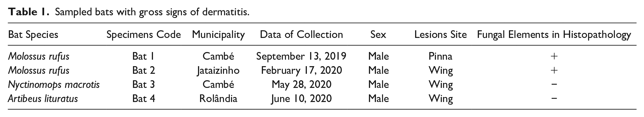

Gross and histologic lesions of bats with fungal infection. (a) M. rufus (bat 1) with hyperkeratosis and crusts (arrow) on the left ear pinnae. (b) Gomori methenamine silver (GMS)-positive hyphae are identified histologically (bat 1). Grocott-Gomori methenamine silver stain; (c) M. rufus (bat 2) with multiple alopecic lesions and skin thickening observed as white patches and macules on the wing membrane (chiropatagium and plagiopathagium), with ectoparasites—arrow. (d) Periodic acid-Schiff (PAS)-positive intralesional hyphae in a thick dermal layer (bat 2). PAS stain.
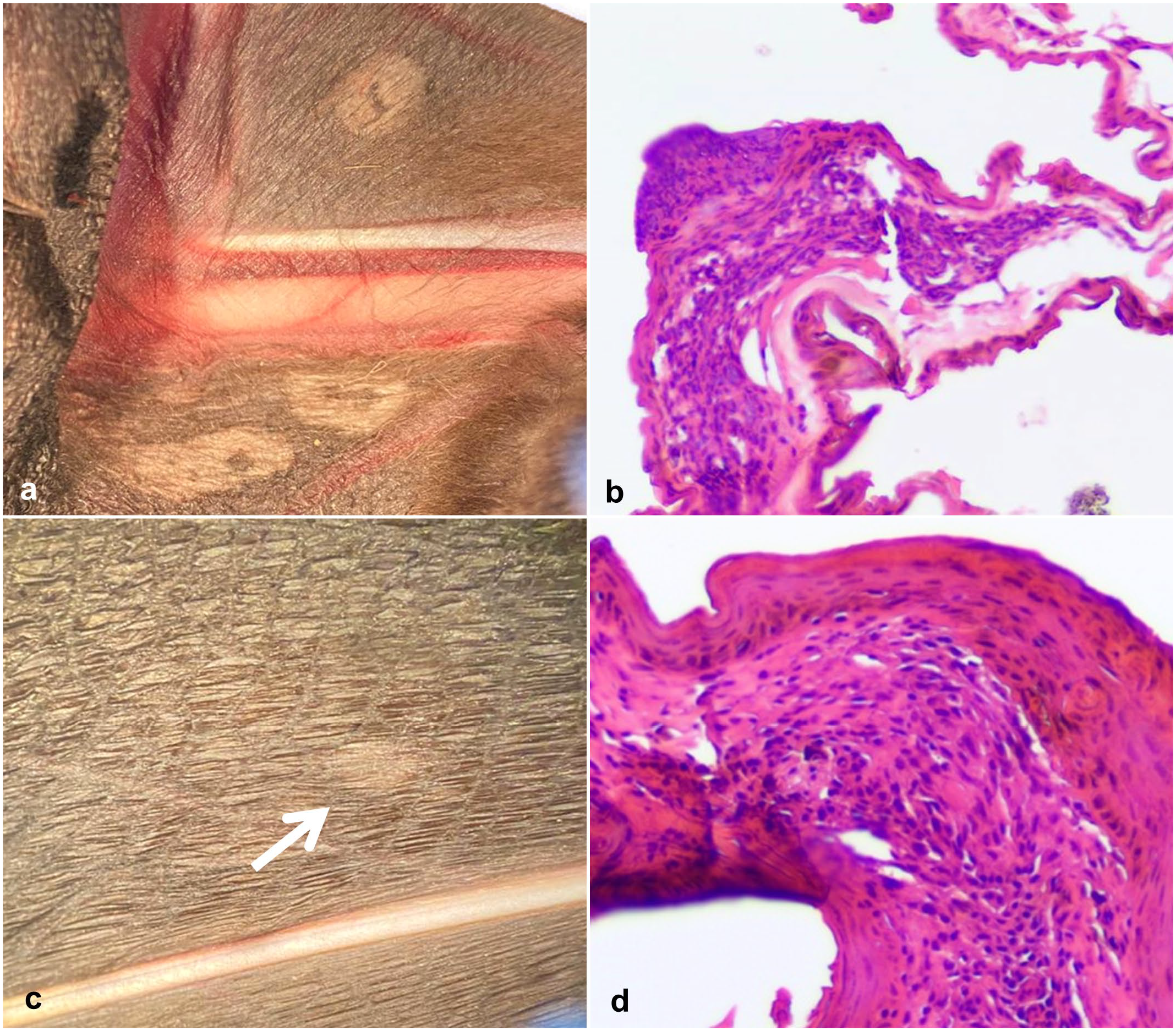
Gross and histologic lesions in bats suggestive of dermatitis. (a) Multiple flat lesions with well-demarcated margins in N. macrotis (bat 3) wing with alopecia and hyperkeratosis. (b) Mild inflammation and absence of fungal figures (bat 3). Hematoxylin and eosin stain. (c) Round patch lesion in an A. lituratus (bat 4) wing. (d) Hyperkeratosis with expansion of the dermis and discrete number of lymphocytes (bat 4). Hematoxylin and eosin stain.
Regarding the gross lesions observed in this study, such as depigmentation, hyperkeratosis, and alopecia, there are reports in the literature of the occurrence of alopecia in free-ranging Artibeus spp. with no determined causes, which histologically correlated with loss of hair follicles, dilation of follicular infundibula, and trichilemmal keratinization. 2 Cat predation, electrocution, and bite wounds are some of the causes of tegumentary lesions in free-ranging bats.7,15 Depigmentation, when not related to primary causes, such as vitiligo, is associated with the skin healing process commonly seen in wing membrane trauma. 7
The majority of the lesions were observed in the wing membranes, plagiopatagium, and chiropatagium. Wing membranes are composed of a scaffolding of a thin layer of epithelial cells, connective tissue, and capillaries (Supplemental Fig. S2), and in cases of dermatitis, dermal thickening is observed with the presence of inflammatory cells. 7 The inflammation observed histologically in this study may be related to different causes. Inflammation is observed in several types of skin lesions in bats including eosinophilic dermatitis secondary to insect bites, necrosis by electrocution in free-ranging Pteropodidae bats, and granulation due to chronic wounds, cellulitis, and abscesses associated to bite wounds, among many others, in which secondary bacterial and fungal infection may be observed. 7
In bats 1 and 2, hyphae were involved locally in dermal thickening and mild inflammation. Skin invasion by non-pigmented hyphae, showing cuplike epidermal erosions with curved conidia, is indicative of WNS lesions, but this is not what was observed in the skin lesions of these bats.14,16 It is also important to point out that the observed infection could have started after the penetration of fungi through a dermal lesion, associated with the presence of macronyssid mites parasitizing bat 2 (Fig. 1c). The low frequency of fungal infection observed in this study impaired a robust epidemiological analysis. However, the diagnosis of presumed lesions is also important, principally because of bats’ susceptibility to fungal diseases such as WNS, but also other mycoses caused by Trichophyton redellii, Hyphopichia burtonii, and Cladosporium sp.1,13,17
To the best of our knowledge, there is no report in the literature of detection of WNS nor P. destructans detection in Brazil. The identification of WNS cases and colonization by P. destructans has been reported in Europe and Asia, but WNS is not an important cause of mortality in these regions.7,12,16 Studies on the occurrence of WNS and P. destructans in neotropical bats are rare. However, the presence of the fungus cannot be discarded in tropical/subtropical regions based solely on the absence of WNS lesions. The fungi could be spread from endemic to non-endemic regions, carried by humans or migrating bats, as some species have long-distance migratory behaviors. 4 Little is known about its possible impact on the neotropical bat population and the host-pathogen interaction between P. destructans and those bats, but its spread remains a concern.
The susceptibility of bats to WNS may differ due to metabolic differences between species from cold and warm climates. One of the factors associated with WNS is the downregulation of the immune system and energetic disbalance caused by hibernation. 14 While hibernation/torpor is often related to bats affected by WNS in the Northern Hemisphere, little is known about the winter bat physiology in tropical and subtropical regions. Torpor is an approach to reduce metabolic rates and is used in bats from warmer climates when the temperature is low or very high. 9 It has been observed in some of the bat species included in this study, both in captivity and under natural conditions. 9 Chiroptera in the tropics and subtropics often show bouts of torpor depending on the ambient temperature and food availability, but they usually roost in more exposed sites than bats from colder climates. 9 We believe that such differences may affect P. destructans transmission and the threat to Brazilian bats by WNS.
In this study, we observed a low occurrence of dermatitis in the sampled neotropical bats. Gross lesions included discoloration, hyperkeratosis, and alopecia of the skin, which were histologically associated with dermal thickening and mild inflammation. Despite observing hyphal colonization of the skin lesions in 2 individuals, none of the affected bats showed histological evidence of WNS. Primary lesions caused by hematophagous acari in M. rufus may be related to fungal invasion of the tegument. The scarcity of data in the literature about dermatitis affecting neotropical bats and the possible spreading of WNS to other regions might be a fertile research field due to the ecological importance of these peculiar animals.
Supplemental Material
sj-pdf-1-vet-10.1177_03009858231155399 – Supplemental material for Histopathological survey of free-ranging neotropical bats with dermatitis
Supplemental material, sj-pdf-1-vet-10.1177_03009858231155399 for Histopathological survey of free-ranging neotropical bats with dermatitis by Igor M. de Souza Suguiura, Ana P. F. L. Bracarense, Giovana G. de Carvalho Ishiuchi, Ayako Sano, Kelvin S. Branco, Eiko N. Itano and Mario A. Ono in Veterinary Pathology
Footnotes
Acknowledgements
The authors thank the CNPq for the productivity fellowship granted to M.A.O. (311922/2018-0) and A.P.F.L.B. (308136/2018-7), the 17th Regional Health District for allowing the sample collection, and Michael Guile for the suggestions on the manuscript.
Declaration of Conflicting Interests
The author(s) declared no potential conflicts of interest with respect to the research, authorship, and/or publication of this article.
Funding
The author(s) disclosed receipt of the following financial support for the research, authorship, and/or publication of this article: CNPq productivity fellowship granted to Mario Augusto Ono (311922/2018-0) and Ana Paula Frederico Loureiro Bracarense (308136/2018-7).
Supplemental Material for this article is available online.
References
Supplementary Material
Please find the following supplemental material available below.
For Open Access articles published under a Creative Commons License, all supplemental material carries the same license as the article it is associated with.
For non-Open Access articles published, all supplemental material carries a non-exclusive license, and permission requests for re-use of supplemental material or any part of supplemental material shall be sent directly to the copyright owner as specified in the copyright notice associated with the article.