
Abstract
Bromethalin is a widely used neurotoxic rodenticide sometimes affecting nontarget wildlife. However, the effects of bromethalin on avian species are largely unknown. Here, we report the neuropathology of 14 feral conures (Psittacara sp.) with bromethalin toxicosis. Clinically, all birds presented with different degrees of paraparesis that sometimes progressed to dysphagia, ataxia, and tetraparesis. Histologically, there was astrogliosis, pallor, and vacuolation of white matter in the brain. This was usually more prominent in the medial longitudinal fasciculus, pons, optic tectum, cerebellar peduncle, and ventral funiculus. In most affected areas, there was loss of oligodendrocytes, and axons had extensive myelin loss or marked intramyelinic edema with splitting of myelin sheaths at the intraperiod line. Conures with bromethalin toxicosis had neuropathological changes similar to those of mammals exposed to bromethalin but with a characteristic distribution, probably related to higher susceptibility to cytotoxic edema in certain regions of the avian brain.
Bromethalin is a neurotoxic rodenticide widely used in North America. 6 Bromethalin is commercially available in a diverse range of bait preparations, making it suitable for domestic, industrial, and environmental use. 4 This versality also makes it a potential cause of poisoning for children, pets, and nontarget wildlife.4,6 Upon ingestion, bromethalin is metabolized to desmethyl-bromethalin, the active compound that impairs mitochondrial oxidative phosphorylation, leading to malfunctioning of ATP-dependent channel pumps.14,15 In mammals, these changes lead to intracellular edema and cell death, particularly in the central nervous system. 14 In birds, 15 little is known about the toxicology and pathology of bromethalin toxicosis, but experimental studies in Northern bobwhite (Colinus virginianus), mallards (Anas platyrhynchos), and chickens indicate that birds are susceptible to this poison. 4 Recently, bromethalin was reported as the cause of a neurological syndrome in an iconic feral population of conures (Psittacara sp.) in San Francisco, California (“The Telegraph Hill conures”). 16 However, our understanding of the most important diagnostic features of bromethalin toxicosis in free-ranging birds is limited, undermining the prompt recognition of this toxicosis in these species. To address this gap, in this report, we describe the neuropathology of bromethalin toxicosis in a group of feral conures.
Between 2013 and 2018, 14 adult (9 male and 5 females) feral conures were euthanized and necropsied. All animals belonged to the same feral population established in Telegraph Hill, San Francisco, California. The flock contains a mix of mitred (Psittacara mitrata), red-masked (also known as red-headed or cherry-head; Psittacara erythrogenys), and red-fronted (also known as scarlet-fronted; Psittacara wagleri) conures, although definitive species identification or confirmation of hybridization is complicated without further phylogenetic studies on this flock. In addition, we examined the central nervous system of 3 green-cheeked parakeets (Pyrrhura molinae) as controls. Tissues routinely collected during necropsy and processed for histopathology included brain, liver, lung, kidney, heart, small and large intestine, adrenal gland, pancreas, proventriculus, ventriculus, esophagus, crop, gonads, bursa, skeletal muscle, and skin. Spinal cord was examined in 5 animals, and eye and optic nerve in 2 birds. Tissues were routinely stained with hematoxylin and eosin and luxol fast blue.1,3,5,7,8,16
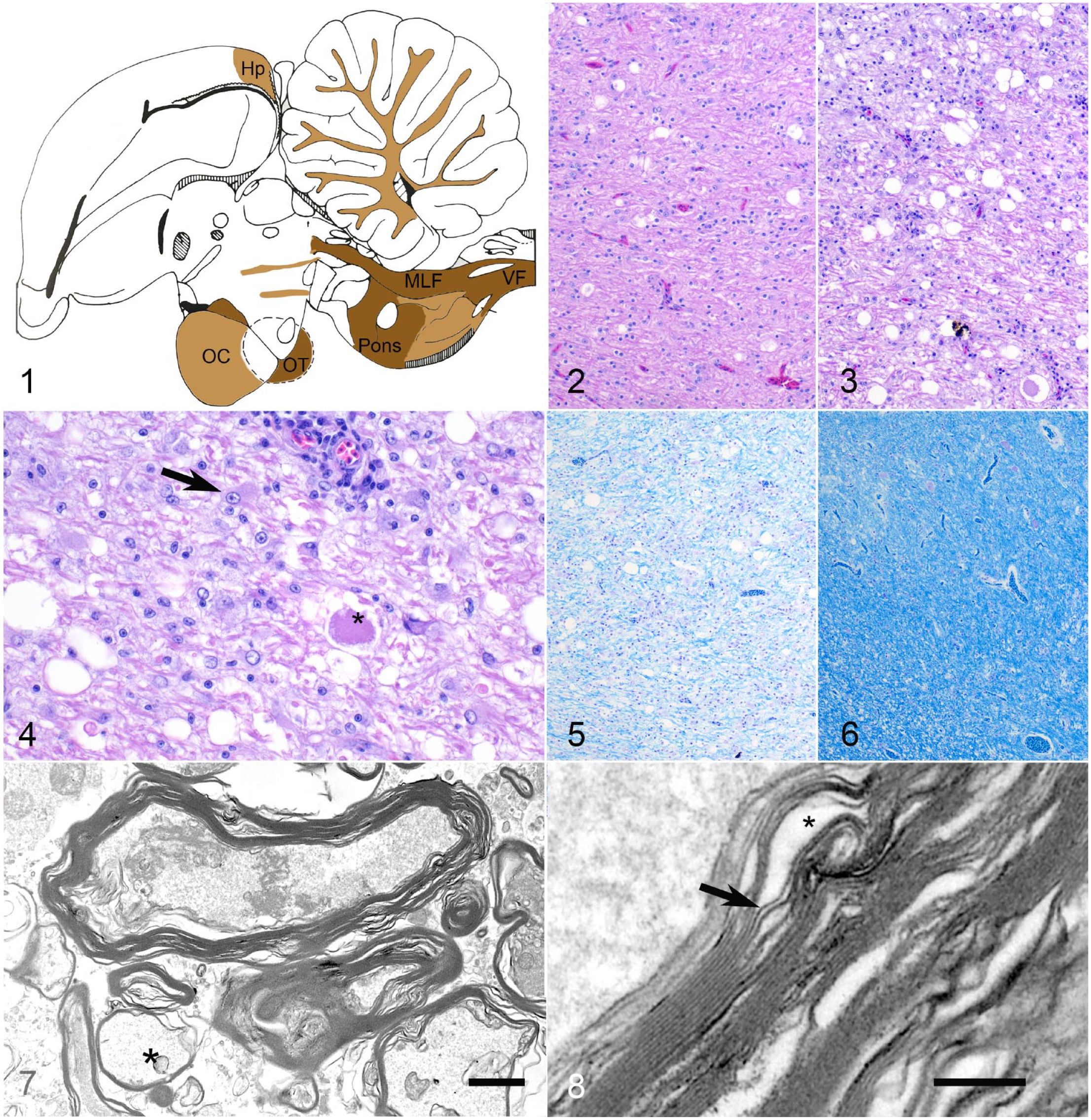
All conures presented clinically with paraparesis that in some progressed to ataxia (n = 12), dysphagia (n = 7), aphagia (n = 4), and tetraparesis (n = 3). Mild to severe emaciation was the most frequent postmortem finding (n = 8) (Table 1). Microscopically, there was mild to severe vacuolation of the white matter tracts in the medial longitudinal fasciculus and pons of the brainstem, cerebellar peduncle and folia, hippocampus, optic tectum, optic chiasma, optic nerve, and ventral funiculus of the spinal cord (Figs. 1–3). In 3 individuals, vacuolation also extended to the telencephalic and thalamic white and gray matter, although this was mild. In the most affected areas, there was also pallor of the neuroparenchyma, increased numbers of glial cells, and small numbers of perivascular leukocytes (Fig. 3). In more severe cases, there were occasional spheroids and higher numbers of prominent glial cells with abundant eosinophilic cytoplasm and an euchromatic nucleus with distinct nucleolus (Fig. 4). In all animals, including those with mild vacuolation, there was significant loss of luxol fast blue staining in the white matter (Figs. 5 and 6). Gray matter changes were limited to rare, scattered, telencephalic neuronal hypereosinophilia, and karyopyknosis.
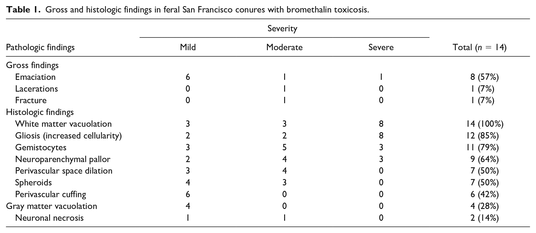
Gross and histologic findings in feral San Francisco conures with bromethalin toxicosis.
Bromethalin toxicosis, brain, feral conures.
In 3 cases and 1 control, we performed immunohistochemistry for CD3, Olig-2, and neuron-specific enolase using commercial antibodies and following standard protocols (Supplemental methods and Supplemental Table S1). Dog and chicken central nervous tissues were used as positive controls. Immunohistochemistry for glial fibrillary acidic protein, Iba-1, and myelin basic protein was attempted but immunoreactivity in conure and parakeet samples was lacking (Supplemental Table S1). The prominent glial cells observed in white matter with hematoxylin and eosin stain were negative for NSE and Olig-2, suggesting an astrocytic lineage. Most white matter glial cells had this immunohistochemistry profile while oligodendrocytes (Olig-2-positive cells) were decreased in number compared with controls (cases = 21 cells/mm2; controls = 76 cells/mm2) (Supplemental Figs. S1 and S2).
We selected paraffin-embedded sections of brain from 1 case with mild vacuolation, 1 case with severe vacuolation, and 1 control for ultrastructural examination. In the mild case, most remaining myelinated axons had undulating, vacuolated myelin sheaths sometimes separated at the intraperiod line (intramyelinic edema) (Figs. 7 and 8). The cytoplasm of a few demyelinated or hypomyelinated axons had swollen mitochondria and loss of microtubule definition. In the case with severe vacuolation, few myelinated axons remained in the medial longitudinal fasciculus, and most axons were swollen and contained scant electron-lucent material and few swollen mitochondria (Supplemental Figs. S3 and S4). The prominent, Olig-2-negative, NSE-negative white matter glial cells had cytoplasmic electron-dense neurofilaments and abundant glycogen and ribosomes, suggesting an astrocyte phenotype (gemistocytic astrocytes).
Toxicological analyses for organic compounds were performed in liver, brain, and feces of all animals using ultraviolet high-performance liquid chromatography (HPLC-UV) (n = 11) or gas chromatography–mass spectrometry (GC-MS) (n = 3). 16 Zinc and lead levels were measured in the liver, spleen, and pancreas of 3 animals through flame atomic absorption spectroscopy (FAAS). 3 There was between 0.03 and 118.4 µg/g of bromethalin in the feces, liver, and/or brain of 12 animals (Supplemental Table S2). In the 3 animals tested, zinc and lead levels were low in the liver and pancreas (zinc= 24–43 ppm dry weight, lead = 0–1 ppm dry weight).
We did not find evidence of Toxoplasma gondii; Sarcocystis sp.; avian paramyxovirus types 1, 2, or 5; Chlamydia sp.; avian bornavirus; West Nile virus; or culturable viruses in any of the animals tested using molecular, antigen detection, or culture-based techniques1,5,7,8 (Supplemental Table S2).
Bromethalin and its metabolites impair ATP production leading to dysfunction of Na+/K+ pumps and intracellular edema. 15 In the brain, osmotic regulation differs from other tissues, making sudden changes in osmolarity particularly detrimental for oligodendrocytes and astrocytes. 9 These glial cells have higher water permeability compared with neurons, 9 probably explaining the preponderance of white matter changes and relative sparing of gray matter in most animal species with bromethalin toxicosis.2,15 In our study, brain lesions were invariably present in the white matter tracts of the pons, medial longitudinal fasciculus, cerebellar peduncle, and optic tectum, suggesting that within the brain, not all white matter is equally affected. 4 In mammals, a few studies mention brainstem, pons, cerebellum, and optic nerve as common sites for vacuolar change.2,12 The preponderance of vacuolar change in the pons, cerebellum, brainstem, and optic white matter is not unique to bromethalin-induced edema and has been described in mammals with edema associated with hyponatremia or hypernatremia, hexachlorophene, and triethyltin toxicosis,9,13 suggesting higher susceptibility of these brain regions to osmotic insult. In birds, aquaporin-4, a water channel that facilitates cellular water intake, 11 is expressed in higher proportions in the pons, medial longitudinal fasciculus, cerebellum, and optic tectum, potentially making these regions more susceptible to cytotoxic edema. 17 All these structures, but particularly the medial longitudinal fasciculus, participate in the integration of spatial and visual information to coordinate head and limb movements. 10 Therefore, their damage could lead to ataxia, the most common clinical sign in the conures with bromethalin toxicosis. This also emphasizes the importance of examining these structures in cases of suspected bromethalin toxicosis in birds.
Vacuolar change, loss of myelin staining, and astrogliosis were the most consistent histological findings in all birds. Ultrastructural examination suggested that vacuolar change was associated with a combination of neuroparenchymal edema, loss of axons, and myelin vacuolation. Similarly, the loss of myelin staining was probably associated with myelin loss and decreased density of myelin sheaths due to splitting at the intraperiod line. In mammals, this type of myelin damage is typical of poisoning with bromethalin or other chemicals that cause brain edema. 13 There is no known direct mechanism by which bromethalin could cause myelin damage; 15 therefore, myelin loss could be a direct effect of edema. In addition, the primary role of oligodendrocytes and astrocytes on myelin production and preservation suggests that myelin degeneration could be secondary to stress and loss of these glial cells. 9 Interestingly, we observed decreased number of oligodendrocytes, even in animals with mild vacuolar change, suggesting that oligodendrocyte loss could be associated with myelin damage or absence of repair. Alternatively, we cannot rule out that oligodendrocyte loss was a consequence of myelin degeneration induced by water overload. Astrocytes were increased in number and were hypertrophied. Although astrocytes are one of the most susceptible cells to osmotic imbalance in the brain, they have a marked capacity for regeneration, contrasting with the minimal regenerative capacity of oligodendrocytes. 9 This suggests that the course of toxicosis was subacute to chronic in all parakeets, giving time for an astrocytic response. However, the lack of oligodendrocyte and myelin recovery may explain the progressive clinical signs for most birds in this study.
Toxicological analyses confirmed the presence of bromethalin in the tissues of all birds tested through HPLC-UV and in 1 out of 3 tested through GC-MS. HPLC-UV and GC-MS should provide enough resolution to detect bromethalin and its metabolites. However, these molecules are photosensitive and inadequate handling of samples can compromise detection. In addition, testing adipose tissue is preferred to other tissues because of the lipophilic nature of these compounds. 15 In our case, tissue storage in transparent plastic bags in early cases could have complicated bromethalin photostability, and lack of adipose tissue in most animals complicated postmortem sampling for toxicological analyses. In addition, the small size of conures complicated ancillary testing in all animals. In these circusmtances, feces are a good alternative for testing for bromethalin since the molecule and its metabolites are probably excreted through the feces. 16
We showed that birds exposed to bromethalin present with different degrees of vacuolar change and myelinopathy, in particular the white matter tracts of the brainstem and optic system. This characteristic distribution of lesions may be related to higher susceptibility to edema in certain regions of the avian brain. The diagnostic features here described and the use of special stains such as luxol fast blue will help to detect bromethalin-induced changes in avian brains.
Supplemental Material
sj-pdf-1-vet-10.1177_03009858221082300 – Supplemental material for Neuropathology of feral conures with bromethalin toxicosis
Supplemental material, sj-pdf-1-vet-10.1177_03009858221082300 for Neuropathology of feral conures with bromethalin toxicosis by Mauricio Seguel, Rita McManamon, Drury Reavill, Fern Van Sant, Sayed M. Hassan, Branson W. Ritchie and Elizabeth W. Howerth in Veterinary Pathology
Footnotes
Acknowledgements
We appreciate the help of Dr Justin Stilwell and Dr Christopher Gregory on conducting necropsies and helping with the processing of diagnostic samples.
Declaration of Conflicting Interests
The author(s) declared no potential conflicts of interest with respect to the research, authorship, and/or publication of this article.
Funding
The author(s) received no financial support for the research, authorship, and/or publication of this article.
Supplemental Material for this article is available online.
References
Supplementary Material
Please find the following supplemental material available below.
For Open Access articles published under a Creative Commons License, all supplemental material carries the same license as the article it is associated with.
For non-Open Access articles published, all supplemental material carries a non-exclusive license, and permission requests for re-use of supplemental material or any part of supplemental material shall be sent directly to the copyright owner as specified in the copyright notice associated with the article.