Urinary system toxicity is a significant concern to pathologists in the hazard
identification, drug and chemical safety evaluation, and diagnostic service industries
worldwide. There are myriad known human and animal urinary system toxicants, and
investigatory renal toxicology and pathology is continually evolving. The system-specific
Research Triangle Park (RTP) Rodent Pathology Course biennially serves to update
scientists on the latest research, laboratory techniques, and debates. The Sixth RTP
Rodent Pathology Course, Urinary Pathology, featured experts from the government,
pharmaceutical, academic, and diagnostic arenas sharing the state of the science in
urinary pathology. Speakers presented on a wide range of topics including background
lesions, treatment-related non-neoplastic and neoplastic lesions, transgenic rodent models
of human disease, diagnostic imaging, biomarkers, and molecular analyses. These seminars
were accompanied by case presentation sessions focused on usual and unusual lesions,
grading schemes, and tumors.
Since 2002, the biennial Research Triangle Park (RTP) Rodent Pathology Course has sought to
provide valuable information about organ system–specific current topics and techniques in
rodent pathology and toxicology to pathologists, trainees, and other involved colleagues of
the scientific community. This course uniquely bridges the rodent toxicologic pathology and
the rodent phenotyping communities. The 2012 program featured experts, mostly from the RTP,
presenting on various contemporary and emerging subjects regarding the pathology of the
urinary system. The first 5 RTP Rodent Pathology Courses focused on reproductive pathology,
neuropathology, cardiopulmonary pathology, immunopathology, and hepatobiliary pathology,
respectively.
In keeping with tradition, the reputation of the speakers attracted a diverse audience from
local institutes such as the University of North Carolina at Chapel Hill, Duke University,
North Carolina State University, the National Toxicology Program (NTP), National Institute of
Environmental Health Sciences (NIEHS), GlaxoSmithKline (GSK), Experimental Pathology Labs
(EPL), Environmental Protection Agency (EPA), the Hamner Institutes of Health Sciences
(previously CIIT), and constituents from other parts of the nation and world. Normal anatomy
and histology of the urinary system were initially presented, followed by 16 presentations on
topics including, but not limited to, renal transporters, neuroendocrine control of renal
function, imaging techniques, biomarkers, carcinogenesis, translational pathology, and
clinical applications. A case presentation intermission, coordinated by Dr Jerrold Ward
(Global Vet Pathology), gave the participants and speakers an opportunity to opine on
difficult cases and discuss popular renal pathology grading schemes.
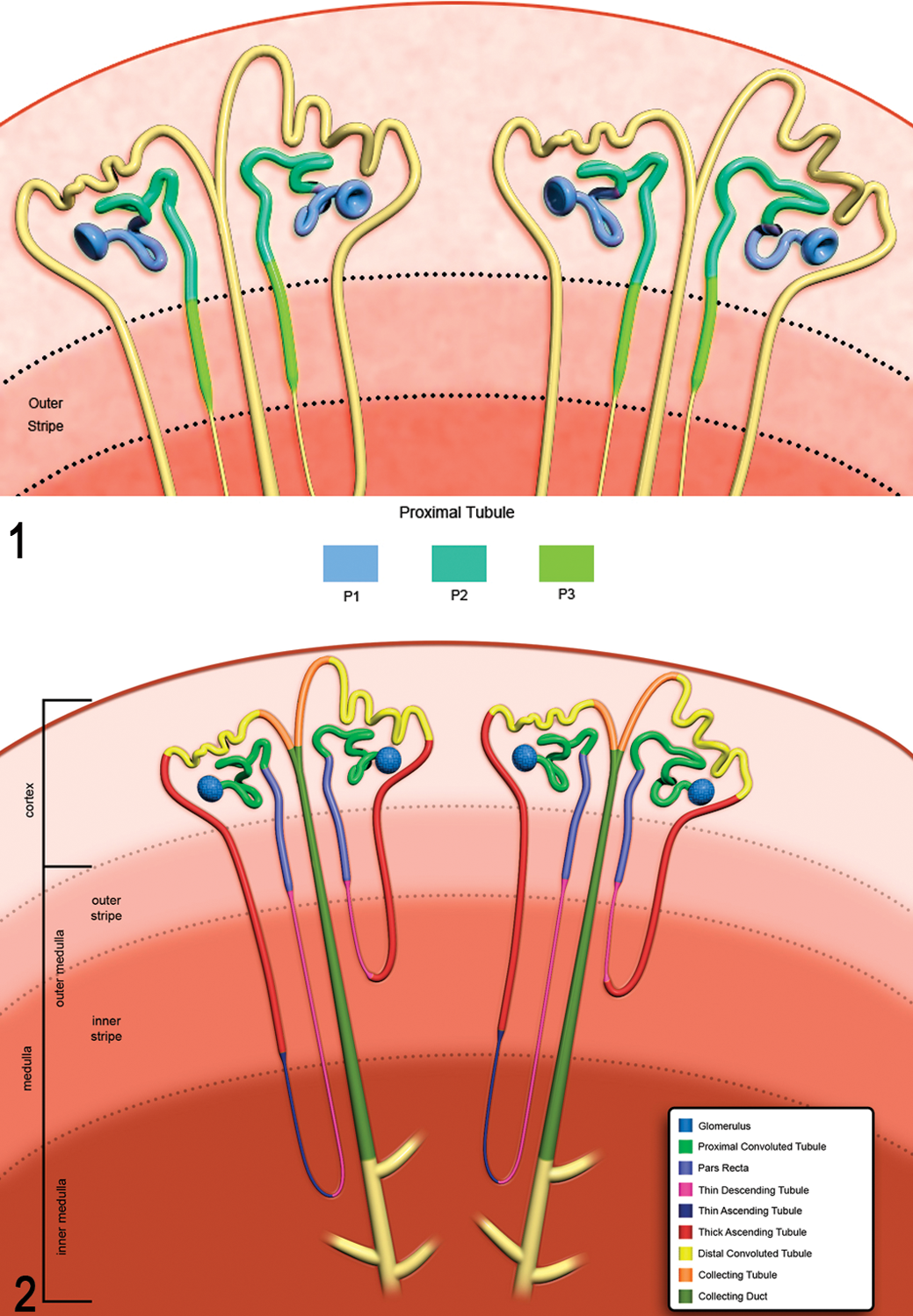
Dr Susan Elmore (NTP/NIEHS) opened the conference with an overview of normal
kidney structure and function. Dr Elmore’s lecture was an educational review from the level of
the budding pathology resident to that of the experienced pathologist. Using diagrams and
photomicrographs, she reviewed the kidney’s role in osmoregulation, waste excretion, hormonal
and metabolic functions, and the subtopographical compartments of glomeruli, tubules,
collecting system, vasculature, and interstitium, all of which should be examined
independently as well as collectively. Particularly instructive were beautiful schematics
differentiating segments P1, P2, and P3 (Fig.
1) and more detailed examination of the corticomedullary renal structure and segment
characterization (Fig. 2). Dr Elmore
reminded the audience to remain cognizant of sexual dimorphisms, such as that found in the
Bowman’s capsular epithelium. An androstenedione-treated female mouse that recapitulated the
cuboidal epithelial morphology found in the male was provided as an example.22
Figure 1.P1, P2, and P3 localization within the proximal convoluted tubule
and pars recta (also known as S1, S2, and S3). Figure 2. Corticomedullary
organization and tubular segment distribution within the nephron.
Dr Jeff Engelhardt (Experimental Pathology Laboratories) expanded on Dr Elmore’s
introductory lecture with his sessions, collectively titled Comparative
Toxicopathology of the Urinary System. The kidney is often a target of xenobiotics
simply due to its receipt of approximately 20% of cardiac output, despite comprising a mere
0.5% to 1% of the body weight in rats. Rats, dogs, and nonhuman primates are generally
apposite comparative species to humans. However, differences in renal pathology due to strains
may be seen, such as the increased sensitivity of Fisher F344 rats to aminoglycoside toxicity
when compared with age-matched Sprague-Dawley rats.26 Dr Engelhardt reviewed the structure and function of the nephron, emphasizing species
variation. As natives to arid environments, hamsters and gerbils have comparatively long loops
of Henle, which help with urine concentration and water conservation.
Familiarity with common background lesions in experimental species (eg, spontaneous
glomerular lipid vacuolation in the Beagle dog; renal amyloidosis in rodents, the most common
cause of death in the hamster) is essential for the toxicologic pathologist. Segment
awareness, as thoroughly reviewed by Dr Elmore, is important when evaluating specific
toxicities such as that observed with cisplatin in the S3 segment.24 Identification of these segments can be accomplished using various methods, including
the periodic acid-Schiff (PAS) reaction for staining the prominent brush border of tubules
within the S1 segment.
Dr Engelhardt discussed contributory aspects of chronic progressive nephropathy (CPN) in
rats, such as its amelioration by diet modification and the uniquity of hypercholesterolemia
due to decreased mevalonic acid feedback to the liver in the CPN-affected rat.23 Interestingly, it is known that tubular epithelial cells that have undergone epithelial
to mesenchyme transition and migrated to the interstitium secrete cytokines that drive matrix
deposition and consequent interstitial fibrosis.20 Of note is that most agents that exacerbate CPN do not necessarily cause nephrotoxicity
in humans. However, certain components of CPN may be aggravated by chemical administration,
such as increased intratubular mineralization in the case of selective estrogen receptor
modulators.
Dr Joseph Polli (GlaxoSmithKline) discussed the importance of drug transporters
in absorption, distribution, metabolism, and excretion (ADME). Drug transporters may act as
effective barriers to drug exposure or, through their induction or inhibition, may lead to
drug-drug interactions, as in the antiretroviral dolutegravir’s potential to inhibit organic
cation transporter (OCT) activity of other drugs.25 Dr Polli presented an exposure paradigm using whole-body autoradiography (WBA). By
identifying the distribution ratios of a compound (eg, liver: blood, brain: blood), it can be
determined where the compound accumulates and, to some extent, potential transport mechanisms.
Despite such tools, however, drug transporters present challenges to toxicologic pathologists.
Drug exposure, as measured by WBA, does not necessarily reflect its route or toxicity target.
Not only are numerous transporters involved in drug ADME, but they also have tissue-specific
distribution patterns and sexual dimorphic expression patterns, and several transporters in
rodents lack human orthologs altogether.1 It is therefore important for drug development to identify key drug transporters and
their genetic variants, elucidate their role in the disposition of a compound, and establish
robust transport assays. In presenting a renal toxicity case study for GSK1, Dr Polli
illustrated that the renal organic anion (Oat) and cation (Oct) transporters, known to be
responsible for uptake of drugs in the kidney, are not the primary drivers of the renal
disposition of GSK1. This was elegantly demonstrated using the cimetidine assay,
autoradiography, and Oct knockout mice, which suggested that active transport is likely not
affecting GSK1’s disposition, and that some other cellular transport mechanism (ie,
phagocytosis) may be at play. Multiple transporters can influence the disposition and
resulting toxicity of a compound, underscoring the challenge of transporter identification for
drug discovery and development programs.
Dr Peter Little (Experimental Pathology Laboratories) dis-cussed the renal
neuroendocrine cross-chatter in blood pressure, fluid, and ion homeostasis. In describing the
function and microanatomical location of the less known and infrequently addressed
circumventricular organs (CVOs) of the brain, he demonstrated the interplay between
juxtaglomerular secretion of renin, plasma osmolality detectors in the brain, and vascular
baroreceptors reporting through the cerebral medullary centers.11 Dr Little highlighted the subfornical organ (SFO), which contains one of the highest
concentrations of atrial natriuretic factor receptors as well as angiotensin II receptors in
the brain. The SFO has long been implicated in the central control of body fluid homeostasis,
blood pressure, and thirst regulation. Dr Little also described the supraoptic nucleus, which
produces antidiuretic hormone for release into the posterior pituitary gland, causing
vasodilation and subsequent blood pressure elevation. After functional descriptions and
physiology reviews, Dr Little gave morphological and pathological correlates to these organs
and systems.28 With light microscopy, secreted antidiuretic hormone is often visible as oval
eosinophilic bodies (Herring bodies) coursing down dendrites. In addition, the audience was
alerted to the fact that the CVO is devoid of a blood-brain barrier and, as Dr Little stated,
an “open physiologic window” to the brain.
Dr Kennita Johnson (University of North Carolina, Chapel Hill) gave an overview
of rodent imaging systems useful for renal analysis. Techniques discussed included those that
provide anatomical and functional information (magnetic resonance imaging, micro–computed
tomography, and ultrasound) and those that reveal functional information only (positron
emission tomography and single photon emission computed tomography). Of particular interest
was a cost-effective, quick, and noninvasive method for quantifying renal perfusion, including
blood flow velocity and blood volume, using microbubble-based contrast media for ultrasound.
With this technique, approximately 6μ bubbles of protein, polymer, or lipid encapsulating air
or a gas are injected into the test subject. With an ultrasound pulse, these microbubbles are
safely destroyed, and the rate at which they refill the ultrasound plane is measured as a
function of perfusion.21
The next presentation, by Dr Sam Cohen (University of Nebraska Medical Center),
discussed the normal structure, toxicity, and carcinogenesis of the lower urinary tract. Dr
Cohen shared comparative urothelial (no longer referred to as “transitional epithelium”)
insight, including the varying thicknesses, lymphocyte populations, and keratohyaline granules
of the primate urothelium. The presence of mucosal lymphocytes in rodents suggests that it
previously suffered ulceration, as erosion alone will not elicit this response. Dr Cohen also
suggested that rodent urothelial papillomatosis may be a sequel of diffuse ulceration and may
resolve in 3 to 4 weeks upon cessation of the stimulus. Human urothelial tumor classification
describes invasive urothelial neoplasia and noninvasive urothelial neoplasia, the latter
further categorized into carcinoma in situ, papilloma, and papillary neoplasms.5 Urinary bladder tumors may morphologically and molecularly be roughly divided into 2
variants: hyperplasia is often the result of deletions on both arms of chromosome 9, whereas
dysplasia and carcinoma in situ result from TP53 alterations. Mutations in FGFR3, TSC1, and
cyclin D have been associated with transformation of hyperplastic lesions to low-grade
papillary urothelial tumors.19 High-grade and invasive urothelial tumors, on the other hand, have been shown to harbor
inactivating mutations of the Rb gene and deletions on chromosome 8p. Dr
Cohen also discussed his research with 2 urothelial tumor–causing agents in rats: sodium
saccharin and melamine.6,9 Dr Cohen concluded by sharing his recommended diagnostic techniques, which include in
situ instillation of Bouin’s fixative, BrdU, Ki-67 or proliferating cell nuclear antigen
staining, and scanning electron microscopy to detect subtle superficial urothelial
changes.
Dr Greg Travlos, a clinical pathologist at the NTP and NIEHS, gave an overview on
renal clinical pathology and biomarkable targets to evaluate the approximately 35 000 nephrons
in the rat kidney. Dr Travlos discussed several urine collection systems and shared studies
showing that β2-microglobulin and γ-glutamyl transferase, among other analytes,
were not stably stored refrigerated or frozen over long periods. For analytes not affected by
refrigeration or freezing for short periods (eg, N-acetyl glucosaminidase), urine must be
brought to room temperature to ensure dissolution of potential precipitates. Regarding
biomarking glomerular filtration rate (GFR), cystatin C has been shown to be more sensitive
than creatinine, as its levels are perturbed with approximately 20% loss of nephron function.15 For segment-specific injury localization, μGST and RPA-1 are indicative of injury to
the distal tubule and collecting duct, respectively, whereas the proximal tubule has many
biomarkers, including albumin, β2-microglobulin, α1-microglobulin, RBP,
glucose, Kim-1, and αGST.13 For tubular epithelial injury localization, Dr Travlos indicated that isoforms of
alkaline phosphatase, aspartate aminotransferase, and N-acetyl-β-D-glucosaminidase were his
preferred analytes to evaluate injury to the brush-border, cytosolic, and lysosomal
compartments, respectively. Dr Travlos concluded with an example demonstrating the critical
importance of timed volume- and creatinine-normalized calculations by comparing raw and
corrected data from control and propylene glycol monobutyl ether (PGMBE)–administered
animals.
No rodent pathology conference on the urinary system would be complete without a discussion
of spontaneous background lesions, including chronic progressive nephropathy (CPN). Dr
Kendall Frazier (GlaxoSmithKline) provided an excellent review of the familiar lesion
that is characterized by tubular basophilia, ensuing thickened basement membranes, nuclear
crowding, and various other sequela.10 Tubular hypertrophy and hyperplasia may both be seen along with CPN. However, tubular
hypertrophy, unlike tubular hyperplasia, is not considered preneoplastic. Dr Frazier indicated
that simple and atypical tubular hyperplasias occur spontaneously as well as with xenobiotic
administration, although atypical tubular hyperplasia is a preneoplastic finding more commonly
seen as test article related. Regarding α2U nephropathy, he cautioned the audience
that although a Mallory Heidenhain stain may differentiate α2u-globulin from other
material, α2u-globulin is rarely irregular or angular. Thus, positively staining
material with such morphology should be investigated further.
Dr Frazier also discussed conventional rodent renal tumor diagnoses. As malignant renal
tumors of the kidney are rare in rats and mice, any increase in numbers in test
article–administered animals is troublesome in a preclinical toxicity study. Typically, males
tend to exhibit greater numbers of renal tumors in a carcinogenicity study, with the exception
of the amphophilic-vacuolar (AV) tumor type.12 Females exhibit a higher incidence of AV tumors because of their longer average life
expectancy; however, when corrected for days on study, these tumors generally also follow the
paradigm of males being more affected.
Dr Frazier concluded his set of lectures by sharing his tiered approach to mechanistic assays
in the urinary system. End points commonly used in toxicity studies (eg, histopathology, organ
weights, routine clinical pathology) comprise the first tier. The second tier is composed of a
combination of electron microscopy, immunohistochemistry, novel urinary system biomarkers, and
WBA. Dr Frazier emphasized that the lack of effective routine clinical biomarkers for
glomerular insult (other than proteinuria) makes special staining techniques and
immunohistochemistry (IHC) particularly useful. For example, IHC antibodies to synaptopodin,
nephrin, von Willebrand factor, and CD90/thy-1 identify podocytes, podocyte slit diaphragms,
endothelial cells, and rat mesangial cells, respectively. Tier III assays include GFR and
renal blood flow measurement, transporter studies such as those shared by Dr Polli, and
imaging studies similar to those presented by Dr Johnson.
Dr Rachel Cianciolo (University of North Carolina, Chapel Hill) opened a set of
lectures regarding animal models of human renal disease. She shared her evaluation of tubules
and interstitium in a doxorubicin nephropathy model using transcriptomics and stereology.
Stereologic evaluation of organs requires strict adherence to sectioning and analysis
protocols to re-create a 3-dimensional evaluation from 2-dimensional tissue sections.2 Using her rat doxorubicin model to study tubulointerstitial renal disease, Dr Cianciolo
discovered altered transcription pathways that led her to understand the interplay between
proteinuria, tubular disease, and peritubular capillary compromise.
Dr Wei Qu (NTP) challenged the current dogma surrounding the carcinogenesis of
metals. It has long been thought that although considered a potent human carcinogen, arsenic
does not cause cancer in rodents.8 Dr Qu’s lab has been modeling metal-induced carcinogenesis and has used in vitro and in
vivo models to study arsenic, lead, nickel, and cadmium carcinogenesis.29 Using a developmental exposure paradigm consisting of fetal exposure in utero via
maternal drinking water coupled with postweaning exposure to dimethylarsinic acid (a potent
promoter), Dr Qu was able to demonstrate renal cell carcinomas in arsenic-exposed animals.29
The keynote address was given by the Chair of Pathology and Laboratory Medicine and Chief of
Service at the University of North Carolina Hospitals Clinical Laboratories, Dr J.
Charles Jennette. Dr Jennette enthralled participants and attendees alike with his
elegant investigations into the pathogenesis of antineutrophil cytoplasmic antibody (ANCA)
glomerulonephritis and vasculitis.16 Dr Jennette described numerous experiments using a variety of mouse models from Rag2
mice immunized against myeloperoxidase (MPO) to mice deficient in critical complement components.17 These experiments were used to posit the following critical criteria for this
devastating human disease: anti-MPO IgG alone may cause disease, the Fcγ receptor is important
in the pathogenesis, neutrophils and the alternative complement pathway are required for
disease, cytokines may exacerbate ANCA, and T cells do not incite the acute inflammatory
component.
Dr Jeffrey Everitt (GlaxoSmithKline) discussed the importance of studies of
hereditary renal cancer for elucidating the signaling pathways associated with cancer
development and opportunities for interventional therapies. He reviewed the current state of
rodent models of renal carcinogenesis and the challenges inherent in the phenotyping of
genetically modified animals. The talk stressed the importance of understanding genotype,
spontaneous lesions, and genetic manipulations when considering phenotypic alterations in
rodent models, as well as the need for a complete necropsy with a thorough macroscopic
examination. Dr Everitt reviewed phenotyping issues using Eker rats that have alteration in
Tsc2 as an example.18 He reviewed how the Tsc2 product tuberin, an mTOR-dependent protein, is involved in
signaling pathways implicated in renal cell carcinoma.14
Dr Susan Gurley (Duke University) continued on the topic of mouse models of human
disease, specifically diabetic nephropathy (DN). For a DN model, criteria including specific
declines in GFR, magnitudes of increase in albuminuria, and characteristic histopathologic
lesions within glomeruli must be present.3 The familiar streptozotocin (STZ)–induced model was compared with the Akita mouse, the
latter of which harbors a mutation leading to an amino acid substitution in mature insulin.4 In addition to lacking the need for toxin administration, the Akita model developed
more pronounced hyperglycemia and albuminuria than did the STZ-treated mice on the same
background. In the human DN population, inhibition of the renin-angiotensin-aldosterone system
prolongs renal function, which Dr Gurley modeled by adding a renin transgene to the mouse.
Dr Jim Swenberg (University of North Carolina, Chapel Hill) closed the Sixth RTP
Rodent Pathology Conference with a historical perspective on α2u-globulin induction
and nephropathy. Dr Swenberg shared years of research, spanning from trimethylpentane to
d-limonene.7 There are many tenets of α2u-globulin in comparative pathology: humans do
not produce it, and the globulins in the human α2u family do not bind chemicals
that induce hyaline droplets in rodents, and this syndrome in male rats should not indicate a
risk of hyaline droplet nephropathy or renal carcinogenesis. However, to definitively
determine a chemical is solely a male rat hyaline droplet nephropathogen, several criteria
must be met: the chemical must be neither directly or indirectly genotoxic, the effect must be
limited to the male rat, and protein droplets must be identified in short-term studies and be
positively identified as α2u-globulin.27
Sixth RTP Rodent Pathology Course Committee
Richard A. Peterson II, Program Co-Chair; GlaxoSmith-Kline
Amera K. Remick, Program Co-Chair; WIL Research
Molly H. Boyle, Integrated Laboratory Systems
Rachel Cianciolo, University of North Carolina, Chapel Hill
Samuel M. Cohen, University of Nebraska Medical Center
Torrie A. Crabbs, Experimental Pathology Laboratories
John M. Cullen, North Carolina State University College of Veterinary Medicine
Susan A. Elmore, National Institute of Environmental Health Sciences/National
Toxicology Program
Jeff Engelhardt, Experimental Pathology Laboratories
Jeffrey Everitt, GlaxoSmithKline
Kendall Frazier, GlaxoSmithKline
Virginia Godfrey, University of North Carolina, Chapel Hill
Susan B. Gurley, Duke University Medical Center
Georgette Hill, Integrated Laboratory Systems
Mark J. Hoenerhoff, National Institute of Environmental Health Sciences/National
Toxicology Program
J. Charles Jennette, University of North Carolina, Chapel Hill
Kennita Johnson, University of North Carolina, Chapel Hill
Peter B. Little, Experimental Pathology Laboratories
Andrew Marias, Experimental Pathology Laboratories
Rodney A. Miller, Experimental Pathology Laboratories
Joseph W. Polli, GlaxoSmithKline
Wei Qu, National Institute of Environmental Health Sciences/National Toxicology
Program
Arlin Rogers, University of North Carolina, Chapel Hill
Brian Short, Allergan
James A. Swenberg, University of North Carolina, Chapel Hill
Gregory S. Travlos, National Institute of Environmental Health Sciences/National
Toxicology Program
We thank Drs Arun Pandiri, Sachin Bhusari, and the presenters of the Sixth RTP Rodent
Pathology Course for their thorough review of this article and David Sabio, Beth Mahler, and
Eli Ney for assistance with figures. This article may be the work product of an employee or
group of employees of the National Institute of Environmental Health Sciences (NIEHS),
National Institutes of Health (NIH); however, the statements, opinions, or conclusions
contained therein do not necessarily represent the statements, opinions, or conclusions of
NIEHS, NIH, or the US government.
Declaration of Conflicting Interests
The author(s) declared no potential conflicts of interest with respect to the research,
authorship, and/or publication of this article.
Funding
The author(s) declared receipt of the following financial support for the research,
authorship, and/or publication of this article: Financial support was provided by
Integrated Laboratory Systems for Molly Boyle to attend the meeting and contribute to this
meeting report.
References
1.
BleasbyKCastleJCRobertsCJ. Expression profiles of 50 xenobiotic transporter genes in
humans and pre-clinical species: a resource for investigations into drug
disposition. Xenobiotica.
2006;36:963–988.
2.
BoyceRWDorph-PetersenKALyckL. Design-based stereology: introduction to basic concepts
and practical approaches for estimation of cell number. Toxicol
Pathol.
2010;38:1011–1025.
3.
BrosiusFC IIIAlpersCEBottingerEP. Mouse models of diabetic nephropathy.
J Am Soc Nephrol.
2009;20:2503–2512.
4.
ChangJHGurleySB. Assessment of diabetic nephropathy in the Akita
mouse. Methods Mol Biol.
2012;933:17–29.
5.
ChengLMontironiRDavidsonDD. Staging and reporting of urothelial carcinoma of the
urinary bladder. Mod Pathol.
2009;22(suppl
2):S70–95.
6.
CohenSMOhnishiTClarkNM. Investigations of rodent urinary bladder carcinogens:
collection, processing, and evaluation of urine and bladders.
Toxicol Pathol.
2007;35:337–347.
7.
DietrichDRSwenbergJA. The presence of alpha 2u-globulin is necessary for
d-limonene promotion of male rat kidney tumors. Cancer
Res.
1991;51:3512–3521.
8.
EatonDLGilbertSG. Principles of toxicology. In:
KlaassenCD, ed. Casarett and Doull’s Toxicology: The Basic Science of
Poisons. 7th ed. New York,
NY: McGraw-Hill;
2008:1310.
9.
EllweinLBCohenSM. The health risks of saccharin revisited.
Crit Rev Toxicol.
1990;20:311–326.
10.
FrazierKSSeelyJCHardGC. Proliferative and nonproliferative lesions of the rat and
mouse urinary system. Toxicol Pathol.
2012;40:14S–86S.
11.
GanongWF. Circumventricular organs: definition and role in the
regulation of endocrine and autonomic function. Clin Exp
Pharmacol Physiol.
2000;27:422–427.
12.
HardGCSeelyJCKisslingGE. Spontaneous occurrence of a distinctive renal tubule tumor
phenotype in rat carcinogenicity studies conducted by the national toxicology
program. Toxicol Pathol.
2008;36:388–396.
13.
HarpurEEnnulatDHoffmanD. Biological qualification of biomarkers of chemical-induced
renal toxicity in two strains of male rat. Toxicol Sci.
2011;122:235–252.
14.
HenskeEP. The genetic basis of kidney cancer: why is tuberous
sclerosis complex often overlooked?Curr Mol Med.
2004;4:825–831.
15.
InkerLAOkparaveroA. Cystatin C as a marker of glomerular filtration rate:
prospects and limitations. Curr Opin Nephrol Hypertens.
2011;20:631–639.
JennetteJCXiaoHFalkR. Experimental models of vasculitis and glomerulonephritis
induced by antineutrophil cytoplasmic autoantibodies. Contrib
Nephrol.
2011;169:211–220.
18.
LapingNJEverittJIFrazierKS. Tumor-specific efficacy of transforming growth factor–beta
RI inhibition in Eker rats. Clin Cancer Res.
2007;13:3087–3099.
19.
LindgrenDFrigyesiAGudjonssonS. Combined gene expression and genomic profiling define two
intrinsic molecular subtypes of urothelial carcinoma and gene signatures for molecular
grading and outcome. Cancer Res.
2010;70:3463–3472.
20.
LiuSFChangSYLeeTC. Dioscorea alata attenuates renal interstitial cellular
fibrosis by regulating Smad- and epithelial-mesenchymal transition signaling
pathways. PLoS One.
2012;7:e47482.
21.
McArthurCBaxterGM. Current and potential renal applications of
contrast-enhanced ultrasound. Clin Radiol.
2012;67:909–922.
22.
National Toxicology Program.
Toxicology and carcinogenesis studies of androstenedione (CAS No.
63-05-8) in F344/N rats and B6C3F1 mice (gavage studies). Natl
Toxicol Program Tech Rep Ser.
2010;(560):1,
7–31, 33–171passim.
23.
OttDBLachancePA. Biochemical controls of liver cholesterol
biosynthesis. Am J Clin Nutr.
1981;34:2295–2306.
24.
PortillaDLiSNagothuKK. Metabolomic study of cisplatin-induced
nephrotoxicity. Kidney Int.
2006;69:2194–2204.
25.
ReeseMJSavinaPMGenerauxGT. In vitro investigations into the roles of drug
transporters and metabolizing enzymes in the disposition and drug interactions of
dolutegravir, a HIV integrase inhibitor. Drug Metab
Dispos.
2012;41:353–361.
26.
SullivanMJRareyKEConollyRB. Comparative ototoxicity of gentamicin in the guinea pig
and two strains of rats. Hear Res.
1987;31:161–167.
27.
SwenbergJA. Alpha 2u-globulin nephropathy: review of the cellular and
molecular mechanisms involved and their implications for human risk
assessment. Environ Health Perspect.
1993;101(suppl
6):39–44.
28.
TakahashiHYoshikaMKomiyamaY. The central mechanism underlying hypertension: a review of
the roles of sodium ions, epithelial sodium channels, the renin-angiotensin-aldosterone
system, oxidative stress and endogenous digitalis in the brain.
Hypertens Res.
2011;34:1147–1160.
29.
TokarEJBenbrahim-TallaaLWardJM. Cancer in experimental animals exposed to arsenic and
arsenic compounds. Crit Rev Toxicol.
2010;40:912–927.