
Abstract
Canine multiple system degeneration (CMSD) is a progressive hereditary neurodegenerative disorder commonly characterized by neuronal degeneration and loss in the cerebellum, olivary nuclei, substantia nigra, and caudate nuclei. In this article, we describe 3 cases of CMSD in Ibizan hounds. All patients exhibited marked cerebellar ataxia and had cerebellar atrophy on magnetic resonance imaging. At necropsy, all cases showed varying degrees of cerebellar atrophy, and 2 cases had gross cavitation of the caudate nuclei. Histologic findings included severe degeneration and loss of all layers of the cerebellum and neuronal loss and degeneration within the olivary nuclei, substantia nigra, and caudate nuclei. Pedigree analysis indicated an autosomal recessive mode of inheritance, but the causative gene in this breed is yet to be identified. CMSD resembles human multiple system atrophy and warrants further investigation.
Keywords
Several forms of hereditary ataxia have been described in the dog, including cerebellar cortical degeneration, cerebellar ataxias without significant neurodegeneration, episodic ataxia, spinocerebellar degeneration, and canine multiple system degeneration (CMSD). 12 Many of these conditions have similarities to human diseases. 11 The pathologic changes observed in these hereditary ataxias always involve the cerebellum and occasionally include the brain stem and spinal cord. 12 The clinical presentation consists of progressive cerebellar ataxia commonly beginning at less than 1 year of age, and clinical diagnostics such as bloodwork, urinalysis, and cerebrospinal fluid analysis are frequently normal in these dogs. 12
Canine multiple system degeneration is a subset of hereditary ataxia that is defined as a progressive neurodegenerative disorder with a distinct pattern of pathologic changes consisting of neuronal degeneration and loss in the cerebellum, olivary nuclei, substantia nigra, and caudate nuclei. 7,12 This disease was first reported in the Kerry blue terrier in 1946 and was later termed hereditary striatonigral and cerebello-olivary degeneration. 4 –6 More recently, a similar condition was described in the Chinese crested dog, and the term canine multiple system degeneration was coined. 7,12 In these 2 breeds, the condition is nearly identical and has been traced to mutations in the SERAC1 gene. 8 This gene is responsible for phospholipid exchange between mitochondria and the endoplasmic reticulum, a process needed for proper function of mitochondria and trafficking of cholesterol within the cell. 13 CMSD pathologically resembles human multiple system atrophy (MSA). 3 In this report, we describe 3 cases of CMSD in a novel breed, the Ibizan hound.
Three related purebred Ibizan hounds, a 9-month-old male (case 1), a 16-month-old female (case 2), and a 4-year-old female (case 3), originating from a single breeder presented individually to the Texas A&M University Veterinary Medical Teaching Hospital neurology service for evaluation of a slowly progressive gait abnormality that began at approximately 6 months of age. A pedigree of affected and nonaffected individuals was constructed from the provided history, revealing what appears to be an autosomal recessive mode of inheritance (Suppl. Fig. S1). Neurologic examination of all cases revealed a pronounced truncal sway, intention tremors, a permanent wide-based stance, and delayed postural reactions, consistent with severe cerebellar ataxia. Bilateral absent menace responses were also noted. Case 3, the oldest dog, was most severely affected and unable to walk without falling to either side. General physical examination, complete blood count, serum chemistry analysis, urinalysis, cerebrospinal fluid analysis, and electrodiagnostic studies to eliminate myopathic disease showed no significant abnormalities in any dog. T2-weighted magnetic resonance imaging of all cases revealed uniform widening of the cerebellar sulci, indicating generalized tissue loss and atrophy of the parenchyma. Cases 2 and 3 also had severe hyperintensity and reduction in size of the caudate nuclei bilaterally.
Following clinical diagnostics and imaging, the animals were humanely euthanized and necropsies were performed. Tissues were fixed in 10% neutral-buffered formalin and processed routinely for histopathology. Sections of caudate nucleus from case 1 were stained with periodic acid Schiff (PAS) and Sudan black.
Tissue sections from each case were immunostained using the streptavidin-biotin-immunoperoxidase method with primary antibodies for ionized calcium binding adaptor molecule 1 (Iba1; 1:500 dilution; Biocare Medical), glial fibrillary acidic protein (1:1500 dilution; GeneTex), and α-synuclein (1:1000 dilution; Abcam). Normal dog brain was used for positive and negative controls. For the negative control, a commercial negative control reagent, was substituted for the primary antibody. Fresh samples of caudate nucleus from case 1 were immersed overnight in fixative (2.5% glutaraldehyde, 2.5% paraformaldehyde in 0.1 M sodium cacodylate buffer), then washed and processed for electron microscopy. Details of the electron microscopy are available in Supplemental File 1.
Buffy coats from 2 affected dogs were shipped to the University of Missouri. DNA was extracted from these buffy coats and genotyped for 2 variants that have been associated with allelic forms of recessive CMSD in Kerry blue terriers and Chinese crested dogs. 7 Details of the genotyping are available in Supplemental File 2.
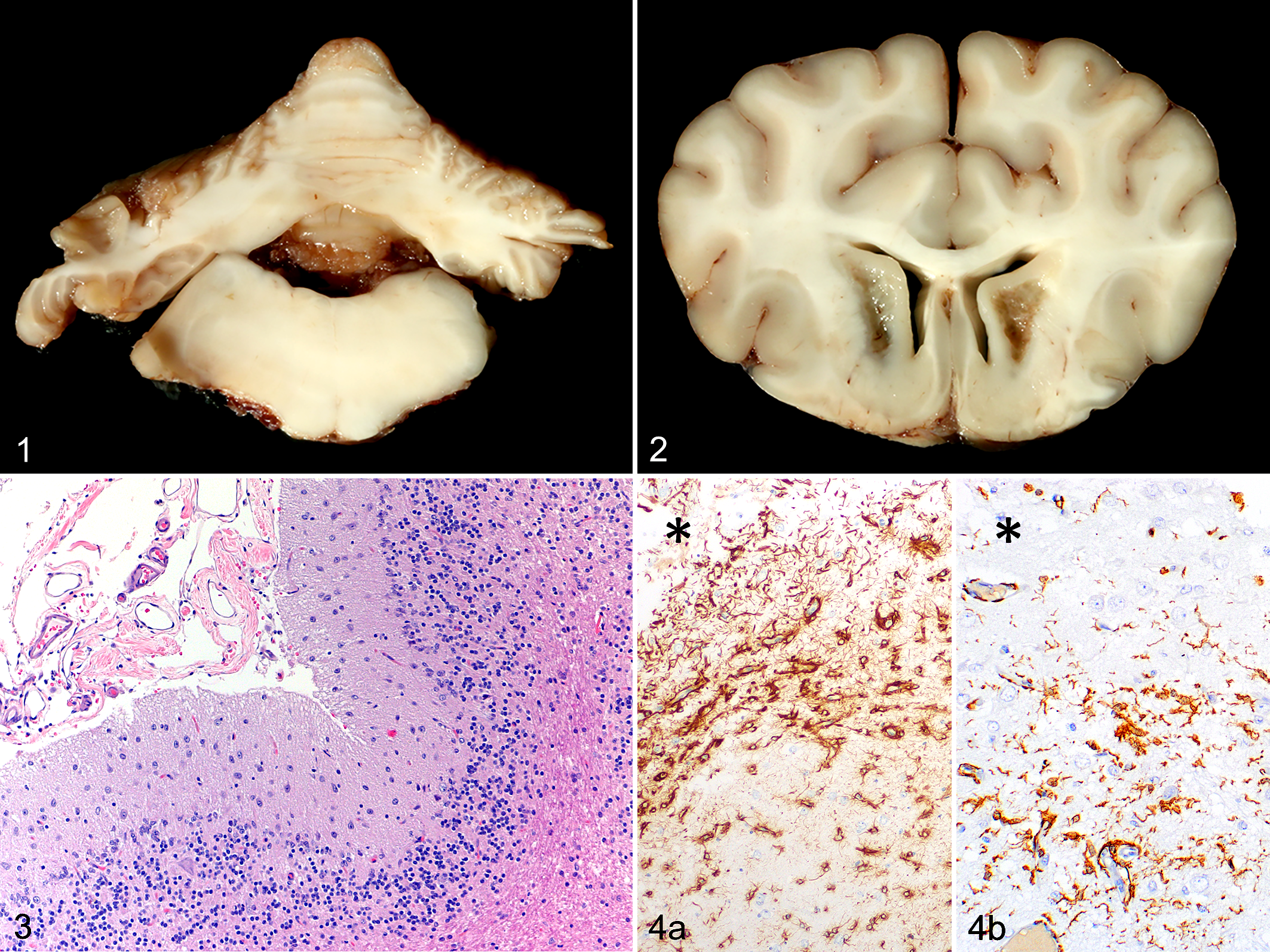
Grossly, all 3 dogs had a reduction in cerebellar size, and case 3 was most severely affected (Fig. 1). Both the vermis and lateral hemispheres were affected, while the flocculus and nodulus were relatively normal. Bilaterally symmetric caudate nucleus lesions were grossly evident in the 2 older dogs. Case 2 had mild dark discoloration of the lateroventral aspect of the nucleus, while case 3 had severe cavitation of the nucleus with relative sparing of the dorsal and medial aspects (Fig. 2).
Canine multiple system degeneration, brain, dog, case 3.
Microscopically, the cerebellum of all dogs was characterized by mild to marked thinning of the molecular and granular cell layers and decreased cellularity of the granular cell layer, most severely in case 3 (Fig. 3). In all cases, there was almost complete loss of Purkinje cells, proliferation of Bergmann’s glia, and scattered white matter spheroids. A few reactive astrocytes were present within the white matter and rarely within the molecular layer of cases 1 and 2. Within the cerebellar nuclei of all dogs were areas of gliosis with neuronal depletion, mild spongiosus, and occasional spheroids (Suppl. Fig. S2). The flocculus, nodulus, and lingula were relatively spared, but the flocculus was more affected in case 3. Alpha-synuclein immunohistochemical stains of cerebellum/medulla oblongata resulted in diffuse cytoplasmic immunopositivity in neurons and glial cells (presumably both astrocytes and oligodendrocytes) of both affected dogs and the control, but distinct inclusions similar to those described in MSA were not evident (Suppl. Fig. S3).
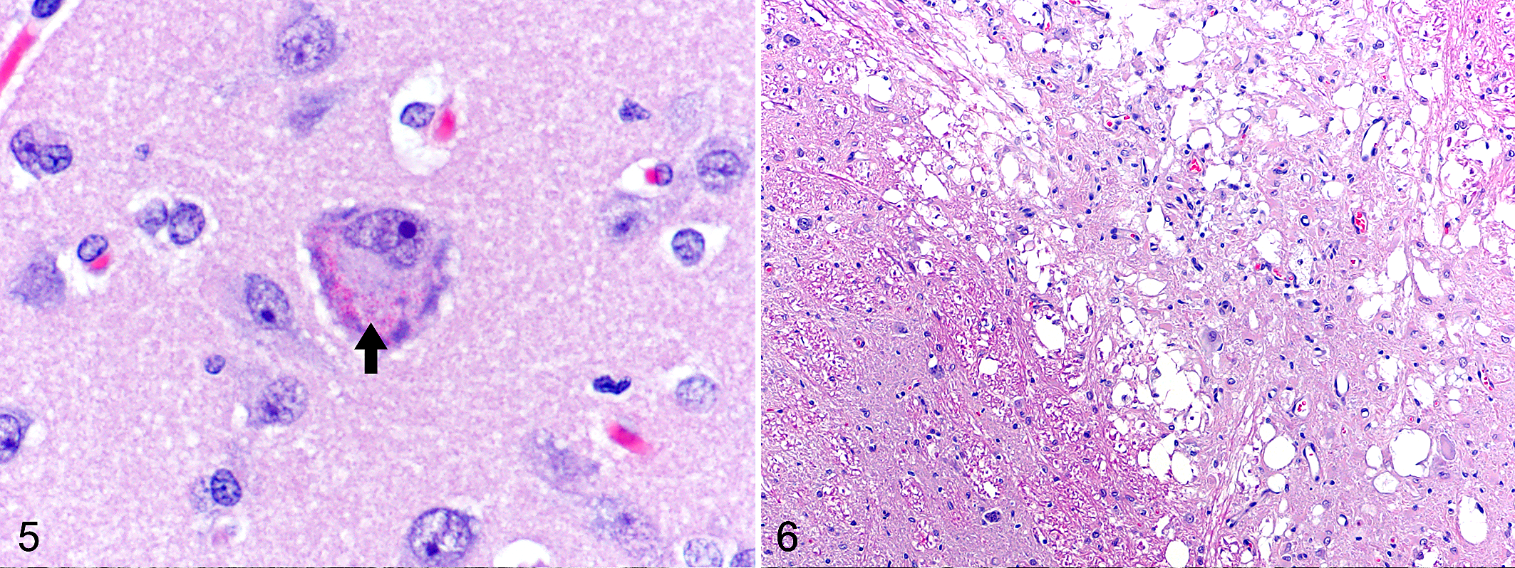
The severity of the lesions within the basal nuclei varied widely between the 3 dogs. The caudate nuclei of case 1 had scattered clusters of glial cells but no obvious neuronal depletion. In cases 2 and 3, the caudate nuclei exhibited areas of cavitation with spongiosus, neuronal loss, and gliosis (Suppl. Fig. S4). These changes were most severe in case 3. A small number of gemistocytes were seen in case 2. Caudate nucleus neurons were decreased in number in case 2 and difficult to identify in case 3. Immunohistochemistry of sections of the caudate nuclei showed that the gliosis included both astrocytes and microglia (Fig. 4). In cases 1 and 3, astrocytes outnumbered microglia, while roughly equal numbers of each cell type were evident in case 2 (Fig. 4). The putamen and globus pallidus exhibited mild to moderate gliosis with rare spheroids, again most severe in case 3. In all cases, a few larger neurons within the caudate nucleus, putamen, and globus pallidus contained approximately 15 to 30 brightly eosinophilic, round, ≤1 µm, intracytoplasmic granules (Fig. 5). These granules did not stain with PAS or Sudan black.
Canine multiple system degeneration, brain, dog, case 1.
Canine multiple system degeneration, brain, dog, case 1.
Within the brainstem, the olivary nuclei and substantia nigra had degenerative changes in all 3 cases. The olivary nuclei exhibited minimal to severe spongiosus that was most pronounced in the youngest dog, case 1 (Fig. 6). Gliosis and neuronal loss were also present with the mildest changes in case 2. Case 3 also had rare spheroids and neurons with intracytoplasmic granules. The substantia nigra of all dogs had mild spongiosus, again most severe in case 1 (Suppl. Fig. S5). Other changes in the substantia nigra included gliosis, neuronal loss, neuronal intracytoplasmic granules, and rare spheroids (Suppl. Fig. S5). The periaqueductal gray matter of cases 2 and 3 contained a few neurons with the previously described granules. Rare spheroids were seen sporadically in other locations throughout the brain stem. Though neuronal depletion was observed throughout the brain, light microscopy showed no clear degenerative changes within neurons.
The spinal cord of cases 2 and 3 featured minimal to mild axonal degeneration within the white matter of the cervical, thoracic, and lumbar sections. Case 1 had only minimal axonal degeneration confined to the thoracic spinal cord. Axonal degeneration was evident in all funiculi. Occasional spheroids were evident in the spinal white and gray matter in case 3. The dorsal root ganglia of case 2 and peripheral nerves of all cases were within normal limits. Incidental findings in case 2 included an eosinophilic granuloma in the cerebrum and rare nematode eggs within the cerebellar meninges. A wide variety of nonneural tissues were examined in case 1, and there were no significant findings.
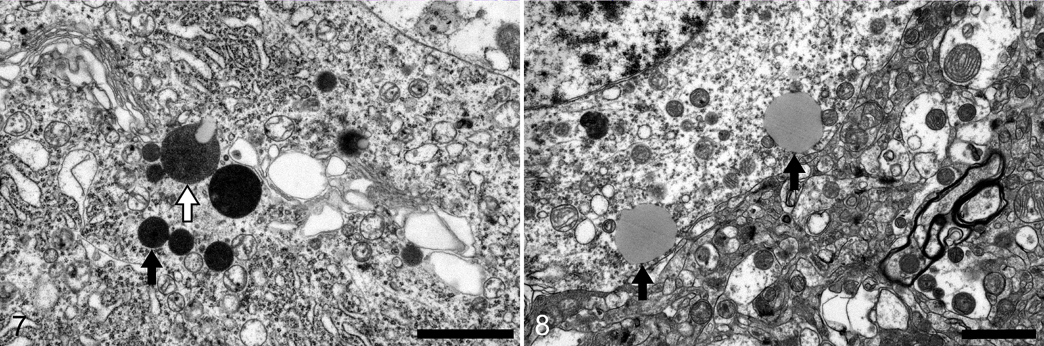
Transmission electron microscopy of the caudate nucleus of case 1 was performed with special focus on large neurons with intracytoplasmic eosinophilic granules. Occasional degenerating and necrotic neurons with pyknotic nuclei were visualized, often associated with swollen astrocyte foot processes. The cytoplasm of a few degenerating neurons contained a small number of round, extremely electron dense, 0.5 to 1 µm diameter structures (Fig. 7). A few of these dense bodies had peripheral vacuoles. A small number of lipid droplets were identified in the perikaryal cytoplasm of several neurons. These lipid droplets were approximately 1 to 2 µm, round, and numbered approximately 2 to 4 per cell (Fig. 8). Genetic analysis revealed no mutations in the SERAC1 gene.
The 3 Ibizan hounds in this report possess lesions similar to those described in both the Kerry blue terrier and Chinese crested dog with CMSD. 5,7 Ante-mortem imaging results supported a severe cerebellar ataxia with diffuse cerebellar atrophy and caudate nuclei degeneration similar to those seen in the imaging performed on the Chinese crested dogs with CMSD. 7 Macroscopic degeneration of the caudate nucleus and cerebellar atrophy were also described in the Kerry blue terrier and Chinese crested dog. 5,7 The microscopic lesions closely mimic the pattern of degeneration described in CMSD with neuronal degeneration and loss in the cerebellum, olivary nuclei, substantia nigra, and caudate nuclei. 5,7,12 The 3 dogs in this study were of different ages, allowing a general evaluation of disease progression. The majority of the macroscopic and microscopic lesions intensified in severity with age with the exception of the spongiosis in the substantia nigra and olivary nuclei. Case 3 was 4-years-old, 2 years older than any previous reported case of CMSD.
Pathologic characterization of CMSD in the Kerry blue terrier was undertaken in the 1980s, and the disease was divided into 3 stages based on chronicity. 5 Cerebellar and caudate nuclei degeneration precedes changes within the substantia nigra and olivary nucleus, which was attributed to transsynaptic neuronal degeneration. 5 In our study, all 3 dogs had lesions in all affected areas, so it could not be determined if the cerebellar and caudate nuclei changes came first. Presumably, the distribution of necrosis and regions spared within the caudate nucleus is related to the target neuron distribution, but further research is warranted. The youngest dog (case 1) appeared to have the most severe lesions in the olivary nuclei. The reason for this finding is uncertain, but it could be related to individual variation in disease progression or differences in the particular part of the olivary nucleus sampled. Lesions described in the Kerry blue terrier that were not observed in this study include gross discoloration of the substantia nigra and olivary nuclei, intense eosinophilia of the cytoplasm of surviving Purkinje cells, the presence of chromatolysis within olivary nucleus neurons, and ischemic neuronal necrosis and gitter cell accumulation within the caudate nuclei. The neuronal intracytoplasmic granules described in these Ibizan hounds were also described in the Kerry blue terrier. These granules were negative with periodic acid–Schiff and Sudan black staining, just as they were in the Kerry blue terrier. Intracytoplasmic granules were not mentioned in the one report of CMSD in the Chinese crested dog, but that report focused on the genetics of the disease and did not include a detailed pathologic description. 7
Ultrastructural lesions within the caudate nucleus were also documented previously in the Kerry blue terrier, allowing comparison to our findings. 6 Both the Ibizan hound and Kerry blue terrier possessed degeneration and necrosis of neurons within this region, as well as reactive swollen astrocytes. The small number of lipid droplets in the perikaryal cytoplasm of neurons is also a shared feature among the 2 breeds. Intracytoplasmic lipid droplets are an uncommon finding during neuronal degeneration compared to other cells of the body, such as hepatocytes. 10 In the human literature, neuronal lipid droplets have been linked to motor neuron disease, but these structures are largely unexplored in veterinary medicine. 10 It is likely that the scattered lipid droplets were not highlighted with the Sudan black stain as they were too small and few in number. Also, it is possible that the stain did not work well on formalin-fixed paraffin-embedded tissues.
The lipid droplets are one possible explanation for the intracytoplasmic eosinophilic granules observed with light microscopy. A more likely explanation for the granules might be the previously described intracytoplasmic dense bodies that are presumed to be a mixture of primary and secondary lysosomes. Lysosomes can impart eosinophilic granularity to the cytoplasm, as evidenced by the appearance of granular cell tumors. 9 Enlarged mitochondria were described in the Kerry blue terrier, and it was speculated that these may have represented the eosinophilic granules, but the ultrastructural imaging in our study did not reveal enlarged mitochondria. The origin of these eosinophilic granules remains unclear.
The mechanism of neuronal degeneration in CMSD is uncertain, but previous studies have suggested alterations in the glutaminergic pathways, or more recently, loss of dopaminergic cells. 5,6,8 Genetic analysis in the Kerry blue terrier and Chinese crested dog has identified mutations in the SERAC1 gene, leading to the development of genetic tests for owners and breeders. 8 These mutations in the SERAC1 gene were not found in this study, and further work to identify the mutation is ongoing.
Neurodegenerative disease involving the basal nuclei, including the caudate nucleus, putamen, globus pallidus, and substantia nigra are uncommon in domestic animals but are well described in humans. Examples include Huntington’s disease, Parkinson’s disease, and MSA. CMSD shares some similarities to MSA, which is divided into 2 categories, MSA-P with predominant parkinsonism and MSA-C with predominant cerebellar ataxia. 3 The defining neuropathology of MSA is similar to dogs with CMSD, including degeneration of striatonigral and olivopontocerebellar structures. Additionally, α-synuclein–positive glial intracytoplasmic inclusions are hallmark of the disease. 3 MSA differs from CMSD clinically as it has an adult-onset that often begins with urogenital dysfunction. Inevitably, the patients develop cerebellar ataxia similarly to dogs with CMSD. 3 Unlike our intraneuronal granules, the inclusions in MSA are not visible on routine hematoxylin and eosin staining and require α-synuclein immunohistochemistry or silver stains to be identified. Studies suggest that this buildup of α-synuclein in patients with MSA may result in the accumulation of abnormal protein forms that may be toxic to the cell and result in direct damage. 1 Another protein, p25α, also accumulates in MSA, and its role in the pathogenesis in unclear. Alpha-synuclein immunohistochemistry did not reveal intracytoplasmic inclusions in these dogs, and the presence of p25α was not assessed. While the differences in pathology between CMSD and MSA could indicate a completely different pathogenesis for the 2 diseases, further investigation into the potential role of α-synuclein, other members of the synuclein family, and p25α in CMSD is warranted.
A previously reported neurologic disease of Ibizan hounds has some clinical similarities to CMSD, but the lesions are very different. 2 Affected dogs develop ataxia at a few weeks of age, and the dysmetric gait includes balance loss and truncal sway. These dogs have loss of the patellar reflex and some have seizures. The lesions are consistent with an axonopathy and are confined primarily to spheroid formation within the trapezoid body, axonal degeneration in the spinal cord, and mild axonal changes in the cerebellum.
In this report, we described 3 cases of CMSD in a novel breed, the Ibizan hound. Given the similarity between CMSD and human MSA, further investigation of CMSD is warranted.
Supplemental Material
Supplemental Material, sj-pdf-1-vet-10.1177_03009858211043088 - Pathologic characterization of canine multiple system degeneration in the Ibizan hound
Supplemental Material, sj-pdf-1-vet-10.1177_03009858211043088 for Pathologic characterization of canine multiple system degeneration in the Ibizan hound by Samantha C. St. Jean, Bernard S. Jortner, Ryan N. Doan, Scott V. Dindot, Gary S. Johnson, Garrett Bullock, Derick B. Whitley, Jonathan M. Levine, Sandra K. Hancock, Andy Ambrus and Brian F. Porter in Veterinary Pathology
Footnotes
Acknowledgements
We thank Cynthia Kittler for help with sample collection and the acquisition of pedigree information. We also thank Ross Payne and Joseph Szule for assistance with electron microscopy, Ralph Storts for help with the ultrastructural evaluation, and Brian Summers for a helpful consultation.
Declaration of Conflicting Interests
The author(s) declared no potential conflicts of interest with respect to the research, authorship, and/or publication of this article.
Funding
The author(s) received no financial support for the research, authorship, and/or publication of this article.
Supplemental material for this article is available online.
References
Supplementary Material
Please find the following supplemental material available below.
For Open Access articles published under a Creative Commons License, all supplemental material carries the same license as the article it is associated with.
For non-Open Access articles published, all supplemental material carries a non-exclusive license, and permission requests for re-use of supplemental material or any part of supplemental material shall be sent directly to the copyright owner as specified in the copyright notice associated with the article.