
Abstract
Canine cutaneous mast cell tumors (ccMCTs) are currently graded according to Patnaik and Kiupel grading schemes. The qualitative and semiquantitative parameters applied in these schemes may lead to inter- and intraobserver variability. This study investigates the prognostic value of volume-weighted mean nuclear volume (
Keywords
Canine cutaneous mast cell tumors (ccMCTs) are among the most frequently diagnosed skin tumors in dogs. 24,41,43 These tumors display a variable and unpredictable biological behavior, ranging from benign behavior to potentially fatal metastatic tumors. 13,27 Tumor grade is widely used for prognostication; 4,13,25 however, no single parameter can accurately predict the biological behavior of ccMCTs. Several additional prognostic factors have been studied to improve ccMCT prognostication, including cytological grade, 6,29 expression pattern of KIT protein, 14 detection of c-KIT mutations, 40,42 proliferation markers, 41,43 and margin evaluation. 29,30
Most histological grading schemes rely on the subjective evaluation of morphologic and cytological parameters, which are prone to personal bias and often lead to intra- and interobserver variability. 1,28 In the case of ccMCTs, the most frequently used grading systems are those of Patnaik and Kiupel. The Patnaik system grades ccMCTs as grade I (G1), II (G2), and III (G3) according to the degree of mast cell differentiation, morphology, cellularity, extent of tissue involvement, stromal reaction, and mitotic count. 25 One of the problems with this grading system is the pronounced interobserver variability, particularly among the grading of G1 and G2 ccMCTs. 13,22,23,27,38,40 To overcome the subjectivity associated with Patnaik grading, a 2-tier grading scheme was developed by Kiupel et al. 13 The latter grades ccMCTs as low-grade (LG) or high-grade (HG) based on semiquantitative parameters, such as number of mitotic figures, multinucleated nuclei and bizarre nuclei per 10 hpf, and the presence of karyomegaly. 13 Several studies have shown the use of Kiupel grading not only significantly decreases interobserver variation but also has a superior prognostic value. 13,27,40,41
This study investigated the advantage of quantifying nuclear size and its variability to overcome the subjectivity associated with ccMCT histological grading. Previous measures of nuclear size have been studied in ccMCT cytological and histological samples.
18,38,39
These studies found associations between either ccMCT nuclear area or perimeter and Patnaik grade. Interestingly, statistical differences were found between G1 and G3 ccMCTs, as well as between G2 and G3, but not between G1 and G2. Two-dimensional morphometrical estimates have the disadvantage that the position and orientation of a section plane across a 3-dimensional object influence the size, shape, and frequency of the 2-dimensional profile.
5,19
Consequently, although there is a correlation between nuclear area or perimeter and the real nuclear size, the true relation between these parameters is uncertain, no matter how elegant the statistical model
33
(Fig. 1). Design-based stereological methods, on the other hand, are based on statistical sampling and geometrical principles to recover 3-dimensional information from 2-dimensional sections. These methods eliminate assumptions about the object’s shape and orientation, thus allowing for precise and reproducible measures of numerous parameters, including nuclear size.
5,19,28
Stereological measures of nuclear size are best performed with the “point-sampled intercept” (PSI) method, which provides a measure of volume-weighted mean nuclear volume (
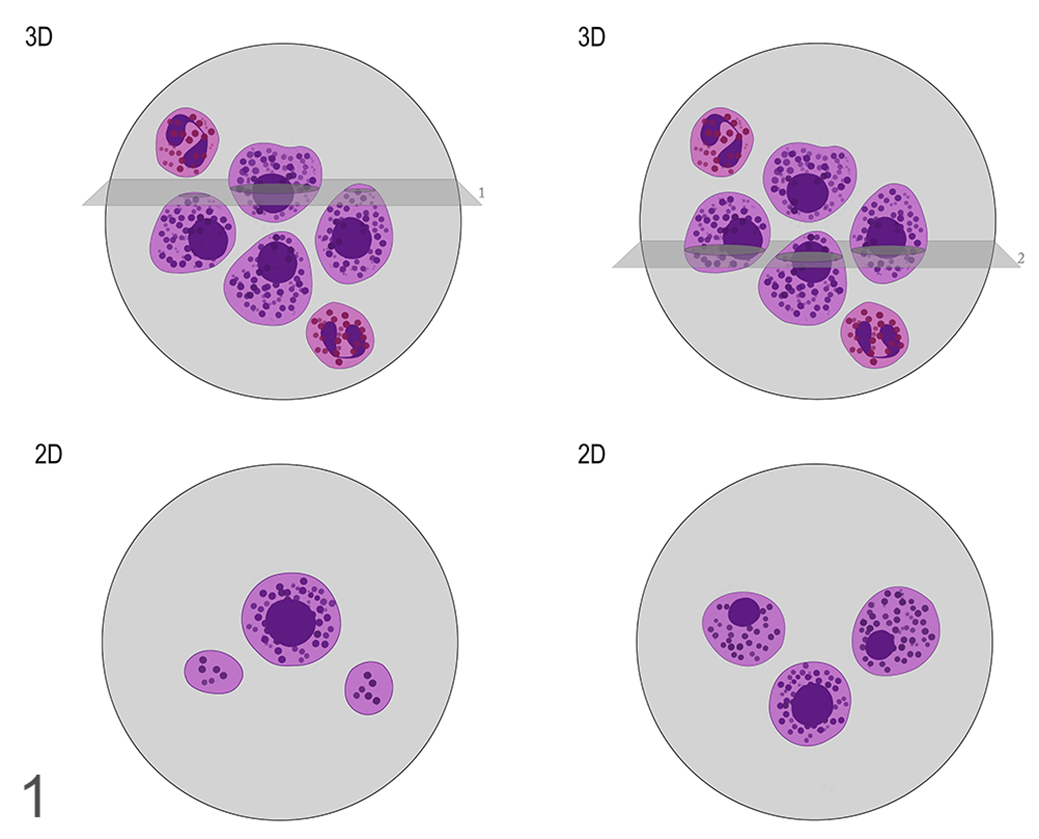
Two-dimensional representations of sections across 3-dimensional mast cells. The orientation of the slice influences not only the frequency with which nuclei are sampled, but also their dimension. Although there is a relation between the nuclear profile area and its true size, this relation is uncertain due to the loss of 3-dimensional information. The right and left panels represent 2 different sections through that same cell population. The top panels are 3-dimensional representations of the cells showing the location of slices. The lower panels are the appearances of the resulting 2-dimensional sections.
Materials and Methods
Case Selection and Histopathology
Fifty-five paraffin-embedded ccMCTs diagnosed in 2017 were selected for this study from the archives of DNATech, Lisbon, Portugal. These cases were selected if the diagnosis had been made at least 1 year before the start of the study (May 2019). Three-micrometer sections were routinely processed and stained with hematoxylin and eosin for tumor grading and stereological estimates. Each tumor was blindly graded according to the Patnaik and Kiupel grading schemes by 3 experienced pathologists (IBV—ECVP board certified, and SB and PF—professors of veterinary pathology with more than 20 years experience). The final diagnosis was established by the consensus of at least 2 observers. Histological grade was assigned according to the criteria described in the original papers. 14,25
Outcomes
Follow-up of dogs treated with surgical excision alone was available for 30 cases. The veterinary clinicians provided clinical data regarding existence of postsurgical local recurrence, metastasis, and/or mast cell tumor-related death (including euthanasia).
The minimum follow-up period was 1 year. Dogs with postsurgical resolution of disease were given an outcome value of 0 (OC0), whereas outcome value of 1 (OC1) included cases that died or were euthanized as a result of local recurrence or development of nodal or visceral metastasis. The lateral and deep surgical margins (cm) were also evaluated at the time of submission and compared between groups.
Stereology
Whole-slide images were obtained (NanoZoomer-SQ Digital slide scanner, Hamamatsu Photonics) with 40× resolution and 800× magnification. Measurements were performed on newCAST stereological software (Visiopharm). In each slide, the region of interest (ROI), that is, the total area of the tumor, was delineated manually at low magnification. Within each ROI, fields of view (1000×, A = 10300.84 µm2) were automatically selected in a systematic random fashion by the software. According to Gundersen and Jensen,
11
approximately 75 nuclei per tumor are needed to accurately estimate
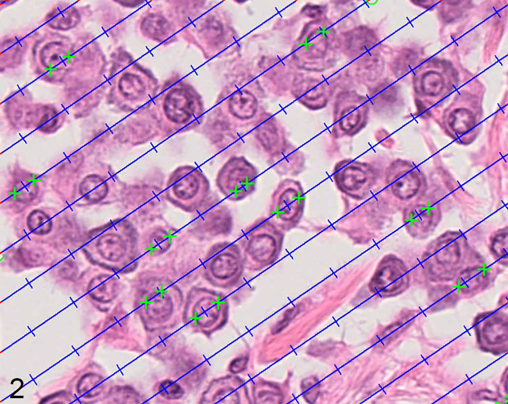
Estimation of mean nuclear volume using the point-sampled intercept method. Fields of view from a mast cell tumor are automatically generated with a constant step size. The nuclear profiles are sampled with points (blue hash marks) and the associated test-lines create linear intercepts across these profiles. The length of the intercepts is traced by marking the nuclear borders in the direction of the line (green hash marks). The goal is to measure ∼75 intercepts per tumor.
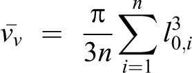
In which
Statistical Analysis
Statistical analysis was performed using R software with the DescTools (version 0.99.37), 31 irr (version 0.81.4), 8 ggplot2 (version 3.2.1), 44 and pROC packages (version 1.14.0). 26
For quantitative variables, such as
Regarding Patnaik and Kiupel grading, the agreement among pathologists was evaluated with Fleiss’ κ statistics. 20
Considering the paired measurements (different measures from the same sample), the differences between
For the variable Outcome, a subset of 30 cases for which we had information on the variables age, sex, breed, and surgical margins was used. To assess if there were any differences between outcome on those variables, as well as differences of
To determine if the
Results
Fifty-five ccMCTs were graded by consensus among 3 pathologists based on Patnaik and Kiupel grading systems. Based on the Patnaik grading system, 39 (71%) ccMCTs were G2 and 16 (29%) as G3. No tumors were diagnosed by consensus as G1, even though 11 tumors were diagnosed as G1 by 1 out of 3 pathologists. The agreement among pathologists was fair in Patnaik grading (κ = 0.32), with 44% concordance in the assignment of G2 and 56% concordance in the assignment of G3 (Table 1). G3 ccMCTs were associated with increased mortality and shorter survival time (Fig. 3). Based on the Kiupel grading system, 35 (64%) ccMCTs were diagnosed as LG and 20 (36%) as HG. There was moderate agreement in Kiupel grading (κ = 0.46), with 66% concordance in the assignment of LG and 55% concordance in the assignment of HG ccMCTs (Table 1). Overall, LG were graded as G2 and HG were graded as G3, except for 4 HG graded as G2. HG ccMCTs were associated with increased mortality and shorter survival time (Fig. 4).
Grade Assignment for 55 Canine Cutaneous Mast Cell Tumors by 3 Pathologistsa. The Data Show the Number and Percentage of Cases.
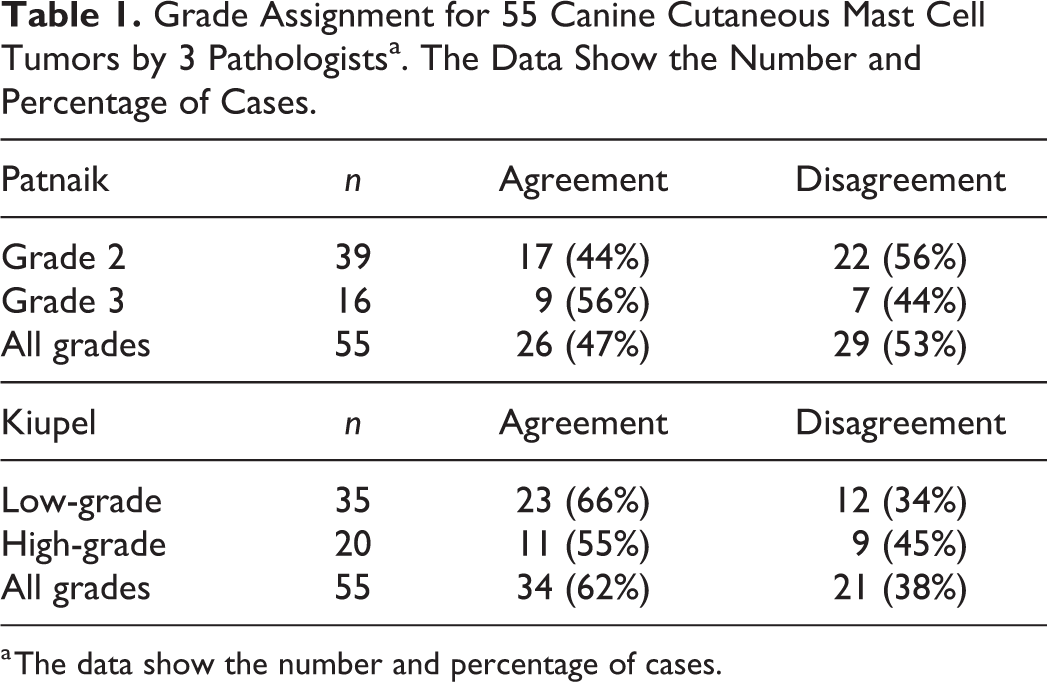
a The data show the number and percentage of cases.
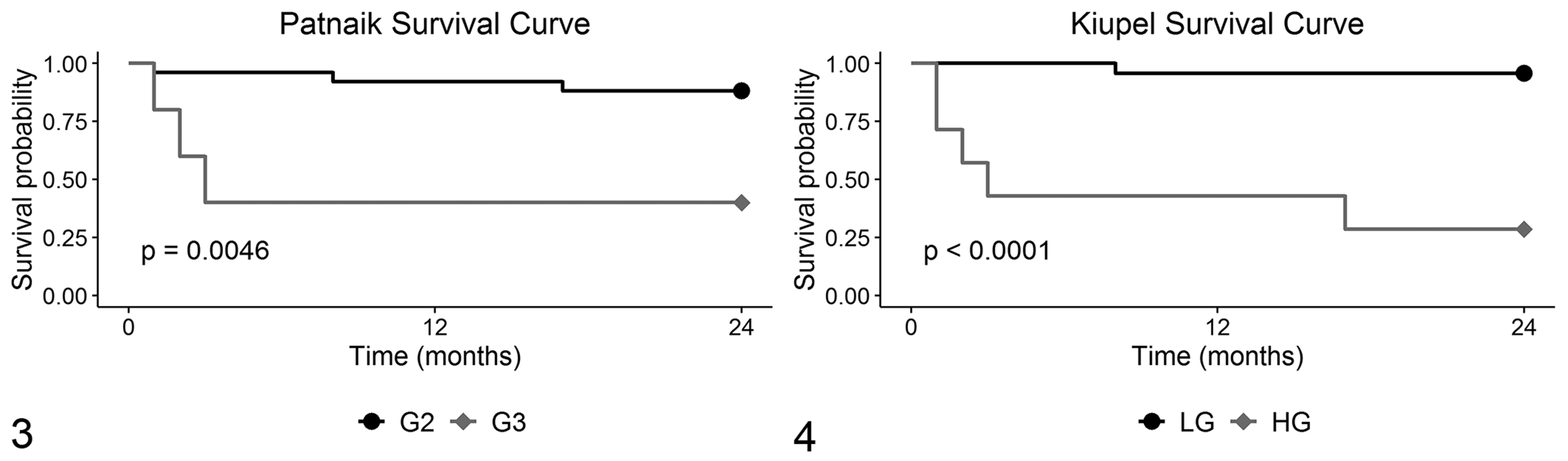
Survival curves for mortality in 30 canine mast cell tumors, graded according to the Patnaik grading system (Fig. 3) and the Kiupel system (Fig. 4). Abbreviations: G2, Patnaik grade 2; G3, Patnaik grade 3; LG, Kiupel low-grade; HG, Kiupel high-grade.
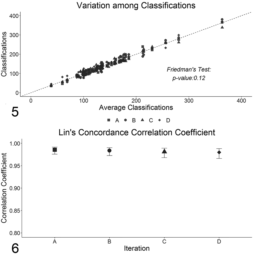
Each tumor was measured by 2 observers, totaling 4 measurements per tumor, and each measurement took approximately 10 minutes. To ease the following analysis, the average of those 4 measurements was calculated and compared with the obtained data. There were no statistical differences among measurements including the average (see Fig. 5) with this yielding concordance coefficients very close to 1 for the 4 original measurements (Fig. 6). Therefore, the average of the measurements was used as a proxy of the
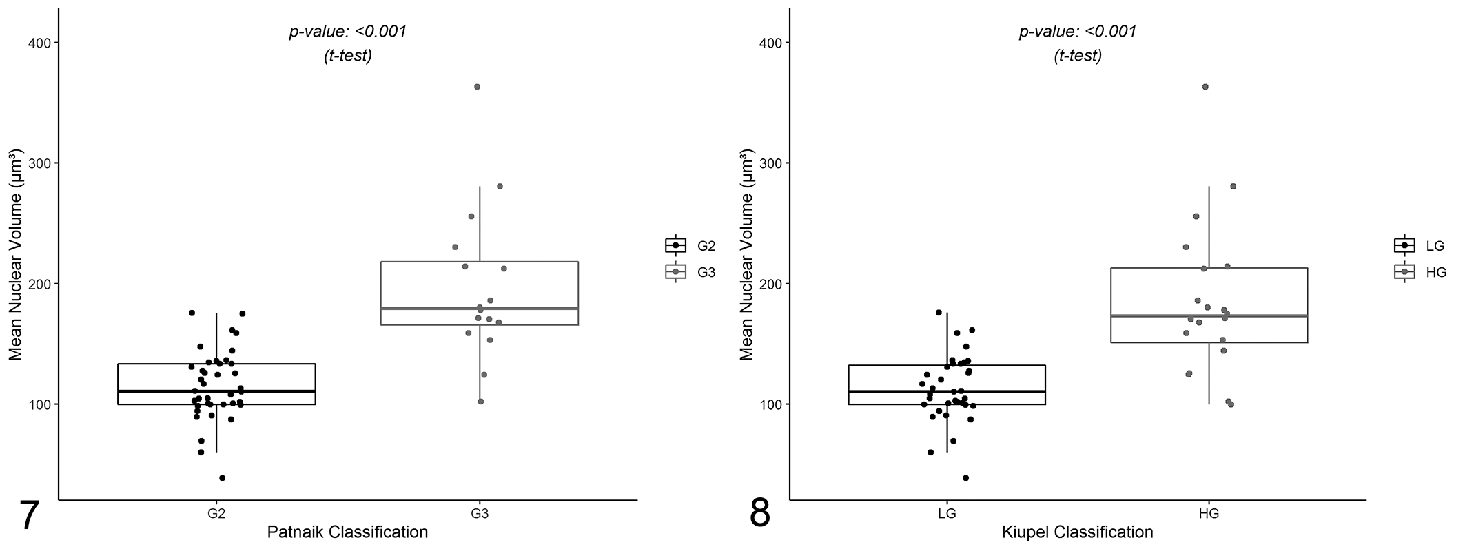
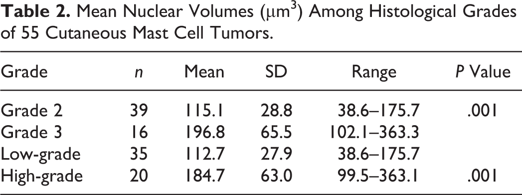
Comparison of the mean nuclear volume values among Patnaik and Kiupel histological grades from 55 cases of canine mast cell tumors. The boxes show first quartile, second quartile (median), and third quartile. The whiskers represent the range (minimum and maximum) values. Mean nuclear volumes are significantly different between grade 2 and grade 3 (Fig. 7), and between low-grade and high-grade (Fig. 8) mast cell tumors (Welch 2-sample t test).
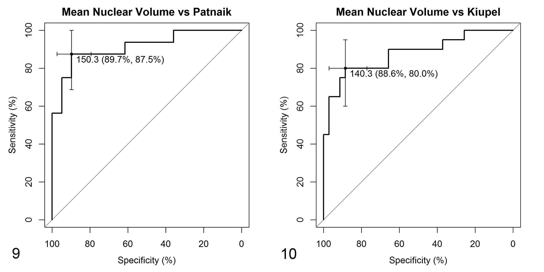
Receiver operating characteristics curves for mean nuclear volume values according to Patnaik (Fig. 9) and Kiupel (Fig. 10) grading systems from 55 cases. For the Patnaik system (Fig. 9), a mean nuclear value ≥150.3 µm3 identifies a grade 3 mast cell tumor with 89.7% specificity and 87.5% sensitivity. For the Kiupel system (Fig.10), a mean nuclear value ≥140.3 µm3 identifies a high-grade mast cell tumor with 88.6% specificity and 80.0% sensitivity. Abbreviations: G2, Patnaik grade 2; G3, Patnaik grade 3; LG, Kiupel low-grade; HG, Kiupel high-grade.
Mean Nuclear Volumes (µm3) Among Histological Grades of 55 Cutaneous Mast Cell Tumors.
To access the differences in
There were no differences between OC0 and OC1
The 24 OC0 cases included 22 G2/LG ccMCTs and 2 G3/HG ccMCTs. The 6 OC1 cases included 1 LG and 5 HG, or 3 G2 and 3 G3.
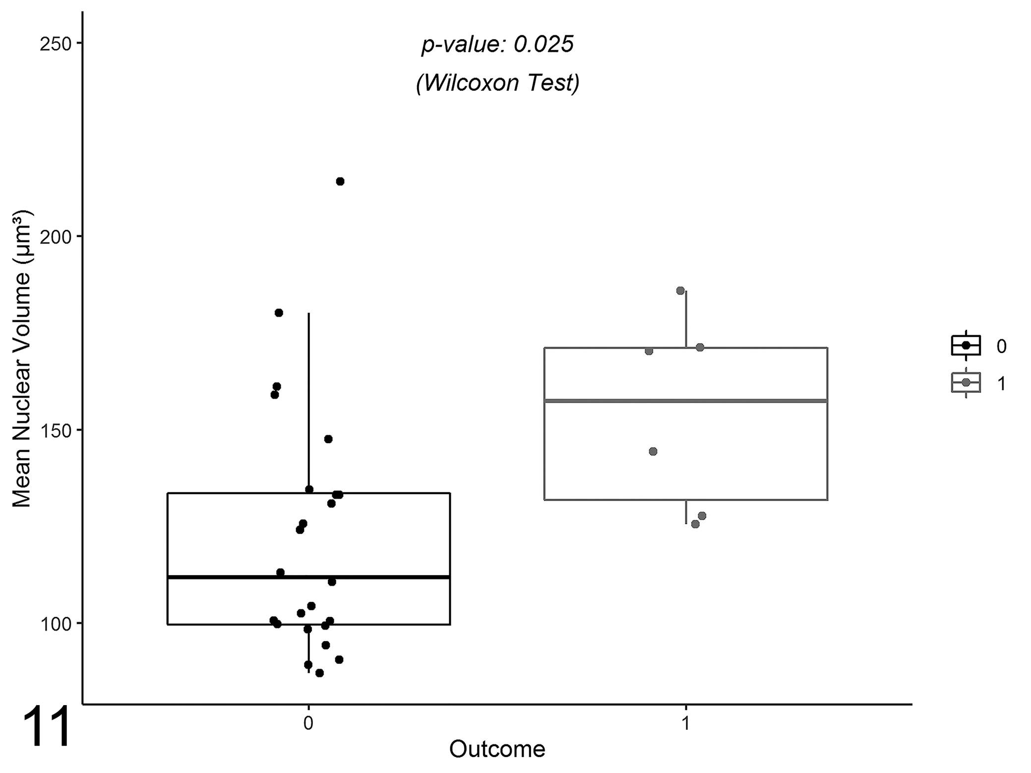
Comparison of the mean nuclear volume values among clinical outcomes from 30 cases of canine mast cell tumor. The boxes show the first quartile, second quartile (median), and third quartile. The whiskers represent the range (minimum and maximum) values. There is a significant difference between outcome OC0 (alive) and OC1 (died) (Wilcoxon’s sum-rank test).
Mean Nuclear Volumes (µm3) Among Clinical Outcomes of 30 ccMCTs.
The potential of
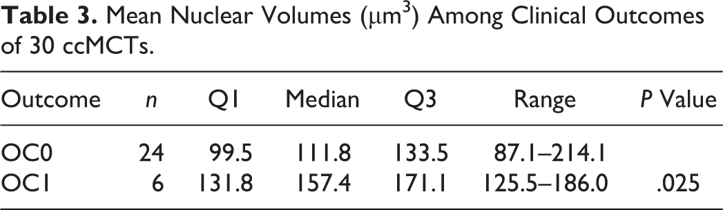
Receiver operating characteristics curve for mean nuclear volume values according to clinical outcome from 30 cases. A mean nuclear value ≥124.8 µm3 is associated OC1 (died) with 58.3% specificity and 100% sensitivity.
By using the cutoff value of 124.8 µm3 to separate cases and assessing their mortality, statistically significant differences were observed between cases, with 100% of the animals with
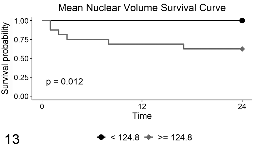
Survival curves for mortality due to mast cell tumors, according to mean nuclear volume values from 30 cases.
Discussion
Our results identified significant variability among pathologists in ccMCT grading. As expected, there was greater consistency in Kiupel grading,
13,27,40,41
and greater inconsistency in Patnaik grading particularly in the assignment of G2. These results are in accordance with previous studies; however, the concordance between pathologists was lower in this study.
13,27,40
Although G1 was assigned to 11 tumors by 1 of 3 pathologists, no ccMCTs were graded G1 by consensus in this study. However, G1 ccMCTs are composed of monomorphic, well-differentiated mast cells, hence these tumors would, in theory, have lower
The PSI method allowed the measurement of
Previous morphometric studies also found an association between Patnaik grade and both nuclear area and perimeter.
18,38,39
In comparison with nuclear morphometry,
In terms of clinical outcome,
Nevertheless, some ccMCTs are diagnosed as HG based on the presence of karyomegaly, regardless of a low mitotic count. Kiupel et al define karyomegaly as “nuclear diameters of at least 10% of neoplastic mast cells vary by at least 2-fold”
13
and this is likely poorly reproducible. In fact, scoring of nuclear pleomorphism has poor agreement between pathologists for other tumors.
7,28
Considering that
Supplemental Material
Supplemental Material, sj-pdf-1-vet-10.1177_0300985820985138 - Stereology in Grading and Prognosis of Canine Cutaneous Mast Cell Tumors
Supplemental Material, sj-pdf-1-vet-10.1177_0300985820985138 for Stereology in Grading and Prognosis of Canine Cutaneous Mast Cell Tumors by Mafalda Casanova, Sandra Branco, Inês Berenguer Veiga, André Barros and Pedro Faísca in Veterinary Pathology
Footnotes
Acknowledgements
Declaration of Conflicting Interests
The author(s) declared no potential conflicts of interest with respect to the research, authorship, and/or publication of this article.
Funding
The author(s) received no financial support for the research, authorship, and/or publication of this article.
Supplemental material for this article is available online.
References
Supplementary Material
Please find the following supplemental material available below.
For Open Access articles published under a Creative Commons License, all supplemental material carries the same license as the article it is associated with.
For non-Open Access articles published, all supplemental material carries a non-exclusive license, and permission requests for re-use of supplemental material or any part of supplemental material shall be sent directly to the copyright owner as specified in the copyright notice associated with the article.