
Abstract
Among 113 feline gastrointestinal epithelial tumors diagnosed between 2006 and 2019, 78 (69%) were detected in the colorectum. Fifty colorectal tumors were selected for further pathological evaluations, of which 9 (18%) were histopathologically diagnosed as adenomas and 41 (82%) as carcinoma. The carcinomas included 33 tubular adenocarcinomas (TAC), 5 tubulovillous adenocarcinomas (TVAC), 2 mucinous adenocarcinomas, and 1 undifferentiated carcinoma. Histopathologically, TAC frequently showed vascular invasion (17/33 cases, 52%). In TAC cases, serosal infiltration (13/15 cases, 87%) and lymph node metastasis (8/9 cases, 89%) were common in bowel resection and lymphadenectomy samples, respectively. Immunohistochemically, the tumor cells of most cases were positive for cytokeratin (CK) 20 (50/50 cases, 100%) and CDX2 (48/50 cases, 96%). Focal immunopositivity for CD10 (11/50 cases, 22%) and CK7 (15/50 cases, 30%) was observed irrespective of the histological subtype. Only a few cases showed diffuse nuclear accumulation of β-catenin (2/50 cases, 4%) and p53 (5/50 cases, 10%). A lack of tubule formation, female sex, and low CDX2 labeling were statistically associated with carcinoma compared to adenoma (ρ = 0.615, P < .001; ρ = 0.279, P = .050; and ρ = −0.265, P = .063, respectively). Other features, including mucin profiles, Ki67 labeling index, and accumulation of β-catenin and p53, were not associated with malignancy. A sequence analysis revealed KRAS mutations in 3/7 TAC cases. These results suggest that KRAS mutations—rather than excessive Wnt/β-catenin signaling and the inactivation of TP53—contribute to the tumorigenesis of feline colorectal carcinoma.
Intestinal neoplasia is a common disease in domestic cats, accounting for 5% to 9.3% of feline tumors. 31 Epithelial tumors, such as adenoma and adenocarcinoma, are the second most common neoplasms in the feline intestine. 31,36 Previous studies reported that feline epithelial intestinal tumors commonly occurred in the small intestine, 10,34 whereas a recent epidemiological study revealed that the large intestine was affected more than twice as often as the small intestine. 36 However, few studies have focused on feline colorectal neoplasms, and their pathological features currently remain unclear.
In humans, it is widely accepted that the adenoma-carcinoma sequence is involved in the development of most colorectal cancers. 24 It represents a stepwise progression from a normal epithelium to adenoma and then carcinoma, which is associated with the accumulation of multiple clonally selected genetic alterations, including APC, TP53, and KRAS. During this process, mutations in the APC and TP53 genes result in the dysregulation of β-catenin and p53 expression, respectively. Recent studies indicated that APC alteration and β-catenin dysregulation are common in canine colorectal tumor. 27,35,39,49 However, it remains unknown whether feline colorectal cancer shares this genetic pathway.
In the present study, we retrospectively examined the frequency of gastrointestinal epithelial tumors in cats. We then described the histopathological findings of feline colorectal epithelial tumors and investigated the relationship between clinical and histopathological features and malignancy. We also discussed the mechanisms underlying the development of feline colorectal cancer based on the immunohistochemical results of β-catenin and p53 and a KRAS mutation analysis.
Materials and Methods
Case Selection
The records of feline gastrointestinal biopsy cases (797 cases) that were submitted to the Laboratory of Veterinary Pathology, the University of Tokyo, between November 2006 and September 2019 were reviewed. Cases diagnosed with epithelial tumors (113 cases) were categorized according to topographical locations. For further histopathological examinations, 50 cases with colorectal epithelial tumors were selected that had sufficient quantities of tumor tissue including at least a portion of muscularis mucosae. Examples of adequate samples are shown in Supplemental Figures S1 to S4. All tissue specimens were obtained through endoscopic biopsy (31 cases) or surgical resection (19 cases). Regional lymph nodes samples (12 cases) were also collected when the cats showed lymphadenopathy.
Histopathology
All tissues were fixed in 10% neutral buffered formalin and routinely embedded in paraffin wax. Consecutive sections (thickness of 4 μm) were stained with Victoria blue-hematoxylin and eosin (VB-HE), Alcian blue (pH 2.5)-periodic acid–Schiff (AB-PAS), and high iron diamine-Alcian blue (pH 2.5) (HID-AB). Immunohistochemistry was also performed.
The histological classification of proliferative lesions was based on the World Health Organization classification and Tumors in Domestic Animals 5th edition. 17,31 Malignancy (adenocarcinoma/carcinoma) was defined by the presence of neoplastic epithelial cells infiltrating the muscularis mucosae or invading the vasculature. Cases without histological evidence of these features were categorized as benign lesions (adenoma) regardless of morphological atypia.
Six histological parameters were also assessed: the degree of differentiation, presence of osseous metaplasia, serrated lesions, vascular invasion, serosal infiltration, and lymph node metastasis. The degree of differentiation was evaluated by gland formation in tumor tissue and scored as well-differentiated (Score 1, >95% with gland formation), moderately differentiated (Score 2, 50% to 90% with gland formation), and poorly differentiated (Score 3, <50% with gland formation) according to the World Health Organization classification in humans. 5 Serrated lesions were characterized by the formation of the stellate or sawtooth-like architecture (slit-like infolding of the epithelium) in >5% of tumor tissue. Serosal infiltration and lymph node metastasis were only assessed in samples obtained from bowel resection and lymphadenectomy, respectively.
Immunohistochemistry
Consecutive sections were labeled immunohistochemically to analyze the characteristics of tumor cells. Endogenous peroxidase in sections was blocked by 3% hydrogen peroxide in methanol for 5 minutes. To unmask antigens, sections were heated in an autoclave (121 °C) for 10 minutes in citrate buffer (pH 6.0) or incubated with 125 μg/ml proteinase K (Wako) solution at room temperature for 30 minutes. After washing with Tris-buffered saline (TBS), slides were treated with 8% skimmed milk in TBS. The primary antibodies used are listed in Table 1. After an incubation with each primary antibody at 4 °C overnight, immunolabeled antigens were visualized using the Dako Envision+ System (Dako) and reacting with 0.05% 3′3-diaminobenzidine plus 0.03% hydrogen peroxide in Tris-hydrochloric acid buffer. Sections were then counterstained with hematoxylin.
Antibodies Used for Immunohistochemistry.
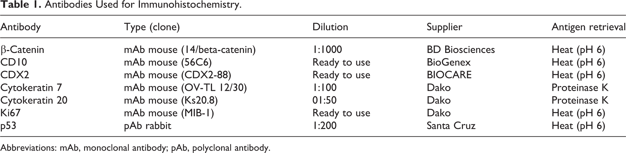
Abbreviations: mAb, monoclonal antibody; pAb, polyclonal antibody.
The Ki67 labeling index was calculated as the average percentage of positive tumor cells under ×400 magnification in 5 randomly selected fields for each case. The immunoreactivity of neoplastic cells for β-catenin, CD10, CDX2, cytokeratin (CK) 7, and CK20 was scored semiquantitatively as follows: 0 (negative), <5% positive cells; 1, 5% to 25% positive cells; 2, 25% to 50% positive cells; 3, 50% to 75% positive cells; 4, >75% positive cells. Nuclear immunoreactivity was interpreted as positive for β-catenin, CDX2, Ki67, and p53, whereas membranous or cytoplasmic reactivity indicated positivity for CD10, CK7, and CK20.
Statistical Analysis
Spearman’s rank correlation coefficient (Spearman’s ρ) was calculated to examine the relationships between clinical profiles, histological parameters, immunohistochemical scores, and malignancy. The Fisher z-transformation was used to calculate the 95% confidence interval and P values. P values <.05 were considered to be significant. Normality tests for “Age” and “Ki67 labeling index” were not performed. All statistical analyses were conducted with software R version 3.5.0 (R Foundation for Statistical Computing).
Analysis of KRAS Gene Mutations
Tissue samples from 9 cats that contained a sufficient quantity of tumor cells were selected for molecular analysis of the KRAS gene. Genomic DNAs were extracted from formalin-fixed paraffin-embedded colorectal tissues using the QIAamp DNA FFPE Tissue Kit (Qiagen). Purified DNAs were used in subsequent PCR (polymerase chain reaction). The feline KRAS gene-specific PCR primer pair, 5′-AGGTGAATTTGCGTTAAAAGGTAT-3′ (forward) and 5′-AAAGAATGGGCCTGCACAAA-3′ (reverse), which amplifies a 286-bp fragment that incorporates exon 1 of the KRAS gene, was used. PCR was performed as follows: 30 cycles at 95 °C for 30 seconds and 53 °C for 30 seconds. PCR products were submitted for a sequence analysis in the forward and reverse directions (FASMAC). Each chromatogram was visually inspected for any abnormalities with particular attention directed to codons 12 and 13. Unaffected feline colorectal tissue was also used to confirm the normal sequences of the feline KRAS gene.
Results
Demograpics and Frequency of Feline Colorectal Epithelial Tumors
Among 797 feline gastrointestinal biopsy cases, including 533 gastric, 528 duodenal, 442 jejunal to ileocecal, and 497 colorectal tissues, 113 (14%) were diagnosed as epithelial tumors. Topographically, the majority of epithelial tumors (78/113 cases, 69%) were located in the colorectum. In the other cases, 11, 10, 7, and 7 tumors were located in the ileocecum, jejunoileum, stomach, and duodenum, respectively (Fig. 1). Among colorectal epithelial tumor cases, there were 42 males (54%) and 36 females (46%). Mean age (±SD) at the time of the diagnosis was 13.0 years (±2.5 years; range: 5–18 years). Cases involved 61 mixed breeds (78%), 4 Abyssinians (5%), and 3 Scottish Folds (4%).
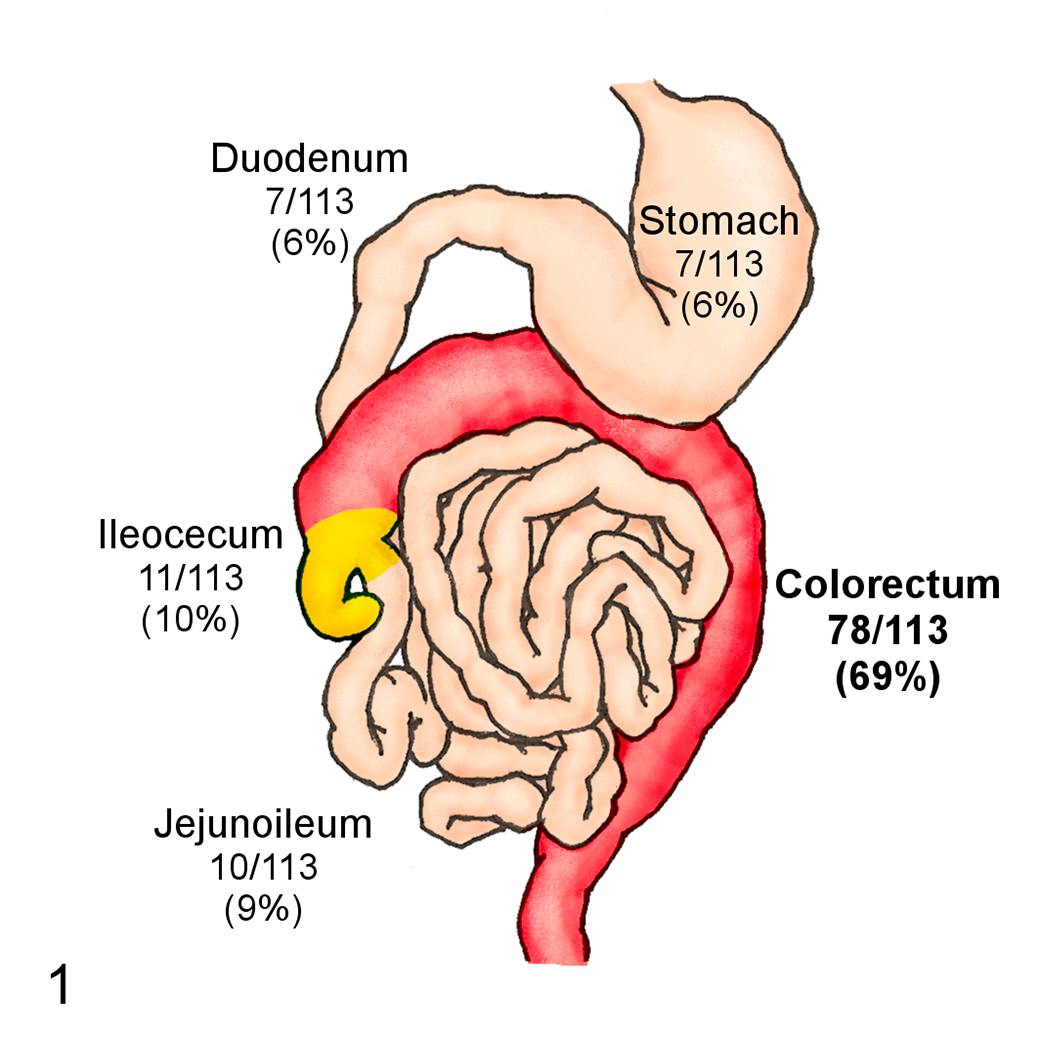
Frequency of feline epithelial tumors in different gastrointestinal segments in this study.
Histopathological Diagnoses
Based on the case selection criteria, 50 feline colorectal tumor cases (27 in colon, 14 in colorectal junction, and 9 in rectum) including 19 surgical and 31 endoscopic biopsy samples were chosen for histopathological examinations. Lymphadenectomy was performed in 9 cases (8 bowel resection and 1 endoscopy case). Information on all examined cases is shown in Supplemental Table S1. The tumors were classified as tubular adenoma (TA; 5 cases, 10%), tubular adenocarcinoma (TAC; 33 cases, 66%), villous adenoma (VA; 2 cases, 4%), tubulovillous adenoma (TVA; 2 cases, 4%), tubulovillous adenocarcinoma (TVAC; 5 cases, 10%), mucinous adenocarcinoma (MAC; 2 cases, 4%), and undifferentiated carcinoma (UDC; 1 case, 2%) according to their histopathological features. No villous adenocarcinoma, signet-ring cell carcinoma, or adenosquamous carcinoma was detected in the present study. The results of evaluations of histological parameters and the Ki67 labeling index of each tumor type are summarized in Table 2. The mean values of the Ki67 labeling index ranged between 16.7% and 54.9%, with no increases in malignant tumors.
Summary of Histological Features of Feline Colorectal Epithelial Tumors.
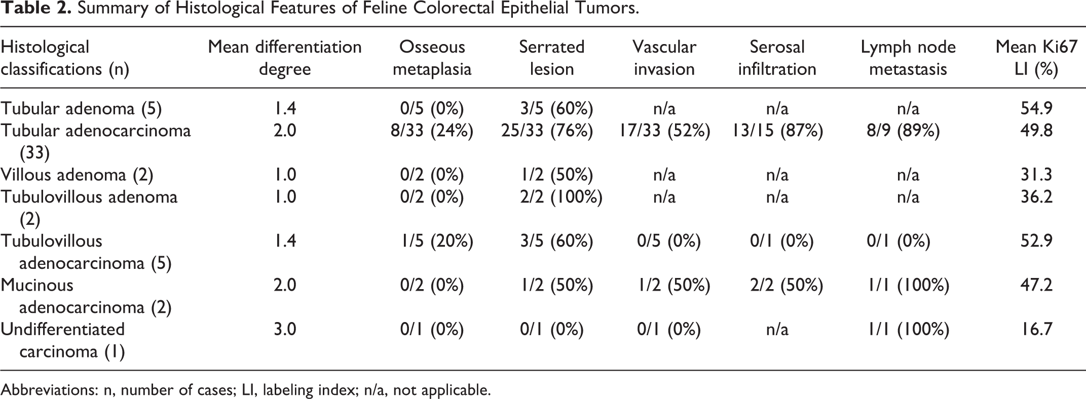
Abbreviations: n, number of cases; LI, labeling index; n/a, not applicable.
Histopathological Features
TA and TAC lesions were characterized by tubular structures composed of columnar to cuboidal cells (Figs. 2, 3). The surfaces of tumors were frequently eroded with infiltration by various numbers of neutrophils or eosinophils. Neoplastic cells had abundant cytoplasm and oval to round nuclei. Nuclear pleomorphism and pseudostratification with the loss of polarity were common. In TACs, invasion by neoplastic ducts with severe stromal desmoplasia was often found (Fig. 3). Osseous metaplasia was more common in TAC (8/33 cases, 24%) than in TA (0/5 cases, 0%). Serosal infiltration (13/15 cases, 87%) and lymph node metastasis (8/9 cases, 89%) were frequently found in TAC (Table 2).
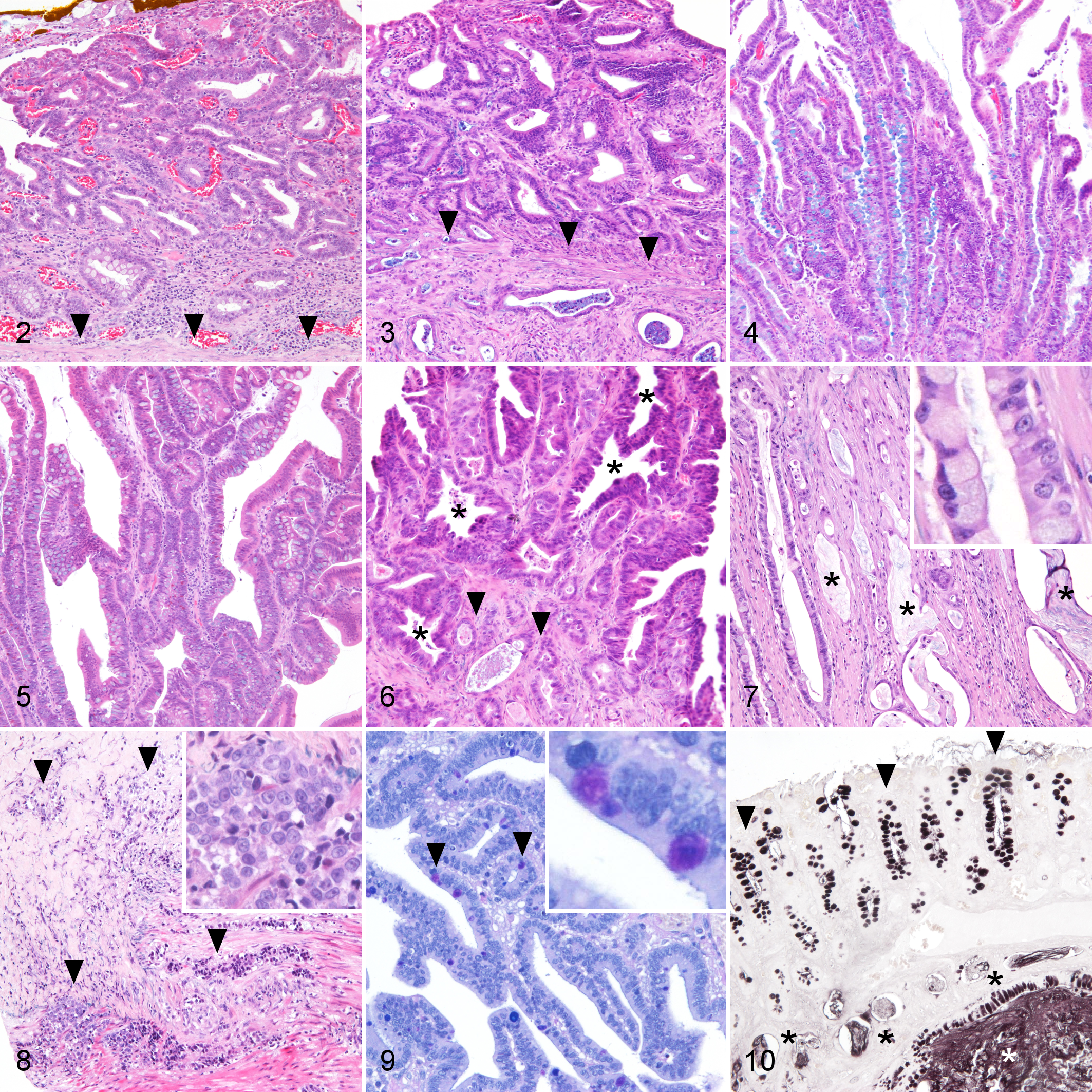
Colorectum, cat.
VA lesions were characterized by elongated tubules with few branches resembling small intestinal villi (Fig. 4). The stroma was scarce and contained small vessels. The morphology of neoplastic cells was similar to that of TA/TAC. In VA, 1 case had a serrated lesion (Table 2), and osseous metaplasia was not observed.
TVA and TVAC lesions showed the characteristics of both TA/TAC and VA (Figs. 5, 6). There were short or branched tubules mingled with a villous structure. The tubular component was dominant in the invading lesions of TVAC (Fig. 5). In TVAC cases, neither vascular invasion (n = 5), serosal infiltration, nor lymph node metastasis (n = 1) was detected (Table 2).
MAC lesions were characterized by dilated ducts containing abundant mucus (Fig. 7). Tumor cells had an enlarged columnar cytoplasm with droplets of intracellular mucin and basally located nuclei, which resembled goblet cells (Fig. 7). These cells were arranged in the tubulovillous structure at the mucosal surface; however, cystic tubules with pools of extracellular mucus were prominent in the submucosa and muscular layer. Of the 2 MAC cases, both showed serosal infiltration, and 1 displayed vascular invasion. A metastatic lesion of the lymph node was also detected in 1 case (Table 2).
The UDC lesion exhibited polygonal epithelial cells invading the muscular layer without the formation of a tubular structure (Fig. 8). These cells had a scant amphophilic cytoplasm and round nuclei with mild anisokaryosis. No vascular invasion was detected, whereas metastatic lesions were found in the mesentery lymph node and omentum.
Histochemically, normal and neoplastic epithelial cells in 41 cases (82%) produced only AB+/PAS+ mucin. On the other hand, 9 cases (18%; 6 TAC and 1 TVA, VA, and TVAC) displayed a small amount of AB−/PAS+ mucus in tumor tissue (Fig. 9). All mucus in 49 cases (98%) was positive for HID (Fig. 10); however, 1 case (2%; case 11, TAC) showed HID−/AB+ mucus in normal crypts.
Immunohistochemical Features
The results of immunohistochemistry are shown in Table 3. Neoplastic cells in all 50 cases (100%) were positive for CK20 to various degrees (Fig. 11). CDX2 was positive to various extents in 48/50 cases (96%), whereas tumor cells in 2/50 cases (4%; 1 VA and 1 TAC) were negative. In 28/50 cases (56%; 1 TA, 1 VA, 21 TAC, 2 TVAC, 2 MAC, and 1 UDC), the immunoreactivity of CDX2 was weaker in neoplastic cells than in normal mucosal epithelial cells (Fig. 12). In total, 11/50 (22%; 7 TAC, 2 TA, and 2 TVAC) and 15/50 cases (30%; 9 TAC, 2 TA, 2 MAC, 1 VA, and 1 TVAC) were focally positive for CD10 and CK7, respectively (Figs. 13, 14). The nuclear accumulation of β-catenin and p53 was detected in 30/50 (60%; 3 TA, 21 TAC, 1 VA, 1 TVA, 3 TVAC, and 1 MAC) and 29/50 cases (58%; 2 TA, 21 TAC, 2 VA, and 4 TVAC), respectively (Figs. 15, 16). However, only 2/50 (4%, 2 TAC) and 5/50 (10%, 3 TAC, 1 TA, and 1TVAC) cases displayed nuclear accumulation of β-catenin and p53 in more than 75% of tumor cells, respectively.
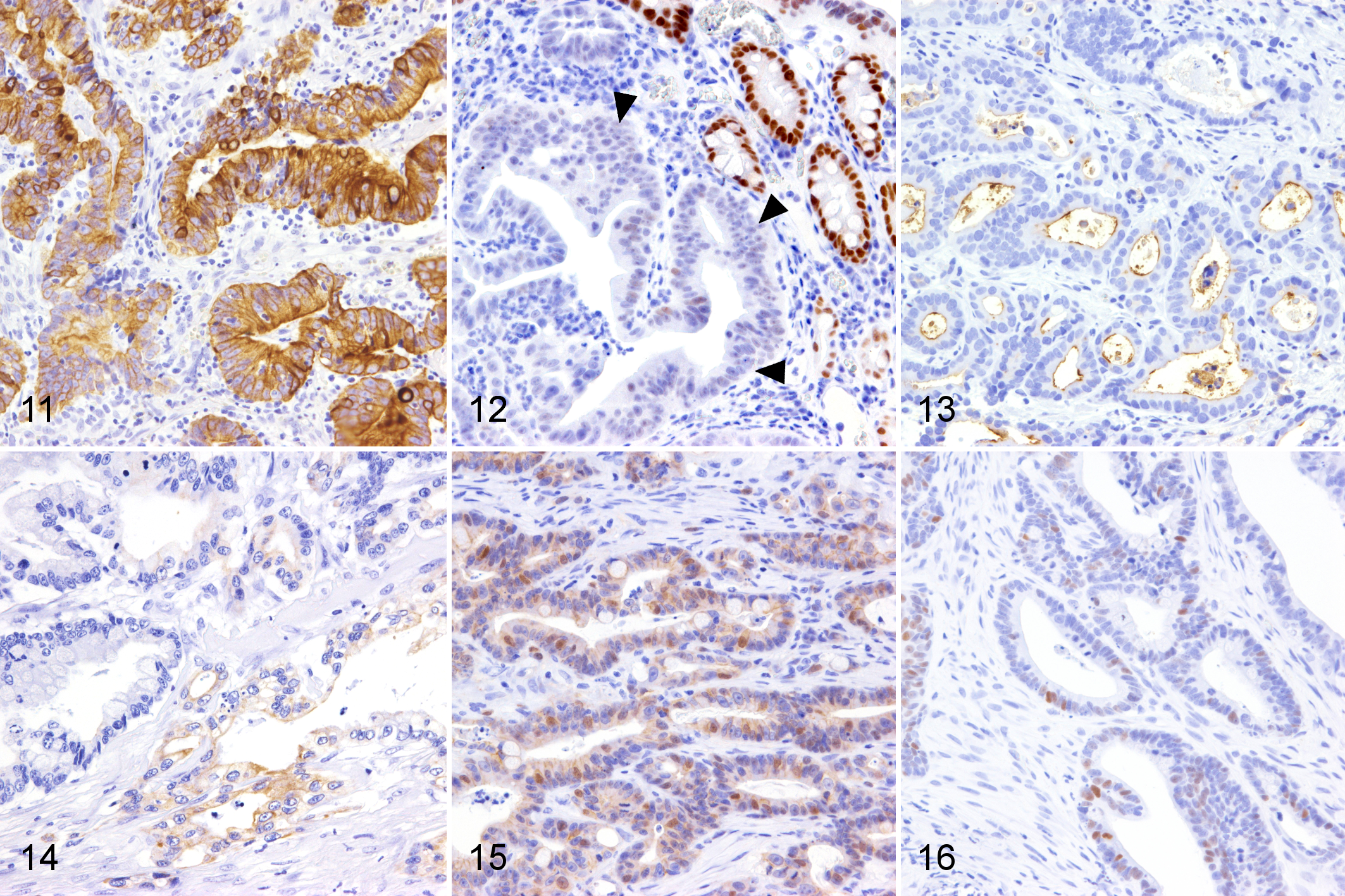
Tubular adenocarcinoma, colorectum, cat.
Results of Immunohistochemical Examinations of Feline Colorectal Epithelial Tumors.

a Benign: tubular adenoma, villous adenoma, tubulovillous adenoma; Malignant: tubular adenocarcinoma, tubulovillous adenocarcinoma, mucinous adenocarcinoma, undifferenciated adenocarcinoma.
b0 (negative), <5% positive cells; 1, 5% to 25% positive cells; 2, 25% to 50% positive cells; 3, 50% to 75% positive cells; 4, >75% positive cells.
c Number of applicable cases (percentage).
d Nuclear immunoreactivity was interpreted as positive.
Statistical Analysis
The 50 feline colorectal tumor cases were divided into benign (9/50 cases, 18%, containing TA, VA, and TVA) and malignant (41/50 cases, 82%, containing TAC, TVAC, MAC, and UDC) lesions based on a 2-tiered stratification for malignancy. Spearman’s ρ indicated a correlation between a low differentiation degree and malignancy (ρ = 0.615, P < .001; Table 4). Sex (female) was also weakly associated with malignancy (ρ = 0.279, P = .050; Table 4). On the other hand, the CDX2 staining score showed a weak and nonsignificant negative association with malignancy (ρ = −0.265, P = .063; Table 4). Other clinical or histopathological features, including the Ki67 labeling index, β-catenin, and p53 staining score, did not correlate with malignancy.
Association of Signalment and Histopathologic Features With Malignant Versus Benign Feline Colorectal Tumors.
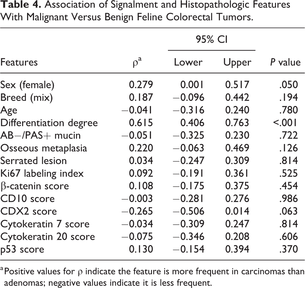
a Positive values for ρ indicate the feature is more frequent in carcinomas than adenomas; negative values indicate it is less frequent.
KRAS Gene Mutation
Extracted genomic DNAs from 7 out of 9 cases (cases 16, 24, 28, 29, 34, 35, and 38, TAC) were successfully amplified; DNA of sufficient quality for analysis was not obtained from the 2 other cases (cases 17 and 44, TAC and TVAC). DNA sequencing identified point mutations in exon 1 of the KRAS gene in 3/7 cases (cases 24, 34, and 38). In codon 13, c.38G>A (p.G13D) and c.38G>C (p.G13A) mutations were found in cases 24 and 38, respectively (Figs. 17–19). The c.109G>C (p.E37Q) mutation was found in case 34 (Figs. 20 and 21). Other mutations, insertions, or deletions were not detected.
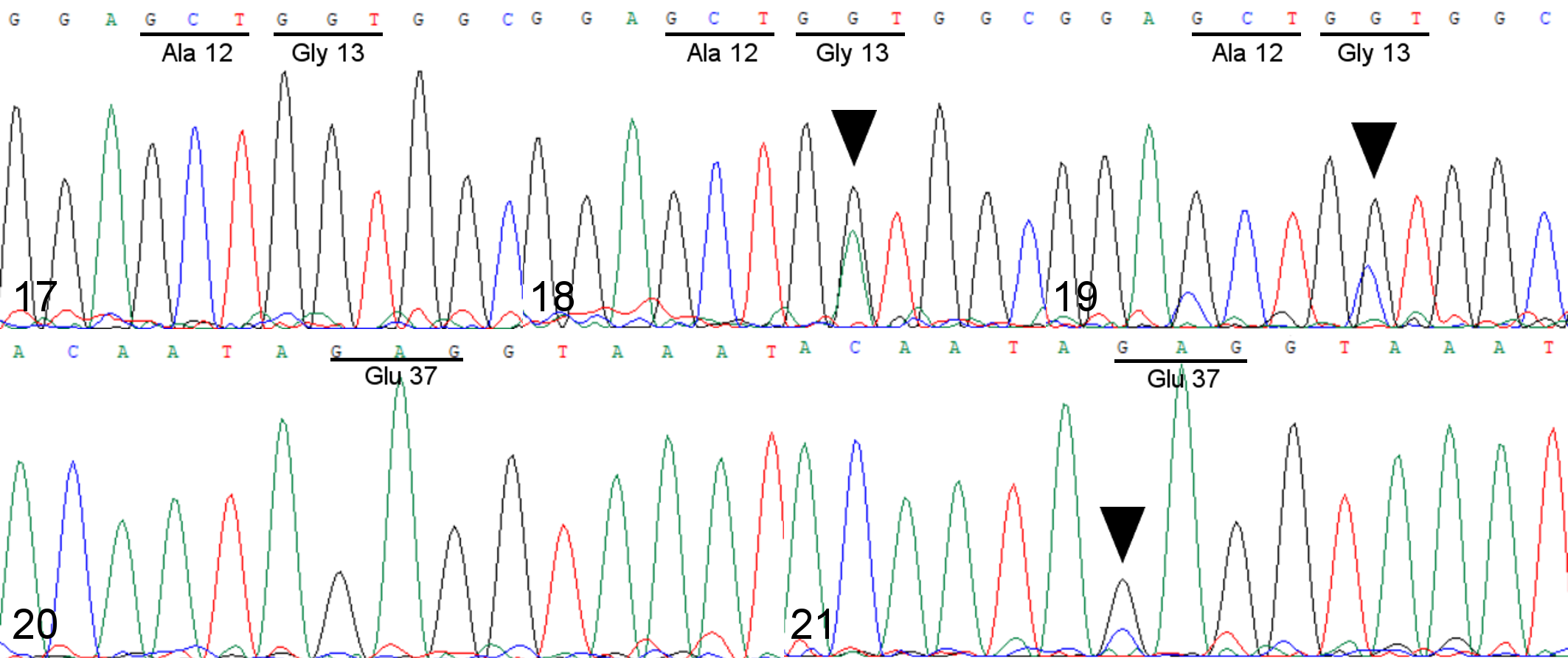
Colorectum, cat. Sequence chromatograms of KRAS.
Discussion
In the present study, colorectal epithelial tumors were detected in 16% of all colorectal biopsy samples and 69% of all gastrointestinal epithelial tumors in cats, and these ratios were markedly higher than those in other gastrointestinal segments. This result is similar to a previous survey in the United States, 36 and supports the high rate of epithelial tumors in the feline colorectum. The majority of feline colorectal epithelial tumor cases involved elderly cats, with a mean age of 13.0 years. This result is consistent with a previous study on colorectal malignant tumors in cats (mean age of 12.5 years), 41 and suggests that senior cats are at a higher risk of developing colorectal tumors. No breed predisposition was indicated in the present study, in contrast to previous findings showing that Siamese, 14,36 Chartreux, 14 and Somali 14 had higher odds of developing intestinal neoplasia. This difference may be attributed to these breeds not being popular in Japan. Furthermore, previous studies displayed no sex predisposition in the occurrence of intestinal tumor, 14,36,43 but did not compare differences in sex between benign and malignant neoplasia. Interestingly, a weak association was observed between being female and colorectal carcinoma in the present study; however, no sex predilection was noted in the occurrence of all colorectal epithelial tumors. This result suggests that female cats are more susceptible to colorectal carcinoma than male cats.
AB-PAS and HID-AB staining methods are used to evaluate mucins in the alimentary tract. Neutral mucins are generally present in the epithelium of the stomach and display AB−/PAS+ staining, whereas acid mucins in the small and large intestines show AB+/PAS+ staining. Acid mucins include sulfated acid mucin (sulphomucin) and non-sulfated mucin (sialomucin), which stain black and blue, respectively, for HID-AB. Goblet cells in the large intestines of humans, rats, mice, dogs, and nonhuman primates produce sulphomucin and sialomucin in varying proportions. 15 Previous studies indicated that mucin profiles are associated with the biological behavior of human colorectal tumors. 4,16,29,45,48 Similarly, the predominance of sialomucin in the colon is a precursor change in the experimental models of colorectal carcinogenesis in rats. 3 However, a relationship was not observed between the presence of neutral mucin and the histological subtype or malignancy of feline colorectal tumors in the present study. Furthermore, sialomucin was not detected in normal mucosal epithelial cells or neoplastic cells in any of the cats examined, except for case 11. This result is consistent with previous findings showing that only sulphomucin was produced in the large intestine of the cat, 40 and suggested that the lack of sialomucin in the feline colon is a species-specific condition. However, the reason why cats lack intestinal sialomucin and why it was detected in case 11 remain unclear.
In humans, the CK7−/CK20+/CDX2+ profile is regarded as a characteristic of colorectal tumor cells. 20,44 A previous study showed that 40% of feline intestinal carcinoma cases were positive for CK7; 13 however, the CDX2 profile was not examined. In the present study, 15/50 (30%), 50/50 (100%), and 48/50 (96%) cases were positive for CK7, CK20, and CDX2, respectively. These results suggest that the CK20+/CDX2+ immunophenotype is a characteristic of feline colorectal tumors, whereas CK7 expression is not. Moreover, CDX2 labeling had a weakly negative association with tumor malignancy. The expression of the CDX2 gene regulates intestinal differentiation and homeostasis; 9 therefore, the lack of CDX2 positivity in feline colorectal tumors may be associated with the dedifferentiation of tumor cells.
CD10 is a cell-surface neutral endopeptidase expressed in the brush borders of the normal small intestine. 7 In human colorectal cancers, a correlation has been reported between CD10 expression and biological aggressiveness, such as liver metastasis. 21,47,48 In the present study, 11/50 cases (22%) showed positive staining for CD10, whereas no correlation was detected between CD10 expression and malignancy. Since information on the presence of liver metastasis was not obtained in the present study, it currently remains unclear whether CD10-positive colorectal cancers in humans and cats share similar clinical behaviors.
β-catenin functions as a cell adhesion molecule and mediator of the Wnt/β-catenin signaling pathway. The abnormal nuclear accumulation of β-catenin is a result of APC or CTNNB1 gene alterations and causes excessive Wnt signaling and tumorigenesis in many organs. 8 In humans, APC gene mutations, which occur in the very early phase of the adenoma-carcinoma sequence, are the most frequent genetic change observed in colorectal tumors. 1,24 Similarly, APC alterations and β-catenin dysregulation are commonly found in canine colorectal tumors. 27,35,39,49 In the present study, nuclear β-catenin accumulation was detected in 30/50 cases (60%), whereas 24 out of those 30 cases (80%) showed only scattered staining. Furthermore, β-catenin nuclear positivity was not associated with carcinoma. These results suggest that the overactivation of Wnt/β-catenin signaling is not likely an important mechanism of tumor development and progression in feline colorectal tumors, which is in contrast to that in humans and dogs. 6,27,49
The TP53 gene is a common tumor suppressor gene, and diffuse nuclear positivity for the p53 protein in tumor cells is associated with the functional inactivation of TP53. 19 The loss of p53 function occurs in the later stage of the adenoma-carcinoma sequence and is considered to contribute to the transition from adenoma to carcinoma and a poor prognosis in human. 24,32 Conversely, a previous study reported that the immunohistochemical detection of p53 was not associated with carcinoma or poor clinical outcomes in canine colorectal tumors. 46 In the present study, only 5/50 cases (10%) displayed diffuse staining for p53, and no relationship was found between p53 positivity and carcinoma. Therefore, these results suggest that p53 dysfunction is not associated with progression of canine and feline colorectal tumors. Taken together with the result that β-catenin dysregulation was rare in feline colorectal tumors, the adenoma-carcinoma sequence may not play a critical role during their development.
Among the 50 cases examined, 41 (82%) were diagnosed as malignant tumors. Due to this high rate of malignancy, it is important to confirm the histopathological features that reflect the invasive potential of tumor tissue. In the present study, serosal infiltration and lymph node metastasis frequently occurred in cats with TAC. In contrast, TVAC, the second most common subtype in the present study, did not show vascular or serosal invasion or lymph node metastasis. Thus, the histological subtype was associated with biological malignancy. Moreover, it is important to note that the degree of differentiation, which was evaluated by gland formation, was moderately associated with malignancy irrespective of the histopathological subtype. On the other hand, the Ki67 labeling index, widely considered to be a prognostic factor in many tumors in humans and cats, 2,22,30,37,38 was not related to the malignancy of feline colorectal tumors. This discrepancy may result from the specific characteristic of intestinal epithelial cells that they have high proliferative activity even in normal tissue. There are conflicting reports of the relation between the Ki67 labeling index and biological behaviors in human colorectal cancer 25,28 ; therefore, it is conceivable that the Ki67 labeling index is difficult to apply as a prognostic factor in colorectal tumors. These results suggest that evaluating the degree of differentiation of tumor cells may be more useful than proliferative activity for distinguishing feline colorectal carcinoma from adenoma when it is difficult to evaluate submucosal invasion.
KRAS is a member of the ras proto-oncogene family, and KRAS mutations are found in approximately 40% of human colorectal carcinoma. 6,42 More than 90% of the KRAS mutations associated with colorectal carcinoma are found in codons 12 and 13. 42 The nucleotide sequence of human KRAS exon 2 containing codons 12 and 13 has >99% similarity to that of feline KRAS exon 1; therefore, KRAS mutations at this region may exert the same effects in humans and cats. 12 In the present study, KRAS mutations at codon 13 or 37 were found in 3/7 cases (42.9%), and were not reported in previous studies on feline KRAS mutations. 11,26 c.38G>A (p.G13D) and c.38G>C (p.G13A) mutations have both been detected in human colorectal carcinoma, and c.38G>A (p.G13D) is the most prevalent mutation at codon 13. 42 Since these missense mutations are regarded as pathogenic mutations in humans, 42 KRAS mutations also appear to contribute to the development of feline colorectal carcinoma. The transversion c.109G>C (p.E37Q) has not yet been reported, 42 and thus, its significance in human or feline tumorigenesis remains unclear.
In humans, neoplasms in the colon and rectum that have serrated morphology have recently been distinguished from conventional adenoma and adenocarcinoma and have a different pathogenesis. 5,18,23,33 The present study revealed that feline colorectal tumors frequently had a serrated structure. Together with the finding that excessive β-catenin and p53 accumulation was limited in feline colorectal tumors, the so-called “serrated pathway” may be involved in feline colorectal tumorigenesis.
In the present study, samples were endoscopically collected in 31 cases (62%) and surgically collected in 19 cases (38%). Serosal infiltration could not be evaluated in endoscopy samples, and lymph node metastasis was evaluated in only one endoscopy case (case 50). This has a potential to introduce a positive bias on the incidence of serosal infiltration and lymph node metastasis in surgical cases, since cats with advanced disease (tumor invasion and/or metastasis) would likely show a more severe clinical presentation and thus be surgically treated. Another limitation of this study is the lack of follow-up data in most of the cases. Further clinical study is necessary to understand the association between histopathological malignancy and clinical outcome.
In conclusion, the present study revealed the histological and immunohistochemical features of feline colorectal epithelial tumors. The majority of feline colorectal epithelial tumors were adenocarcinomas, and TAC was the most common subtype. TAC frequently showed serosal infiltration and lymph node metastasis, whereas TVAC, which was the second most common subtype, did not. The degree of differentiation negatively correlated with malignancy, and lower immunolabeling for CDX2 was a common feature of carcinoma. Other clinicopathological and immunohistochemical parameters, including mucin profiles, the Ki67 labeling index, and the accumulation of β-catenin and p53, did not correlate with malignancy. KRAS mutations at codon 13 or 37 were identified in 3/7 TAC cases (42.9%), suggesting that KRAS mutations may be associated with the development of feline colorectal tumors. Further investigations are needed to obtain a more detailed understanding of tumorigenesis in the feline large intestine.
Supplemental Material
Supplemental Material, Combined_supplemental_materials-Uneyama_et_al - Histological Classification and Immunohistochemical Study of Feline Colorectal Epithelial Tumors
Supplemental Material, Combined_supplemental_materials-Uneyama_et_al for Histological Classification and Immunohistochemical Study of Feline Colorectal Epithelial Tumors by Mizuho Uneyama, James K. Chambers, Ko Nakashima, Kazuyuki Uchida and Hiroyuki Nakayama in Veterinary Pathology
Footnotes
Declaration of Conflicting Interests
The author(s) declared no potential conflicts of interest with respect to the research, authorship, and/or publication of this article.
Funding
The author(s) received no financial support for the research, authorship, and/or publication of this article.
Supplemental material for this article is available online.
References
Supplementary Material
Please find the following supplemental material available below.
For Open Access articles published under a Creative Commons License, all supplemental material carries the same license as the article it is associated with.
For non-Open Access articles published, all supplemental material carries a non-exclusive license, and permission requests for re-use of supplemental material or any part of supplemental material shall be sent directly to the copyright owner as specified in the copyright notice associated with the article.