
Abstract
Using immunohistochemistry, 170 canine mammary carcinomas were evaluated for p53, ER (estrogen receptor), and Ki67. Of the 170 tumors, 89 were grade I (52.3%), 36 were grade II (21.2%), and 45 were grade III (26.4%). Eight cases (0.5%) were positive for p53 and 69/170 cases (40.5%) were positive for ER. Ki67 values were 24 ± 18% (mean ± SD). Using a cutoff value of 33.3% Ki67-positive neoplastic nuclei, 38/159 (23.8%) were classified as high proliferative and 121/159 (76.2%) as low proliferative. p53-positive cases had significantly higher Ki67 expression and higher histological grade. ER expression was not correlated with p53 expression but was significantly related to low Ki67 values and low histological grade. Moreover, ER-positive cases had significantly longer survival compared to ER-negative tumors, and ER expression had better correlation with tumor-related survival than histological grade. In summary, p53 accumulated in a small subset of canine mammary tumors and was associated with higher proliferative activity and higher histological grade. ER expression was confirmed as a differentiation marker associated with more favorable prognosis and biological behavior. The combined use of these 3 markers could be used in addition to histological grade to predict the biological behavior of canine mammary carcinomas.
Canine mammary tumors (CMTs) are the most common neoplasms in female dogs. 14 The prognosis of CMTs depends on several factors, of which many are related to pathological variables, such as histological grade 7 and subtype 28 and molecular profiles. 22,2 However, the role of p53 has been poorly documented in CMTs. p53 is known as the guardian of the genome as it detects DNA damage triggering apoptosis in damaged cells. In humans, the role of p53 in carcinogenesis is well established; it is in fact lost or mutated in approximately half of human tumors, and in those tumors where it is not mutated, its activation is prevented. 27 In breast cancer, p53 mutations reach a 30% frequency and are generally associated with the most aggressive subtypes, such as the triple-negative, and high expression of p53 correlates with a worse prognosis and shorter survival times. 3 Its role is either well established or presumptive in several canine neoplasms, 29 but its involvement in the progression and behavior of CMTs is poorly documented. Previous studies have investigated the expression of p53 in CMTs by PCR (polymerase chain reaction) and/or immunohistochemistry, but its association with prognosis and malignancy is still controversial. 5,16,17,22 Earlier research found that p53-positive status was significantly related to poor tumor differentiation, higher mitotic count, invasive growth, and necrosis. 5,31 However, other studies on TP53 gene expression found that its expression profile could not be considered a marker for tumor aggressiveness in CMTs. 12,22
By contrast, tumor proliferative activity, assessed by Ki67, is a well-recognized prognostic element as demonstrated in several studies on canine mammary carcinomas. In fact, a high proliferative index has been associated with disease progression, poor prognosis, and shorter survival times in dogs with mammary carcinomas. 4,25,30 Estrogen receptors (ER) have also been extensively investigated in CMTs, and although many authors agree that ER expression is decreased in malignant CMTs, 13,15,21 there is no consensus on their role in behavior and prognosis. 24
Therefore, the aims of the present studies were (1) to analyze the expression of p53 in CMTs in relation to proliferative activity and ER expression profile; (2) to investigate the correlation of p53, Ki67, and ER with histological features and grade and their impact on survival.
Materials and Methods
Case Selection, Histological Classification, and Grading
The study was based on 170 mammary carcinomas in 143 dogs identified between 2012 and 2019 in the archives of the Anatomic Pathology Section of the Department of Veterinary Medical Sciences of the University of Bologna and of the Veterinary Hospital “Portoni Rossi,” Bologna, Italy, for which formalin-fixed paraffin-embedded specimens were available. Hematoxylin and eosin–stained histological sections were reviewed by 2 board-certified authors (BBr, LVM) and cases were classified according to the most recent classification for CMTs, based on tubule formation, nuclear pleomorphism, and mitotic count. 33
Tissue Microarrays (TMAs)
TMAs were created following previously published procedures. 18,19 In brief, for each case, areas of interest were selected based on higher proliferation and avoiding areas with normal tissue, inflammation, fibrosis/desmoplasia, and necrosis. Core samples were taken from donor paraffin block with a 4 mm skin biopsy punch and inserted into the recipient paraffin block. Each recipient block contained 12 cases and controls. A map of each recipient block was created. A total of 26 recipient paraffin blocks were built for 170 samples.
Immunohistochemistry
Immunohistochemistry for ER, Ki67, and p53 was performed in each case. Validation of these antibodies for TMA has already been documented in previous published work. 10,19,29 For each tumor, 4-μm-thick consecutive sections were used. Sections were dewaxed in diaphane and rehydrated. Endogenous peroxidase was blocked by immersion in 3% hydrogen peroxide for 30 minutes. Antigen retrieval was achieved with citrate buffer pH 6.0, and heating for two 5-minute cycles in a microwave oven at 750 W. Primary antibodies were p53 (monoclonal, clone PAb240, BD Pharmigen, 1:200 dilution), ER (polyclonal, ThermoFisher Scientific, dilution 1:100), and Ki67 (monoclonal, clone MIB-1, Dako Denmark, dilution 1:600). All antibodies were incubated with the tissue sections overnight at 4 °C. The reaction was amplified by the avidin-biotin method (ABC kit elite, Vector) and visualized with 3,3′-diaminobenzidine (0.04% for 4 minutes). Sections were counterstained with Papanicolaou’s hematoxylin, rinsed in tap water, dehydrated, and coverslipped. Sections of canine mammary carcinoma in which the expression of p53 was known were used as positive controls. p53 antibody, clone PAb240, recognizes both wild-type and mutated p53, and its cross-reactivity with canine tissues has been already demonstrated. 1,11 Positive controls for ER consisted of canine uterus and internal normal mammary gland. For Ki67, normal canine intestine was used as positive control. Negative controls comprised slides incubated with omission of the primary antibody.
p53 labeling was considered positive when >10% neoplastic cell nuclei exhibited positivity, as previously published. 29 ER positivity was assessed following the Allred score, in which both the labeling score and labeling intensity were evaluated. 24
The Ki67 index was determined by counting the number of Ki67-positive cells per 500 neoplastic cells. Cell count was performed using a digital cell counter (Image J, National Institutes of Health). A cutoff of 33.3% positive neoplastic nuclei was used to separate high versus low proliferative tumors. 20
Follow-up
Follow-up data were analyzed following published guidelines. 32 Follow-up information was retrieved by telephone interview with the animal owners or referring veterinarians. Overall survival (OS) was calculated including all cases in which death was due to the tumor or not known; cases in which death was not tumor-related were censored. Tumor-specific survival (TSS) was calculated as the time elapsed from the date of diagnosis to death due to the tumor (including natural death and euthanasia due to advanced disease). Cases that were still alive at the date of the study were censored.
Statistical Analysis
Statistical analysis was performed with GraphPad 8 and IBM SPSS Statistics 21 (IBM Corporation). Normality was evaluated with D’Agostino and Pearson test. Mean and standard deviation (SD) were calculated for normally distributed data, while median (min-max) were reported for nonnormally distributed data. Correlations were performed with χ2 tests for categorical variables and Kruskall-Wallis for continuous variables. Survival curves were generated using the Kaplan-Meier product-limit estimates method, with log rank (Mantel-Cox) tests being used to estimate the differences in survival fractions. Values <.05 were considered statistically significant.
Results
Histopathology and Grading
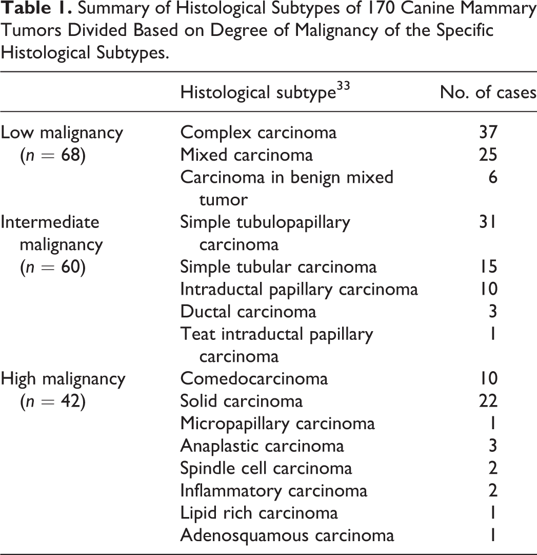
Histological classification of 170 CMTs is reported in Table 1. Cases were grouped into 3 malignancy groups 28 as follows: (1) “low malignancy” including complex carcinomas, mixed carcinomas, and carcinomas in benign mixed tumors; (2) “intermediate malignancy” including simple tubulopapillary, papillary, and tubular carcinomas as well as intraductal papillary carcinomas, and ductal carcinomas; (3) “high malignancy” including comedocarcinoma, solid, micropapillary, anaplastic, spindle cell, inflammatory, lipid-rich, and adenosquamous carcinomas. In this respect, 68 cases (40%) were low malignancy, 60 cases were intermediate malignancy (35%), and 42 were high malignancy (25%). Histological grade 7 was I in 89 cases (52.3%), grade II in 36 cases (21.2%), and grade III in 45 cases (26.4%).
Summary of Histological Subtypes of 170 Canine Mammary Tumors Divided Based on Degree of Malignancy of the Specific Histological Subtypes.
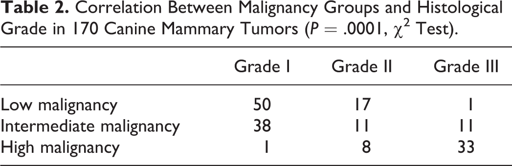
There was a statistically significant correlation between histological malignancy groups and histological grade, with a majority of grade III tumors being in the high malignancy groups and grade I tumors being in the low malignancy group (χ2test, P = .0001; Table 2).
Correlation Between Malignancy Groups and Histological Grade in 170 Canine Mammary Tumors (P = .0001, χ2 Test).
Immunohistochemistry
Immunohistochemical results are summarized in Table 3. p53 nuclear positivity was observed in 8/170 cases (0.5%), of which 2 were overexpressing (>50% positive neoplastic cell nuclei; Fig. 1). There was a significant correlation between histological grade and p53 positivity with 7/8 p53-positive cases being high grade (Mann-Whitney, P = .0001), while no association was found between p53 and malignancy groups.
Summary of Immunohistochemical Positivity for p53, ER, and Ki67 in Relation to Grade and Survival in 170 Canine Mammary Carcinomas.
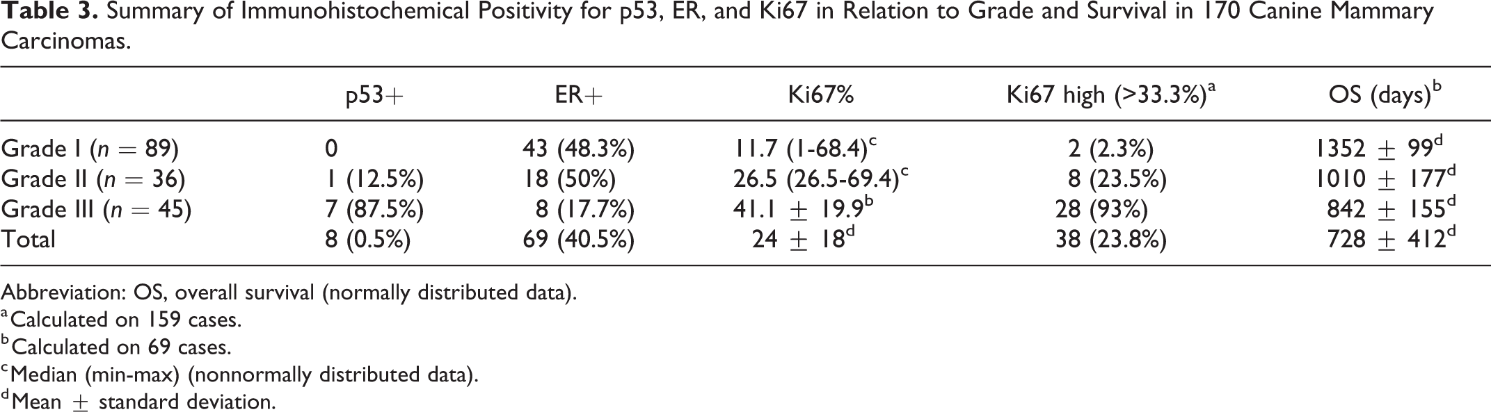
Abbreviation: OS, overall survival (normally distributed data).
a Calculated on 159 cases.
b Calculated on 69 cases.
c Median (min-max) (nonnormally distributed data).
d Mean ± standard deviation.
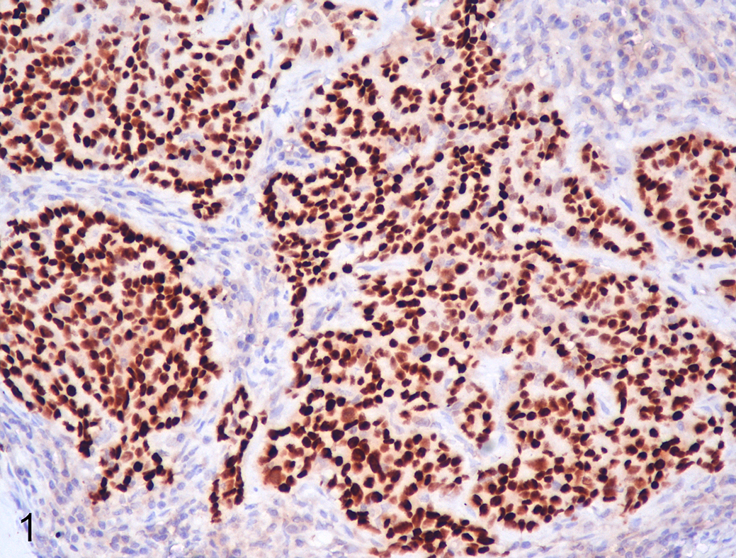
Comedocarcinoma (grade III), mammary gland, dog. Intense p53 nuclear immunolabeling of neoplastic cells. Immunohistochemistry.
ER was positive in 69/170 samples (40.5%) and exhibited nuclear immunolabeling. Using the Allred score, 24 27 tumors had a score of 3, 14 had score of 4, 11 had a score of 5, 14 had a score of 6, 2 had a score of 7, and 1 had a score of 8. ER was expressed in 43/89 (48.3%) grade I tumors, 18/36 (50%) grade II tumors, and 8/45 (17.7%) grade III tumors. ER expression was significantly associated with low histological grade (Mann-Whitney, P = .002; Fig. 2) and low malignancy group (χ2 test, P = .001; Fig. 3).
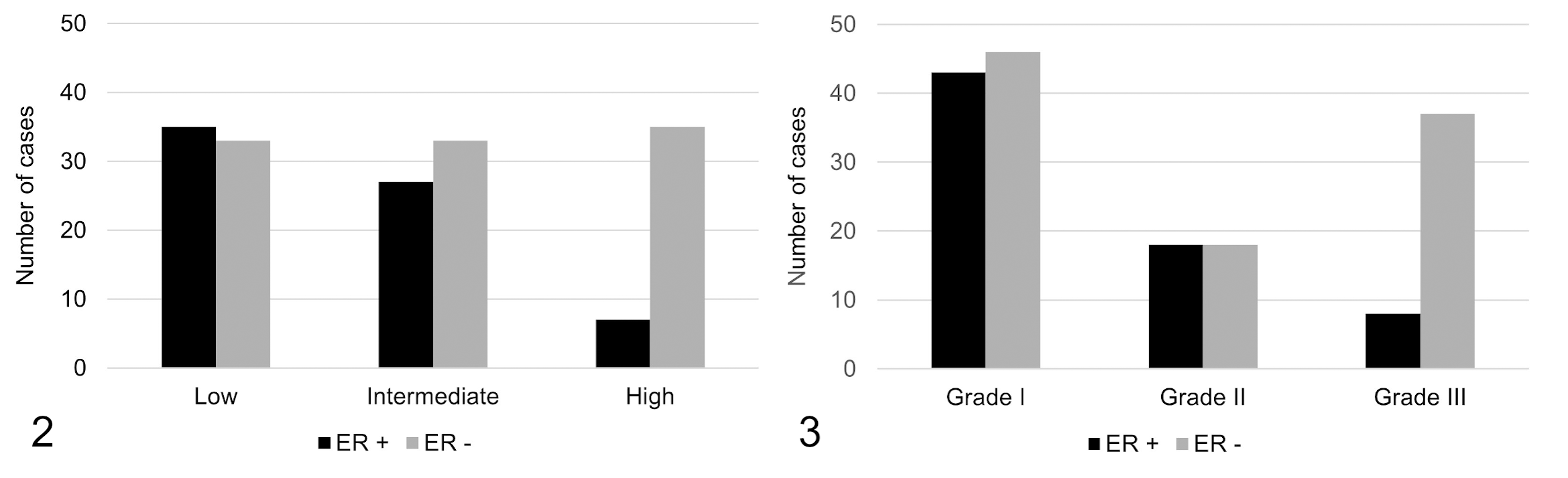
Estrogen receptor (ER) expression is significantly correlated with malignancy groups (P = .001, χ2 test). A higher percentage of ER-negative cases have increased malignancy.
Ki67 positivity was evaluated in 159/170 cases (in 11 cases Ki67 was not immunoreactive). Ki67 positivity in all samples was 24 ± 18% (mean ± SD). Using a 33.3% cutoff rate, 38 (23.8%) were classified as “high proliferative” and 121 (76.2%) as “low proliferative.” High Ki67 values (high proliferative tumors) were significantly correlated with higher histological grade (Kruskal-Wallis test, P = .001; Fig. 4), but not with malignancy groups. Moreover, high Ki67 values were significantly associated with p53 positivity (Fig. 5).
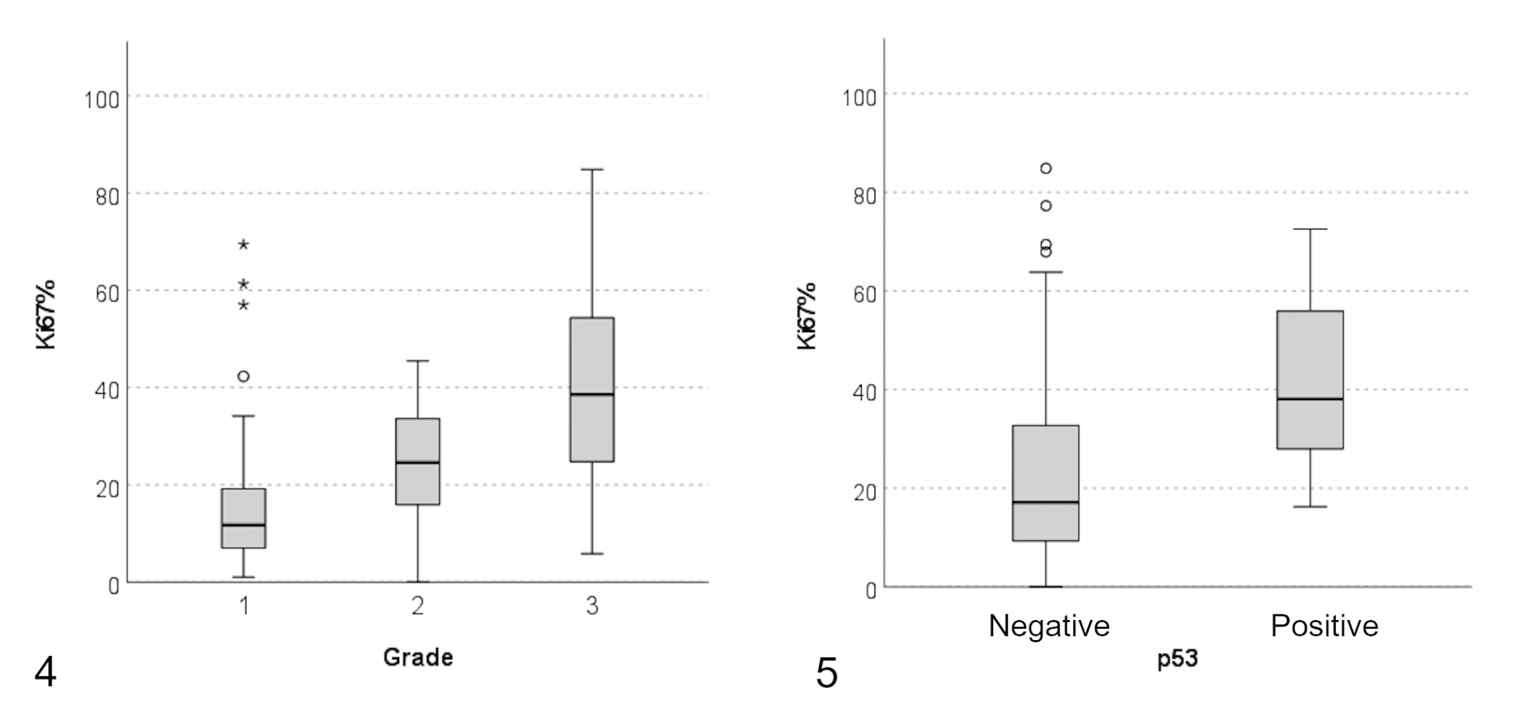
High Ki67 values (high proliferating tumors) are significantly correlated with higher histological grade (P = .001, Kruskal-Wallis test).
Five of 8 p53-positive cases were observed in high proliferative, high grade, and high malignancy group tumors. Correlation between p53 and ER was found not to be significant (Fisher test, P = .097), but a significant association was observed between ER positivity and low Ki67 values (Mann-Whitney test, P = .001).
Follow-up and Survival
Follow-up information was available in 69/143 cases. Of these, tumor-specific death was recorded in 10 cases in which the dogs were known to have died due to the mammary tumor, while death for either tumor-specific or unknown causes was recorded in 22 cases. Forty-seven cases were alive at the end of the study.
Median survival was 686 days (range 59-1659 days). Mean survival was 728 ± 412 days. According to histological grade, mean (± SD) survival time was 1352 ± 99 days for grade I carcinomas, 1010 ± 177 days for grade II carcinomas, and 842 ± 155 days for grade III tumors. Histological grade was correlated with OS (P = .007) but not with TSS (P = .3; Fig. 6). Histological malignancy groups (ie, high, intermediate and low malignancy 28 ) were also significantly correlated with OS (P = .01) but not with TSS (P = .1).
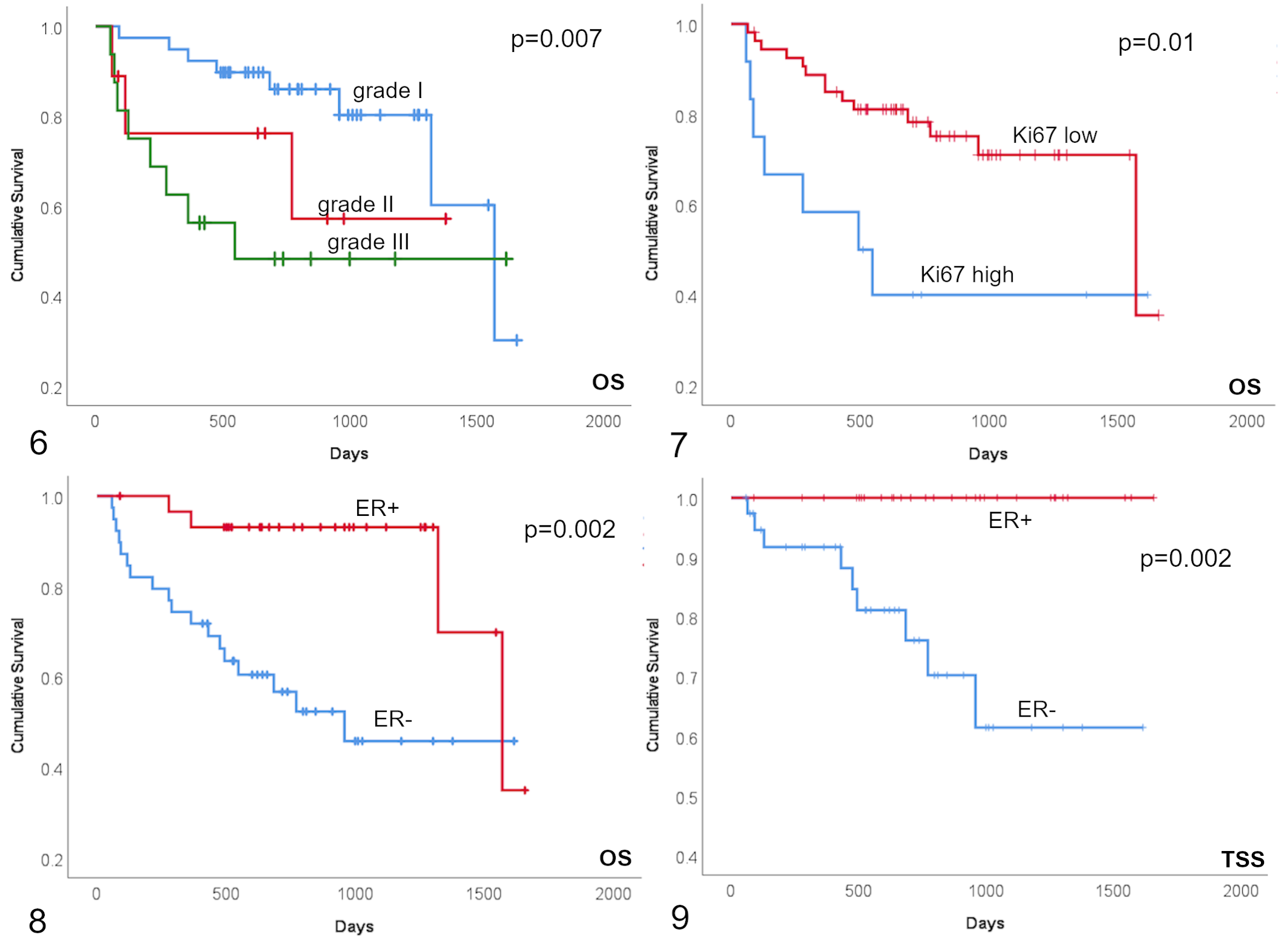
Kaplan-Meier survival curves of 69 dogs with mammary carcinomas. In each curve the y-axis represents the survival probability. Vertical marks represent censored cases.
High-proliferative versus low-proliferative tumors were also compared to survival times and high-proliferative tumors had significantly shorter OS compared to low-proliferative neoplasms (log rank test, P = .01; Fig. 7). However, TSS was not significantly correlated with Ki67 values (log rank test, P = .4).
Moreover, all 10 cases that experienced tumor-related death were ER-negative. In fact, ER-positive cases had significantly longer OS and TSS than ER-negative tumors (log rank test, P = .002 for both; Figs. 8, 9).
Discussion
The results of this study indicate that p53, while being expressed in a minority of CMTs, is correlated with malignant behavior. In our study, p53 was expressed in cases with high histological grade and high proliferative activity, suggesting that p53 may be involved in the progression of CMTs. This aspect was previously documented in other studies, where p53 expression was found to be correlated with grade and with increased malignancy of histological subtypes. 16 p53 mutation has been also documented in breast cancer, where it is mutated in 30% to 35% of invasive primary tumors including 80% of patients with triple negative carcinomas. 6,23 The role of p53 in carcinogenesis is well known: p53 mutation inhibits the development of cell death through apoptosis, thus allowing neoplastic cells to survive. However, there are conflicting opinions regarding the reliability of immunohistochemistry to detect p53 anomalies. The mutant p53 protein has a significantly longer half-life and greater stability than the wild-type counterpart and is therefore detectable by immunohistochemical methods. 6 Indeed, studies suggest that p53 immunoexpression indicates the presence of missense mutations that have resulted in the accumulation of non-functional but stable protein. In parallel, the absence of p53 immunostaining can either indicate normal p53 function or a complete lack of p53 protein due to the presence of nonsense mutations in the p53. 9,23 On the other hand, it is now known that certain types of mutations, such as truncating mutations, do not result in protein stabilization. Furthermore, it appears that wild-type p53 can also be stabilized in the absence of mutations. 26 Despite this, early studies found an association between immunohistochemically detected p53 and poor outcome. 23 Therefore, while immunohistochemistry for p53 is a useful and rapid tool to detect p53 accumulation, further analysis by gene sequencing is needed to confirm the mutational status and to correlate it with tumor biologic behavior and prognosis.
Here, ER was also investigated to identify any associations with p53 mutation and proliferative activity. ER was found to be expressed in a high percentage of cases and was correlated with low proliferative activity. However, no association with p53 expression was proven. As expected, ER-positive cases had a lower risk of tumor-related death compared to ER-negative cases by survival analysis. In fact, in our case series ER expression had better correlation with tumor-related survival than histological grade.
The involvement of ER and PR in the development of CMTs has been extensively investigated in the literature both independently and in relation to molecular subtypes. Most studies agree that the expression of ER is decreased in malignant CMTs, albeit with extremely variable percentages of positivity that range from 10% to 92%. 24 However, in the published literature, there is no consensus regarding the degree of ER expression in relation to prognosis. 24 In women, ER and PR expression are routinely evaluated to assess response to endocrine therapy, 8 but this does not apply to dogs. In this respect, results of our study show a strong correlation of ER with survival, thus suggesting that routine evaluation of ER expression may be useful in predicting prognosis of CMTs.
Proliferative activity was also investigated through Ki67 immunohistochemistry. Ki67 is a non-histone protein expressed during G1, S, G2, and M phases, but it is not found during G0; hence, tumors with higher proliferation rate show greater Ki67 expression. Our results are in agreement with the literature, indicating that CMTs with higher grade are associated with increased proliferative activity. Moreover, high Ki67 index was correlated with p53 expression and inversely correlated with ER positivity. The inverse correlation of ER and proliferation is well documented in the literature and is confirmed in this work. 21 Several studies have investigated proliferation rates of mammary tumors to evaluate their impact on prognosis; however, a standard cutoff rate for Ki67 counts in CMTs has not yet been defined. In our study, a cutoff of 33.3% was used, following recently published results. 20 Our results suggest that this cutoff value is appropriate and can be routinely used to distinguish “high proliferative” versus “low proliferative” CMTs and that this cutoff value defines tumors groups that directly correlate with survival.
Survival times were analyzed for each of the histological and immunohistochemical variables. Grade, malignancy groups, and Ki67 were associated with OS but not with TSS; the lack of significance of the latter value may be due to the low number of cases in which TSS was available. These results may suggest that in addition to grade, Ki67 and histological subtypes can predict prognosis, but further confirmatory studies with higher number of cases are needed. Instead, ER proved to be a good prognostic indicator even if a low number of cases with survival was analyzed.
In conclusion, p53 is accumulated in a subset of canine mammary carcinomas, in which it correlates with proliferative activity, histological grade, and prognosis. ER is confirmed as a valid differentiation marker associated with a better biological behavior, lower proliferative activity, and more favorable outcome. These results further knowledge on the markers of prognosis in CMTs and suggest that, in addition to histological grading and molecular subtyping, further prognostic information can be obtained through the analysis of proliferative activity, estrogen receptor status, and p53 evaluation. These markers could be included in routine evaluation of CMTs for a more accurate exchange of information between clinicians and oncologists on prognosis and response to therapy.
Footnotes
Declaration of Conflicting Interests
The author(s) declared no potential conflicts of interest with respect to the research, authorship, and/or publication of this article.
Funding
The author(s) received no financial support for the research, authorship, and/or publication of this article.