
Abstract
Mammary tumors of all types are rare in herbivores, and there is a particular paucity of reports in sheep. The present report describes a case of mammary carcinoma in a 6-year-old uniparous ewe. The tumor was present for at least 18 months, during which time the ewe remained clinically well. At postmortem examination, the mass was found to be multilobulated with occasional cysts. Histologically, the lobules consisted of tubules lined by cuboidal to low columnar epithelium with loss of polarity and moderate anisokaryosis within a moderately extensive fibrous stroma. It was classified as a low-grade carcinoma. The histologic classification and lack of evidence of invasion correlated well with the slow clinical progression.
Mammary tumors are common in carnivores but very uncommon in herbivores. 7 Although occasional cases have been reported in mares, 6,10,11 cows, 3,9 and goats, 12 reports of mammary tumors are rare in sheep. Two cases of ovine mammary neoplasia, one of epithelial origin and one of mesenchymal origin, both malignant, have previously been cited, but no further details are available (Laffolay B: 1946, Les tumeurs de la mamelle chez les herbivores [Mammary tumors in herbivorous animals]. Thesis, Alfort. Cited by Cotchin E: 1950, Vet Bull, Abstract 427, p. 95). The only descriptions of ovine mammary tumors found in the veterinary literature were of a mammary adenoma in a 3-year-old ewe 1 and a fibroadenoma in a lamb. 5 In a survey of 673 tumors of all types in sheep over a 40-year period in South Africa, no mammary tumors were recorded. 2 The rarity of mammary neoplasia in sheep renders it important to report the present case of mammary carcinoma in a ewe.
The 6-year-old Suffolk-cross ewe was maintained as part of a flock on a research farm. The ewe had not been involved in any research projects. Despite her age, the ewe was uniparous and had reared twins almost 2 years previously. After the lambs had been weaned, the udder was noted to be hard and swollen, and a tentative diagnosis of chronic mastitis was made. Although the udder remained swollen, the ewe was bright, alert, and nonfebrile and retained a normal appetite. At a veterinary clinical examination 18 months later, a diagnosis of mammary tumor was made, and a decision was made to euthanize the ewe.
The ewe was euthanized immediately before necropsy. There were adequate reserves of body fat, and the uterus was fully involuted. No significant pathologic changes were observed on macroscopic examination other than in the left mammary gland, which was misshapen, indurated, and markedly enlarged (approximately 18 cm × 15 cm × 15 cm). On cross-section, it consisted mainly of pale, firm nodules intermixed with occasional cysts; some of the cysts were filled with brownish fluid (Fig. 1). Histopathologically, the affected gland was composed of lobules separated from each other by fibrous trabeculae. Lobules were composed of tubular and occasional tubulopapillary structures. Tubules were lined by one to several layers of cuboidal to low columnar epithelium with large round to oval vesicular nuclei and moderate amounts of pale eosinophilic cytoplasm. There was loss of polarity and moderate anisokaryosis (Fig. 2). Mitoses were rare. Some tubules contained eosinophilic fluid. Within some lobules, the fibrous stroma was abundant and scirrhous in nature. No evidence of infiltration or lymphatic invasion was seen. Multifocal, random aggregates of lymphocytes were present within the stroma. Based on the size of the mass, the degree of the fibrosis, the loss of polarity of the cells, and the anisokaryosis, a diagnosis of low-grade mammary carcinoma was made.
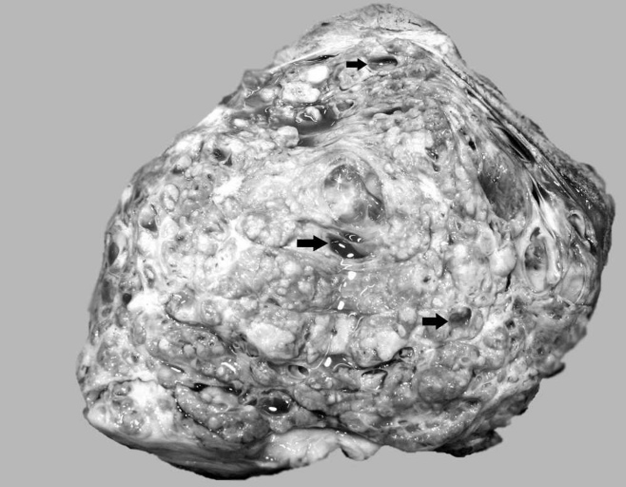
Cross-section of the left mammary gland (18 cm × 15 cm × 15 cm) showing the lobulated appearance and occasional cysts (arrows).
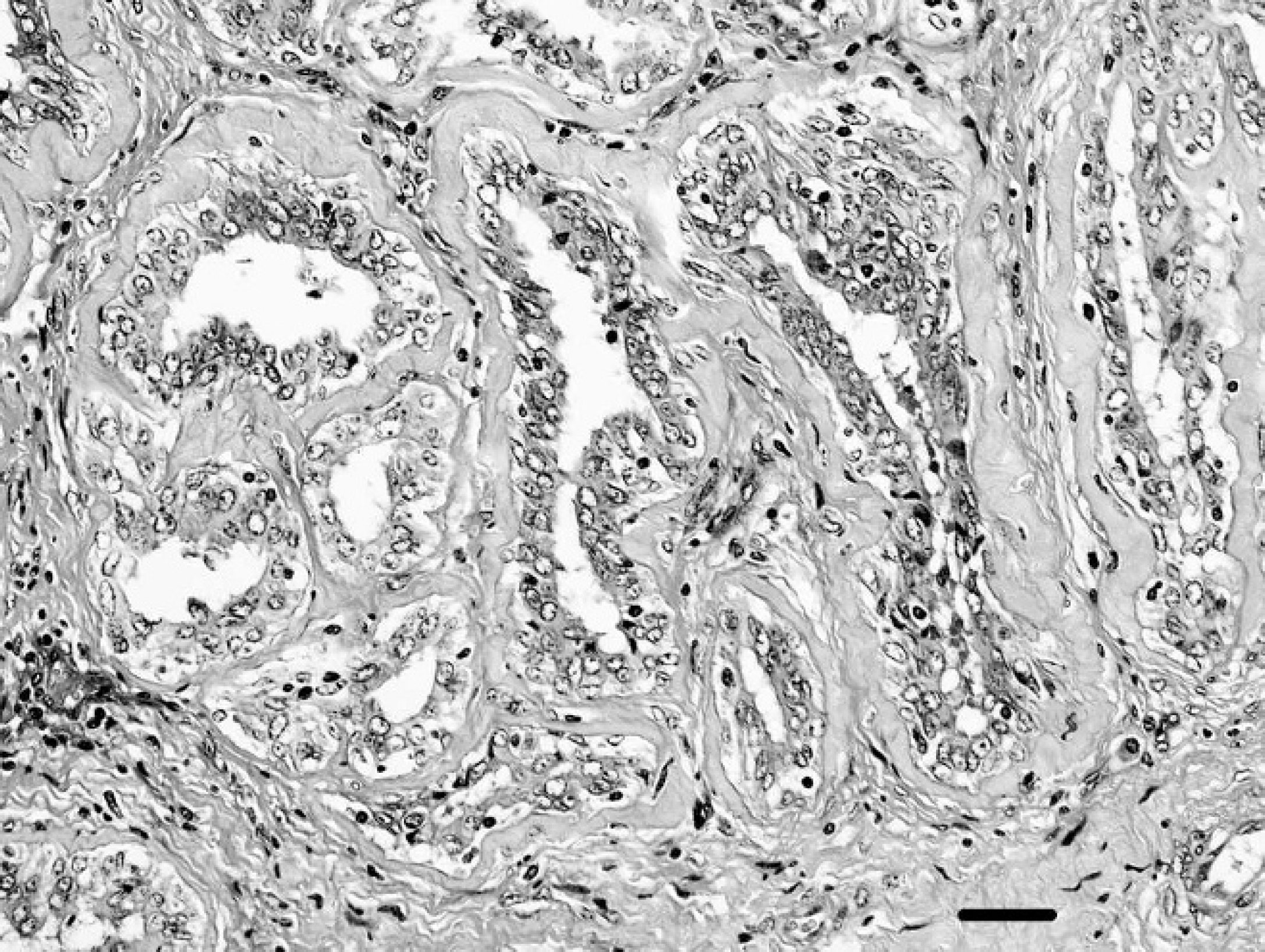
Mammary carcinoma in a ewe. Tubular structures lined by cells showing poor polarity and moderate anisokaryosis within a fibrous stroma. Hematoxylin and eosin. Bar = 50 μm.
The reasons for the rarity of mammary neoplasia in sheep and other herbivores are unclear. It has been proposed that diet may play a role. Herbivores generally have a low-fat, high-fiber diet, whereas carnivores generally have a high-fat, low-fiber diet. In addition, because carnivores are at the top of the food chain, they may have diets that biomagnify exposures to environmental carcinogens or contain high levels of harmful hormones. 8 However, diet alone would not explain the low incidence of mammary tumors in herbivores. The management conditions that the ewe in the current case had been kept under were unusual in that she had only been bred once and thus only had 1 lactation in her 6-year life. Terminal differentiation of mammary gland alveoli, which is necessary for lactation, may be protective across all species, 8 but many domestic ungulates, including mares and zoo ungulates, are not bred, so other factors must contribute to the low incidence in herbivores. The factors that increase risk of mammary neoplasia in dogs and cats, in which the disease is common, are multiple and complex and include the levels and patterns of steroid hormones, as well as growth factors and their receptors and genetic factors. 7 The expression of genes involved in the regulation of lactation maintenance and apoptosis in ovine mammary glands has been studied, 4 and these data may contribute to elucidating why mammary neoplasia is rare in sheep.
A histologic grading system, based on assessing the degree of tubule formation, mitotic activity, and nuclear pleomorphism, has been used for canine and feline mammary carcinoma. 7 The histologic grade of malignancy is determined by the sum of individual scores for these features and is of prognostic significance. Through the use of this system, the mass in the current study was categorized as low grade. The low-grade malignancy and the lack of evidence of infiltration correlate well with the slow clinical progression in this ewe.
Mastitis is a common reason for culling ewes. If this sheep had been part of a commercial flock, it is highly probable that she would have been culled without veterinary examination in the mistaken belief that she had chronic mastitis, and the tumor would not have been identified. Thus, it is likely that mammary neoplasia, although undoubtedly rare, is underreported in sheep. An abattoir survey of enlarged mammary glands in culled ewes may reveal further cases.
Acknowledgements. Technical assistance was provided by Brian Cloak and the staff in the veterinary histopathology laboratory, School of Agriculture, Food Science and Veterinary Medicine, University College Dublin, Ireland.