
Abstract
A 6-year-old, spayed female Labrador/Weimaraner cross-breed dog that had previously lived in Arizona presented in Montana for an annual examination with an incidentally enlarged popliteal lymph node, which was subsequently biopsied. Histologically, the lymph node was expanded by eosinophil-rich granulomas with both extracellular and intrahistiocytic green algae. These algae had intracytoplasmic, birefringent, and refractile granules; readily formed 2 to 3 mm green colonies on Columbia blood agar medium; and ultrastructurally had a multilayered cell wall and intracytoplasmic chloroplasts. Amplified product from the internal transcribed spacer and D1/D2 regions of the 28S ribosomal RNA gene had high sequence identity to Scenedesmus sp. Despite similar infection in the retropharyngeal lymph node 1 year later, the animal remained otherwise healthy with no clinical signs. To the authors’ knowledge, this is the first case of Scenedesmus species infection in a dog and is a differential diagnosis for Coccidioides immitis.
Keywords
A 6-year-old spayed female Labrador/Weimaraner cross-breed dog presented for a routine annual physical examination to a general practitioner in Montana. The animal had spent the first 3 years of its life in Arizona before moving to Montana. The practitioner found a firm mass in place of the popliteal lymph node that, according to the owner, had been growing slowly for several months with no associated clinical signs. The physical examination was otherwise unremarkable, as were a complete blood count and serum biochemistry performed at the same appointment. An incisional biopsy of the enlargement was submitted in 10% neutral-buffered formalin, processed routinely, and stained with hematoxylin and eosin, periodic acid–Schiff, and Gomori methenamine-silver. Histology and polymerase chain reaction (PCR) with genomic sequencing from the fixed tissue on this initial biopsy allowed for an initial diagnosis of Scenedesmus sp. algal lymphadenitis, as described below. Two months after the initial submission, the lymph node had enlarged to 5.0 × 2.5 × 1.3 cm and was completely excised. Half was fixed in 10% neutral-buffered formalin and processed as above, and half was submitted fresh for culture, cytology, and transmission electron microscopy.
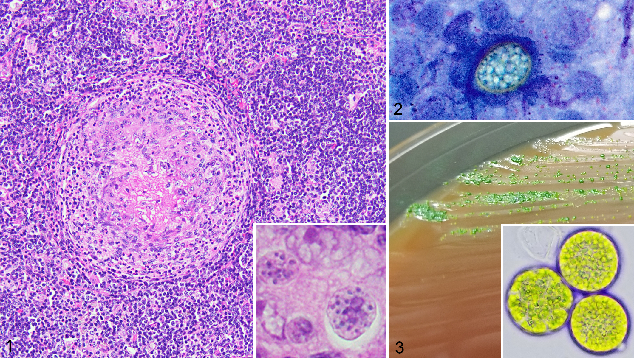
In both the incisional and excisional biopsy specimens, the lymph node was expanded by numerous randomly distributed, multifocal to coalescing granulomas up to 800 µm in diameter. The granulomas were composed of epithelioid macrophages and Langhans-type multinucleated giant cells organized around a variably necrotic center and were surrounded by eosinophils, lymphocytes, and, in larger granulomas, an outer ring of fibrosis (Fig. 1). Scattered individual Langhans-type multinucleated giant cells were also distributed throughout the lymph node in addition to the granulomas. Moderate numbers of round organisms 10 to 20 µm in diameter were free within the lymph node, within the giant cell cytoplasm, and within the necrotic centers of some granulomas. These organisms had a thin, clear, refractile cell wall, and all organisms seen in the initial examination had eosinophilic cytoplasm without any discernible organelles. Infection by Coccidioides immitis was suspected based on these features, the size, and the history of residence in Arizona, and a presumptive diagnosis of fungal lymphadenitis was made pending sequencing from the formalin-fixed, paraffin-embedded tissue. After the sequencing indicated high sequence identity with Scenedesmus sp. (see below), the slides were reexamined to detect any features specific to algae. While the majority of organisms had no distinguishing features beyond eosinophilic cytoplasm as noted previously, several scattered organisms were filled by intracytoplasmic vacuoles containing pinpoint birefringent and refractile granules which surrounded a central, 4 to 8 µm nucleus (Fig. 1, inset). These granules and the outermost contour of the cell wall stained strongly magenta with periodic acid–Schiff. With Grocott’s methenamine silver, the granules stained strongly black while the outer cell wall had variable, weak staining.
Scenedesmus sp. algal infection, lymph node, dog.
Impression smears and scrape preparations from the unfixed specimen were stained with Wright-Giemsa. The algae were evident as relatively frequent oval to round structures ranging from 10 to 20 µm in diameter with a green to pale green, granular cytoplasm and a thin, refractile wall (Fig. 2). Occasionally, they also contained a variable number of deep purple, variably sized, cytoplasmic granules.
DNA was extracted from the formalin-fixed paraffin-embedded popliteal lymph node tissues and a cultured isolate from the same tissue using the QIAamp DNA Mini-Kit (Qiagen Valencia). Pan-fungal primers ITS4 and ITS5 were used to amplify the ITS (internal transcribed spacer) region (ITS1, 5.8S, and ITS2) of the ribosomal RNA gene as previously described. 3 A second PCR was employed using pan-fungal primers targeting the D1/D2 region of the 26S ribosomal RNA gene as previously described. 1 Nucleotide sequencing of amplicons was carried out by a commercial company (GENEWIZ). Forward and reverse sequences were aligned using ClustalW, 5 and the sequences were compared to those in the GenBank nucleotide sequence database (Nucleotide Basic Local Alignment Search Tool; National Center for Biotechnology Biotechnology Information, National Institutes of Health, Bethesda, MD; www.ncbi.nlm.nih.gov/blast/blast). The ITS region sequences obtained for both specimens most closely matched that of Scenedesmus species with 99.76% sequence identity (411/412 bp) to GenBank accession number KY655009. The next closest match was to Scenedesmus species with 99.03% sequence identity (407/411 bp) to GenBank accession number KJ676125. The D1/D2 region sequences most closely matched that of Scenedesmus (Tetradesmus) obliquus, with a 98.78% sequence identity (569/576 bp) to GenBank accession number KY741858. The next closest match was to Scenedesmus rotundus, with a 98.6% sequence identity (562/570 bp) to GenBank accession number KC145438.
A sample of the unfixed specimen was directly inoculated onto Columbia blood agar and chocolate agar (Hardy Diagnostics) and incubated at 35 °C in 5% CO2 atmosphere. Growth was observed on both media after 48 hours incubation, developing a vibrant green colony morphology over approximately 7 days (Fig. 3).
Several 1-mm cubic sections were trimmed from the unfixed specimen with a razor blade and fixed for approximately 24 hours in 2.5% glutaraldehyde with 0.1 M cacodylate buffer, stored for 12 days in 3.5% sucrose solution with 0.1 M cacodylate buffer, postfixed in 1% osmium tetroxide, and embedded by infiltration of Spurr’s Resin using propylxene oxide as a transition solvent. Samples were sectioned at approximately 80 nm, stained with 2% uranyl acetate followed by Reynold’s lead, and examined with an FEI Tecnai G2 transmission electron microscope. Ultrastructurally, occasional individual algae 5 to 6 µm in diameter were seen free and within macrophages. The algae had an undulating, simple cell membrane and a microfibrillar cell wall (Fig. 4). Within the cytoplasm were many electron-lucent starch granules surrounded by thylakoid stacks (Fig. 4) and several round, osmiophilic lipid globules (Fig. 5). No nuclei or pyrenoids were identified.
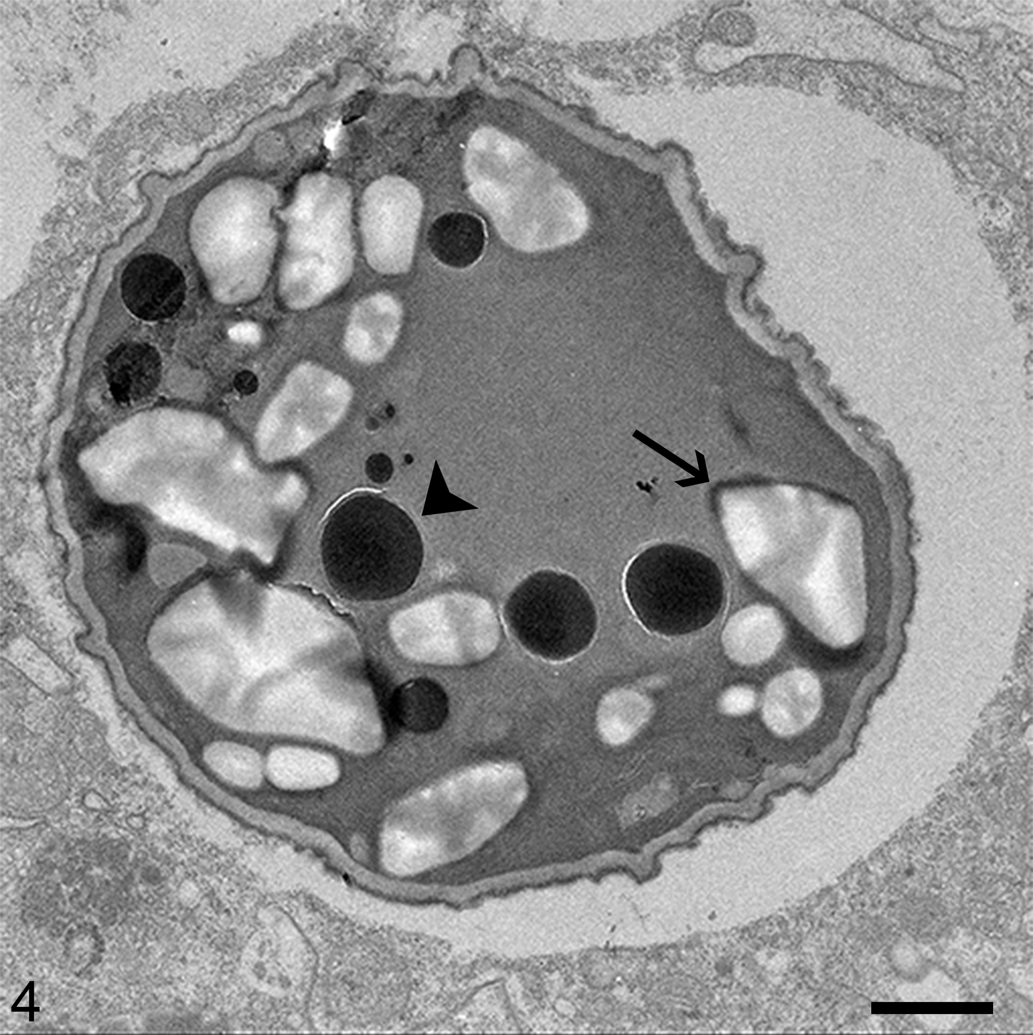
Scenedesmus sp. algal infection, lymph node, dog. Transmission electron microscopy.
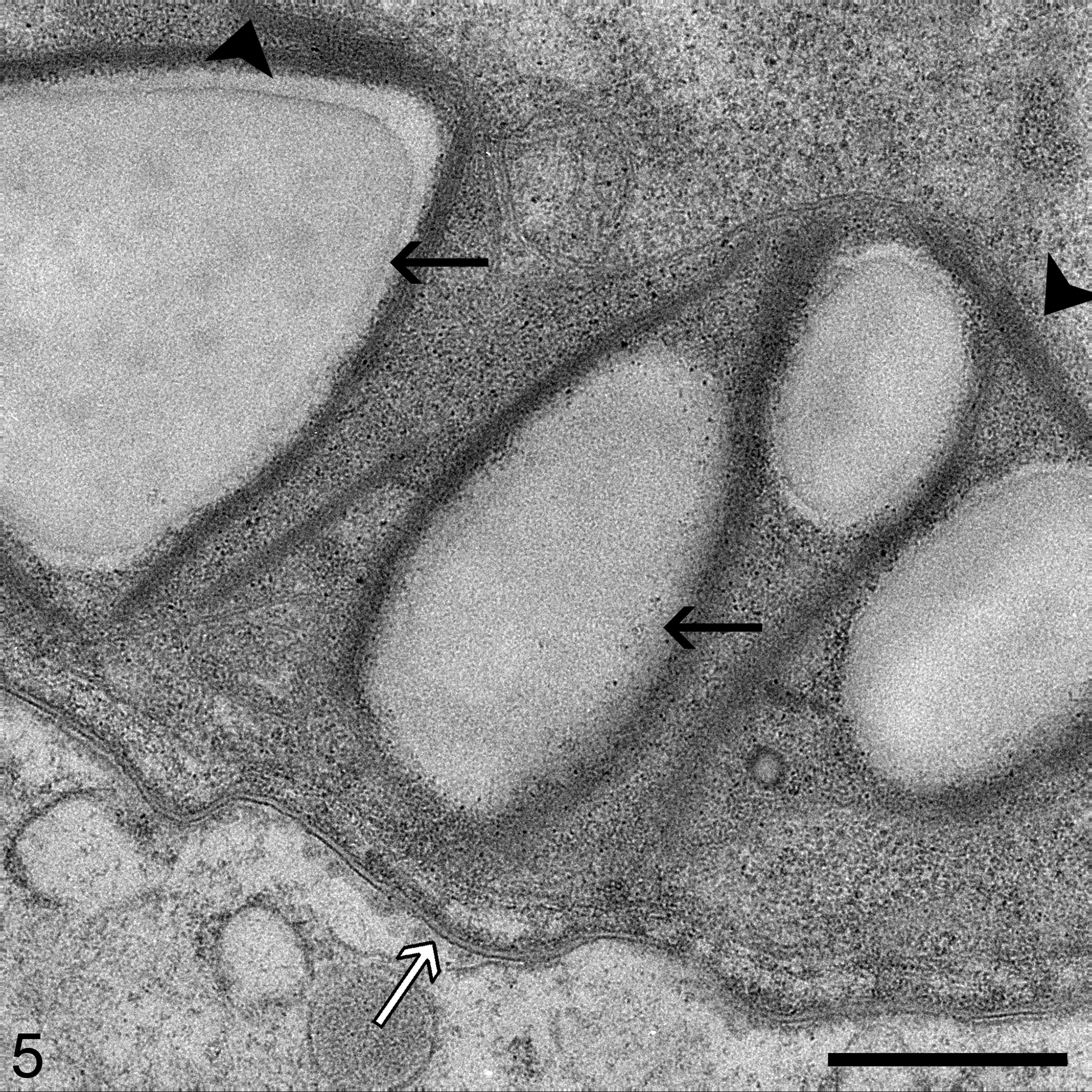
Scenedesmus sp. algal infection, lymph node, dog. The algae have a trilaminar cell wall (white arrow), and the cytoplasm contains numerous starch granules (black arrows) within thylakoid stacks (arrowheads). Bar = 500 nm.
The Scenedesmus species algae seen in this case are noteworthy in that most algal infections in veterinary medicine are limited to the green algae genera Chlorella and Prototheca, 7,8 both of which are in the phylum Chlorophyceae, class Trebouxiophyceae, order Chlorellales, and family Chlorellaceae. Scenesdesmus, while also in the phylum Chlorophyceae, is in order Sphaeropleales and family Scenedesmaceae. To the authors’ knowledge, this is the first confirmed case of infection by a Scenedesmus species in a dog and only the second in mammals. Green algae with high sequence identity to Scenedesmus species have been associated with peritonitis in 2 cattle at slaughter, 2 and green algae attributed to either Chlorella or Scenedesmus have been associated with lymphadenitis in 8 cattle at slaughter. 9 Algae consistent with Scenedesmus species on wet mount were seen in skin ulcers and systemic granulomas in one group of Maylandia species cichlids. 11 Desmodesmus armatus, another alga in Scenedesmaceae, has been associated with infections in 2 human patients following traumatic injuries in fresh water. 10
A source of the infection was not apparent on the veterinarian’s examination or the client history. However, given the slow growth of the lesion and the size at presentation, the initial exposure may have occurred a long time prior to the enlargement noted by the owner. The enlargement of the popliteal lymph node suggests a route of entry through the distal hind limb drained by this node. A myriad of sources are possible as many Scenedesmus species are distributed among soil in the western United States, 4 and they are among the most common groups of algae in fresh water worldwide. 6 The owners had resided in Arizona for the first 3 years of the dog’s life before moving to Montana, and the animal may have been infected in either location due to the widespread distribution of Scenedesmus algae. The organism’s ubiquity in the environment is at odds with the relative paucity of recorded Scenedesmus sp. infection, and the specific aspects of the host-pathogen interaction that result in infection by Scenedesmus species remain elusive. No evidence of immunocompromise was noted clinically, as a complete blood count performed at the time of examination was normal and no other illnesses had been reported in the animal.
A lymph node reported to be the retropharyngeal lymph node by the clinician became enlarged 1 year after submission of the popliteal lymph node. While this node was not biopsied, it was aspirated and algae identical to those previously described were found on cytology. Despite the apparent spread of the organism, the owner had not reported any novel clinical signs in follow-up calls extending to 1 year after the initial submission.
This case was a diagnostic challenge as many of the algal organisms lacked the algae-specific features described due to the plane of section, artifact, and cell death. In many foci, the size, refractile wall, and staining properties resembled those of fungi. Of particular concern initially was Coccidioides immitis, which overlaps in size with the observed algae and was a major differential diagnosis given the previous history of residence in Arizona. Had the organisms been less dense within the lymph node and if molecular diagnostics had not been performed, this case may have been easily confused with infection by C. immitis. This finding underscores the need for routinely verifying light microscopic findings with additional diagnostics, particularly for infectious diseases.
Footnotes
Acknowledgements
We are grateful to Dr Kelly Rankin for her information and help in initially working up the case, and to Dr Allan Pessier for help in planning and sampling. We also thank the researchers and staff of the Franceschi Microscopy and Imaging Center at WSU for assistance in transmission electron microscopy, image generation, and ultrastructural interpretation. We appreciate the contributions of the technical staff at WADDL including the Histology, Molecular Diagnostics, Bacteriology, and Sample Receiving departments.
Declaration of Conflicting Interests
The author(s) declared no potential conflicts of interest with respect to the research, authorship, and/or publication of this article.
Funding
The author(s) received no financial support for the research, authorship, and/or publication of this article.