
Abstract
A wildlife sanctuary presented an adult female cottontail rabbit (Sylvilagus spp.), age unknown, to the Colorado State University Pathology service for postmortem examination. Gross examination revealed numerous pigmented wartlike lesions arising from the skin of the head surrounding the ears, eyes, nares, mouth, and dorsum. Masses were firm, friable, and easily detached from the underlying skin. Differential diagnoses included Cottontail rabbit papillomavirus, Rabbit fibroma virus, and Myxoma virus. Histological examination revealed multiple papillary masses lined by stratified squamous epithelial cells with central cores of fibrovascular connective tissue and parakeratotic hyperkeratosis. Cells of the Stratum spinosum were frequently swollen with abundant perinuclear, cytoplasmic, clearing, and occasional intranuclear basophilic, glassy, spherical inclusions up to 3 μm in diameter. The lesions were consistent with Cottontail rabbit papillomavirus infection. Papilloma virus antigens were identified by immunohistochemistry. In addition, papillomavirus particles were identified by transmission electron microscopy within Langerhans cells of the epidermis, suggesting a unique mechanism for systemic dissemination of the virus. The present case report highlights the finding of viral particles within the Langerhans cells and suggests a novel mechanism of pathogenesis.
A wildlife sanctuary presented an adult female cottontail rabbit (Sylvilagus spp.), age unknown, to the Colorado State University Pathology service (Fort Collins, CO) for necropsy. The available history on this animal was limited, but it was reported that at least 1 other Sylvilagus spp. rabbit in the same area exhibited similar gross cutaneous lesions.
On gross examination, the animal had numerous pigmented wartlike lesions arising from the skin of the head surrounding the ears, eyes, nares, mouth, and a few areas along the dorsum (Fig. 1A). The animal's body condition was poor, and the remainder of the gross examination was unremarkable. Affected cutaneous tissue was fixed in 10% neutral buffered formalin and routinely processed for histologic examination.
Prior to further evaluation, 3 viral agents were considered as likely etiological agents for the cutaneous lesions on this animal, including 1) Cottontail rabbit papillomavirus (CRPV; commonly known as Shope papilloma virus), a nonsegmented double-stranded DNA virus in the family Papovaviridae 26 ; 2) Rabbit fibroma virus (commonly known as Shope fibroma virus), a large DNA in the family Poxviridae and the genus Leporipoxvirus 15 ; and 3) Myxoma virus, another poxvirus in the Leporipoxvirus genus 8 that is morphologically impossible to differentiate from Vaccinia virus. 22 Gross diagnosis was most consistent with CRPV as lesions arose from the haired skin, were heavily keratinized, and projected outward. Cottontail rabbit papillomavirus is distinguishable phenotypically (since haired skin is affected) and genetically from the Rabbit oral papillomavirus 5,16,26 and can be transmitted to Oryctolagus spp. experimentally or naturally via arthropods that feed on the lesions. 22,25 Lesions may regress via immune-mediated processes or may develop into carcinomas. 5,21,22 Notably, a high proportion of these tumors in Oryctolagus spp. undergo conversion to carcinoma. 5 Furthermore, CRPV infection has served as a standard in vivo preclinical model for the development of high-risk—type Human papillomavirus (HPV)-induced malignancy in humans. 3,5,12,25 However, there is difficulty in studying the naturally occurring disease in the laboratory as there are limited commercial supplies available of Sylvilagus spp., and this species does not generally adapt well to captivity. 11
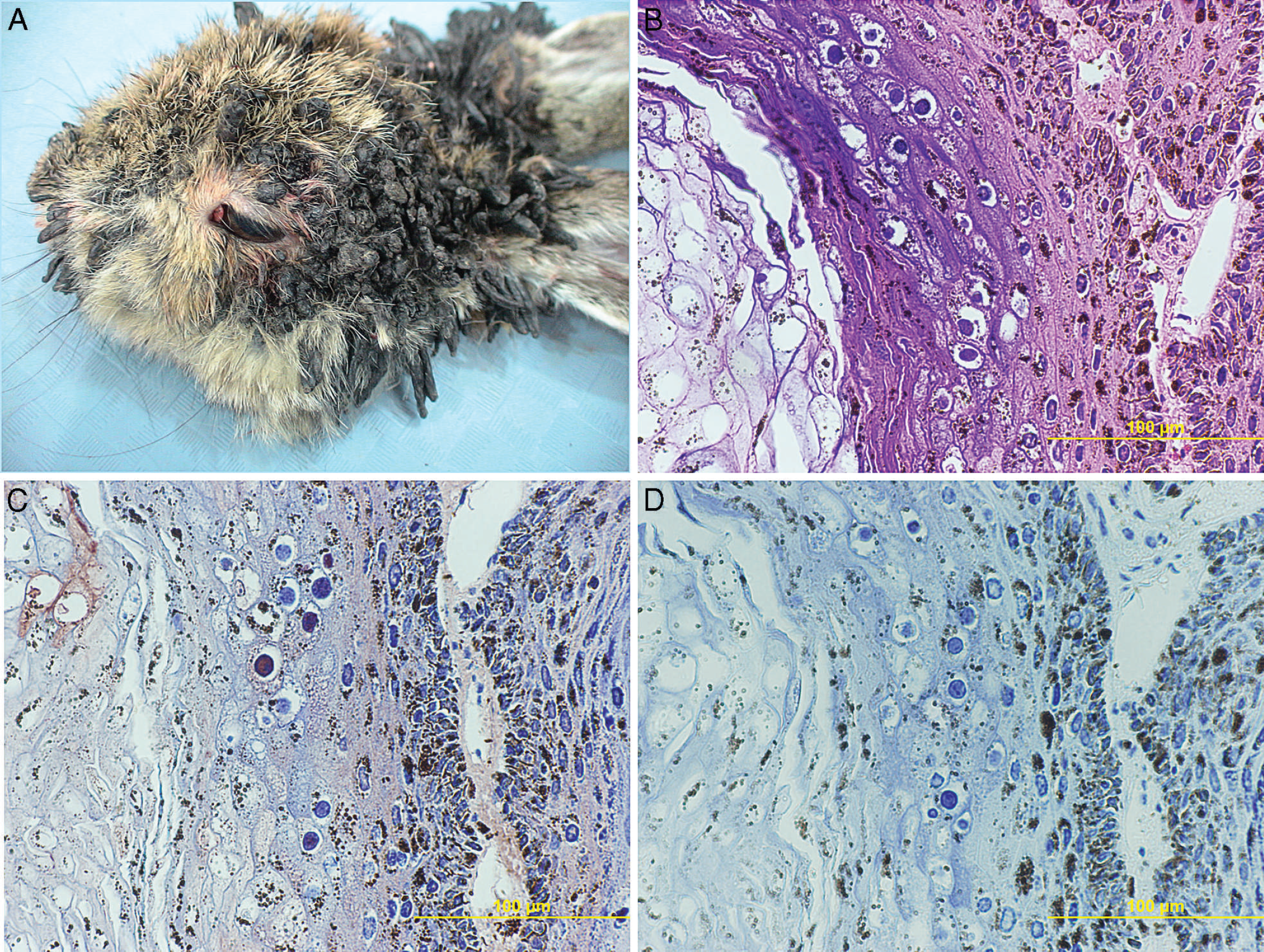
A photograph of gross specimen. There are many wartlike structures extending from the haired skin.
Microscopic examination of affected areas revealed multiple papillary masses lined by stratified squamous epithelial cells with central cores of fibrovascular connective tissue arising from the epidermis as well as paraker-atotic hyperkeratosis. Cells of the Stratum spinosum were frequently swollen with abundant perinuclear, cytoplasmic, clearing, and occasional intranuclear, basophilic, glassy, spherical inclusions up to 3 μm in diameter. These findings were consistent with lesions caused by papillomavirus.
To confirm the etiologic diagnosis, tissue sections were submitted to the University of Illinois at Urbana—Champaign Veterinary Diagnostic Laboratory (Urbana, IL) for immunohistochemistry. A 2-step polymer method a was used without antigen retrieval for immunohistochemical staining. A ready-to-use antibody generated against chemically disrupted Bovine papillomavirus-1 b was used. The positive control samples were from a canine specimen known to contain papillomavirus. Negative control samples included omission of the primary or secondary antibody, respectively. The resulting positive identification of papillomavirus antigens in the lesions stained brightly red with Nova red chromogen and are shown in Figure 1B—D. The data, coupled with gross and histologic pathology, were consistent with a diagnosis of CRPV.
As an additional diagnostic measure, affected tissue fixed in formalin was processed for transmission electron microscopy. Briefly, the tissue was sliced into 2–3-mm 3 pieces, fixed in 4% (v/v) glutaraldehyde in 0.1 M sodium cacodylate, and then postfixed in osmium tetroxide and embedded using a resin test kit. c Thin sections (60–80 nm) were cut and stained with uranyl acetate and lead citrate and examined using a transmission electron microscope. d
As shown in Figure 2, viral particles were identified in the nucleus of degenerate epithelial cells (Fig. 2A, B), as well as in intravascular Langerhans cells (Fig. 2C-E). A total of 5 Langerhans cells found in 3 blood vessels were examined and contained mature virions. Langerhans cells were located within the lumen of blood vessels lined by endothelial cells. Langerhans cells were identifiable by the presence of the characteristic Birbeck granules (Fig. 2D), which are rodlike structures with a median striated line and regular cross-striations. 2 Although different types of papillomavirus cannot be distinguished by traditional ultrastructural methods, the gross and histopathologic evidence supports a diagnosis of CRPV as the etiologic agent for the lesions seen in the rabbit in the current study. Polymerase chain reaction evaluation would be an additional test to confirm the identity of CRPV but was not conducted in the present case.
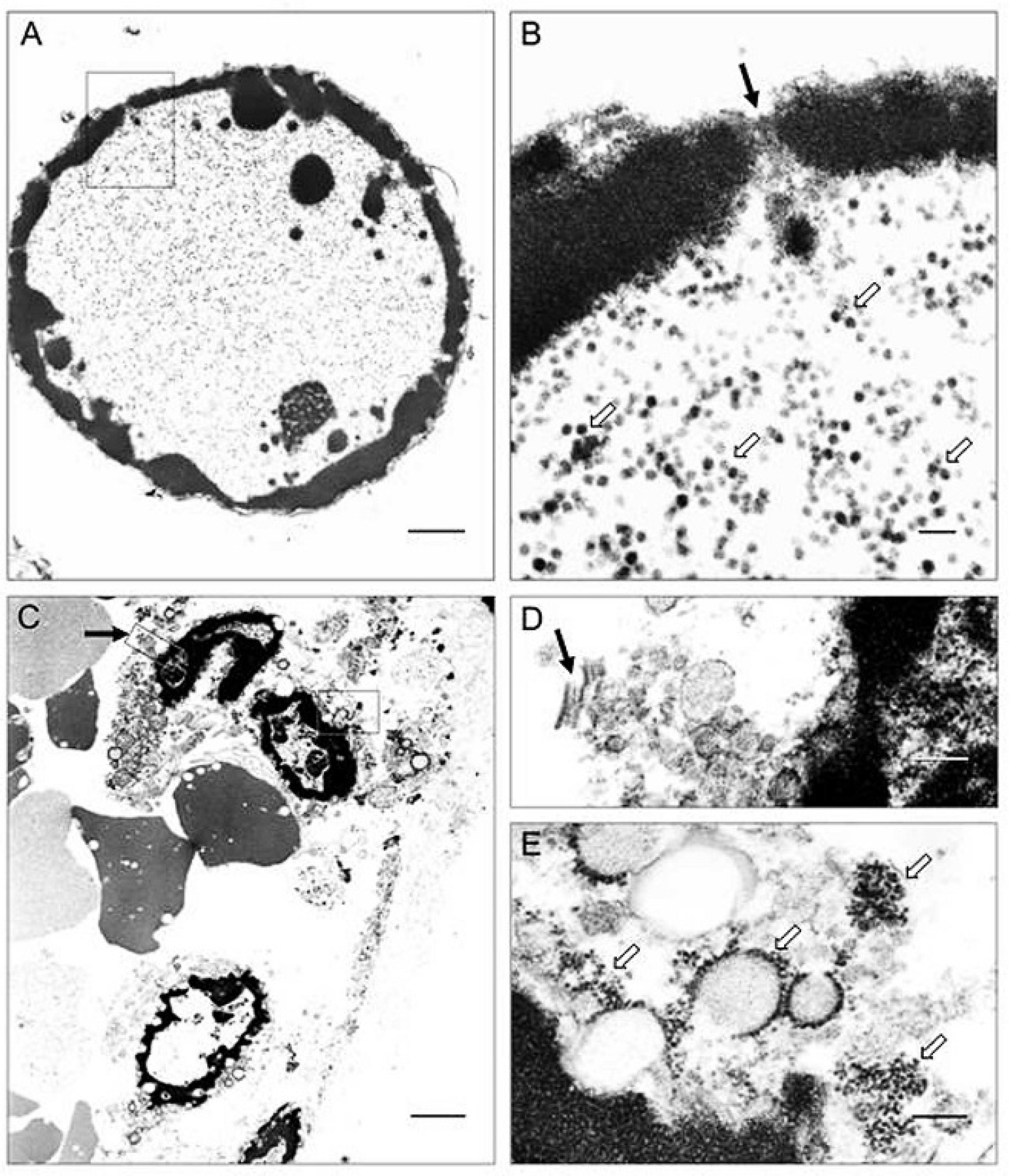
Transmission electron micrographs.
Electron microscopy of specimens from a variety of species has documented papilloma virus particles in many cell types of the dermis and epidermis. 1,13,20 Historically, the ultrastructure of papilloma virus at various stages of development in the epidermal cells of the rabbit was described in detail as early as 1959. 19,24 Furthermore, virus-induced papillomas have been noted to occur in the oral and genital mucosa as well as the skin in many species. 5,20,23 The pathology caused by papillomavirus infection is dependent on many factors such as host cell cycle stage and viral genome insertion position. 4,7,18,27 Infection with papillomavirus can lead to 2 alternate outcomes for the target cells. Cellular degeneration and cell death may result following viral replication in the cell nucleus, or infection may induce mitosis and proliferation in epithelial cells, resulting in hyperplasia and hyperkeratosis. 18 The difference in these 2 courses of action is the direct result of the papillomavirus life cycle. In the current case, a likely outcome might be an abortive replicative cycle, antigen processing, and presentation.
The identification of active infection of Langerhans cells is noteworthy. Langerhans cells are the antigen-presenting dendritic cells of the epidermis, and as such they play an important role in regulation of the skin-associated immune system and defense. 10,14,17 A previous study documented sparse hypofunctional Langerhans cells early in the proliferative phase of Equine papillomavirus-1 infection but an increase in number during papilloma maturation. 10 It was hypothesized that expansion of the Langerhans population enhanced cell-mediated immunity, leading ultimately to tumor regression. 10 In human cervical cancer, manipulation of Langerhans cells has been implicated as a means by which HPV escapes immune clearance, but a definitive mechanism of pathogenesis has not been described. 6 In addition to playing a role in the immune response to papillomavirus infection, the findings in the current report suggest a potential role for Langerhans cells in transmission of the virus to other organs. However, the role for Langerhans cells in the pathogenesis is unknown in cottontail rabbits. While no other lesions ascribable to the viral infection were found in the animal examined herein, the ultrastructural finding nevertheless suggests that spread to internal organs may occur via Langerhans cell dissemination. At a minimum, the Langerhans cells would appear to transport virus and viral antigens to the draining lymph nodes for presentation to T cells, potentially boosting virus-specific immune response. 9,27
The current report describes a disease with different pathological spectra following cross-species transmission. The electron microscopy findings also suggest a potential novel pathogenesis pathway and suggest a possible mechanism for papilloma virus dissemination.
Acknowledgements The authors would like to thank the Colorado State University Diagnostic Histopathology Services for their assistance in processing the tissues, and in particular, the authors would like to thank Jennifer Palmer and Carol Moeller for their assistance with the transmission electron microscopy.
Footnotes
a.
BioGenex Laboratories Inc., San Ramon, CA.
b.
DAKO N 1547 Rabbit polyclonal anti-papilloma virus (Bovine), Dako North America Inc., Carpinteria, CA.
c.
Poly/Bed® 812, Polysciences, Warrington, PA.
d.
JEOL-1200EX, JEOL USA Inc., Peabody, MA.