
Abstract
A 10-year-old female Eurasian river otter (Lutra lutra) died after prolonged anorexia and weight loss in the Seoul Grand Park Zoo, Seoul, Republic of Korea. On necropsy, the liver was found to be swollen and friable with 1 lobe enlarged and necrotic. The other organs showed no significant alterations except for mild atrophy of the right kidney. Microscopically, there was multifocal hepatic necrosis. The hepatocytes around the necrotic areas were swollen and contained large basophilic intranuclear inclusions. Periportal infiltration by plasma cells and lymphocytes was also evident. Transmission electron microscopy revealed characteristic hexagonal virus particles sized approximately 70 nm in diameter in the nuclei of the hepatocytes, which were consistent with an adenovirus. Polymerase chain reaction of the formalin-fixed, paraffin-embedded liver sections was used to determine whether the virus was either the canine adenovirus type 1 (CAV-1), canine adenovirus type 2 (CAV-2), or some other viral agent. The results of these tests showed that the virus was CAV-1. To our knowledge, this is the first report on a CAV-1 infection in an otter.
Canine adenovirus type 1 (CAV-1), which is also known as infectious canine hepatitis, can cause a lethal hepatic infection in young Canidae and Ursidae. 12, 19 The disease is characterized clinically by its rapidly fatal progression. 19 CAV-1 is both endotheliotropic and hepatotropic. In addition to acute hepatocellular necrosis, severe acute hemorrhages are observed on the serosal surfaces, within the lymph nodes and the liver. 12 Antibody to CAV-1 was not detected in a recent serologic study of selected viral agents in free-ranging North American river otters. 13 To the best of our knowledge, there are no reports of CAV-1 in otters. Here, we describe a case of a CAV-1 infection in a Eurasian river otter (Lutra lutra).
A 10-year-old female Eurasian river otter (L lutra) died after prolonged anorexia and weight loss in the Seoul Grand Park Zoo, Seoul, Republic of Korea. The otter was held on its own with no companions in an outdoor pen. The otter had a 3-day-history of lethargy and anorexia prior to death. The necropsy showed that the animal was very thin (body weight, 4.4 kg; normal body weight, 7–8 kg). Focal alopecia was observed in the skin of the dorsum, and the liver was swollen and friable. One lobe was enlarged, mottled, and necrotic. The necrotic area was yellow and fragile. Multifocal necrotic areas were present in the other lobes (Fig. 1). The other organs did not show any significant alterations except for mild atrophy of the right kidney. Samples of liver and other organs were fixed in 10% neutral buffered formalin and embedded in paraffin. The sections were stained with HE. Microscopically, the liver sections revealed severe congestion, hemorrhages, intermittent areas of fatty changes, and multifocal hepatic necrosis. The necrosis of hepatocytes was prominent around the central veins. These hepatocytes had a granular acidophilic cytoplasm indicating coagulative necrosis. Swelling of individual hepatocytes resulted in reduction of the sinusoidal space. Hepatocytes around the necrotic areas were profoundly swollen and contained large basophilic intranuclear inclusions (Fig. 2). The inclusion bodies were surrounded by a clear zone separating them from margins. There was severe multifocal chronic lymphoplasmacytic periportal hepatitis. Microscopically other organs did not reveal morphologic changes indicating viral infection.
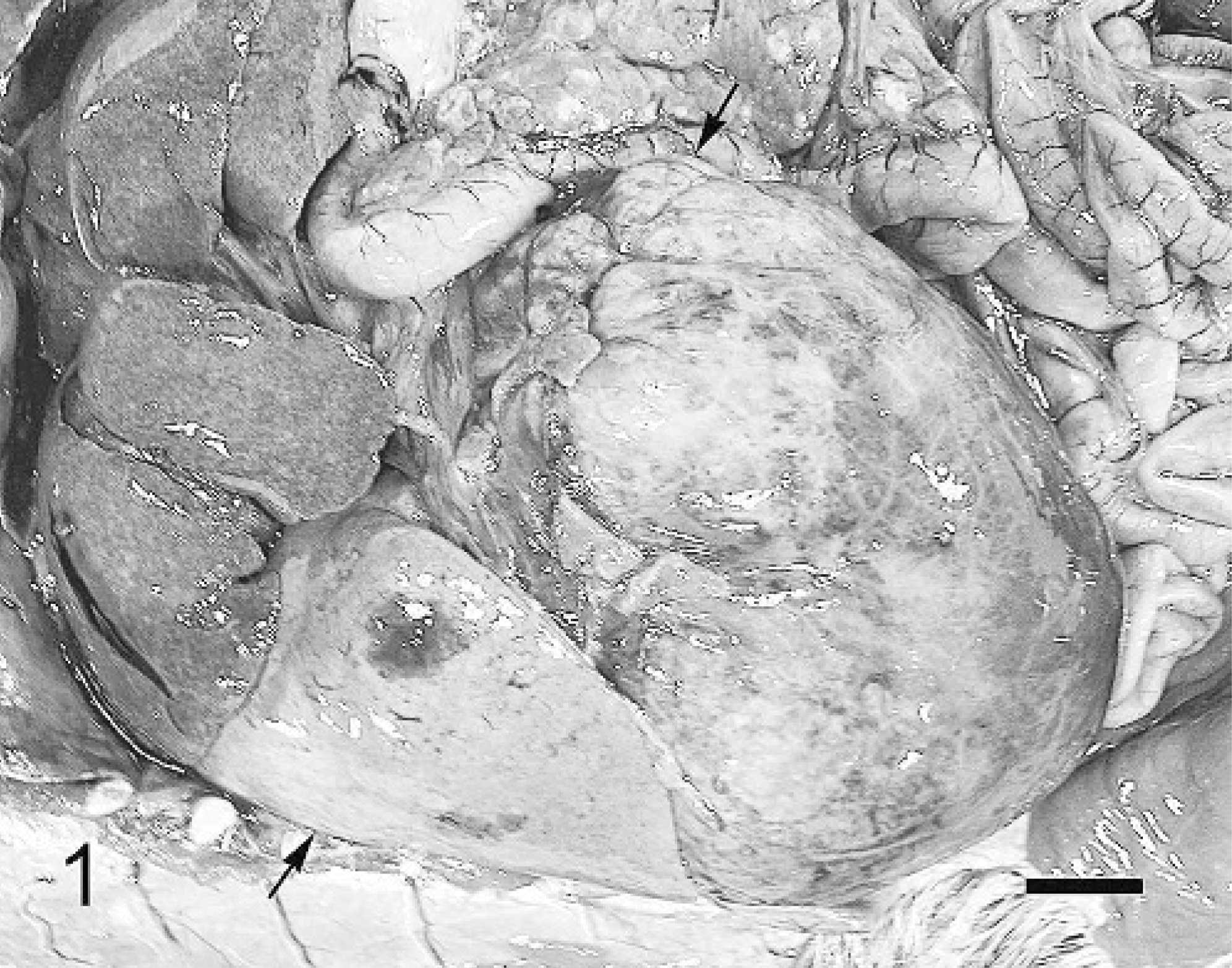
Liver; otter. Enlarged, fragile liver has a large necrotic lobe (arrows). Bar = 2 cm.
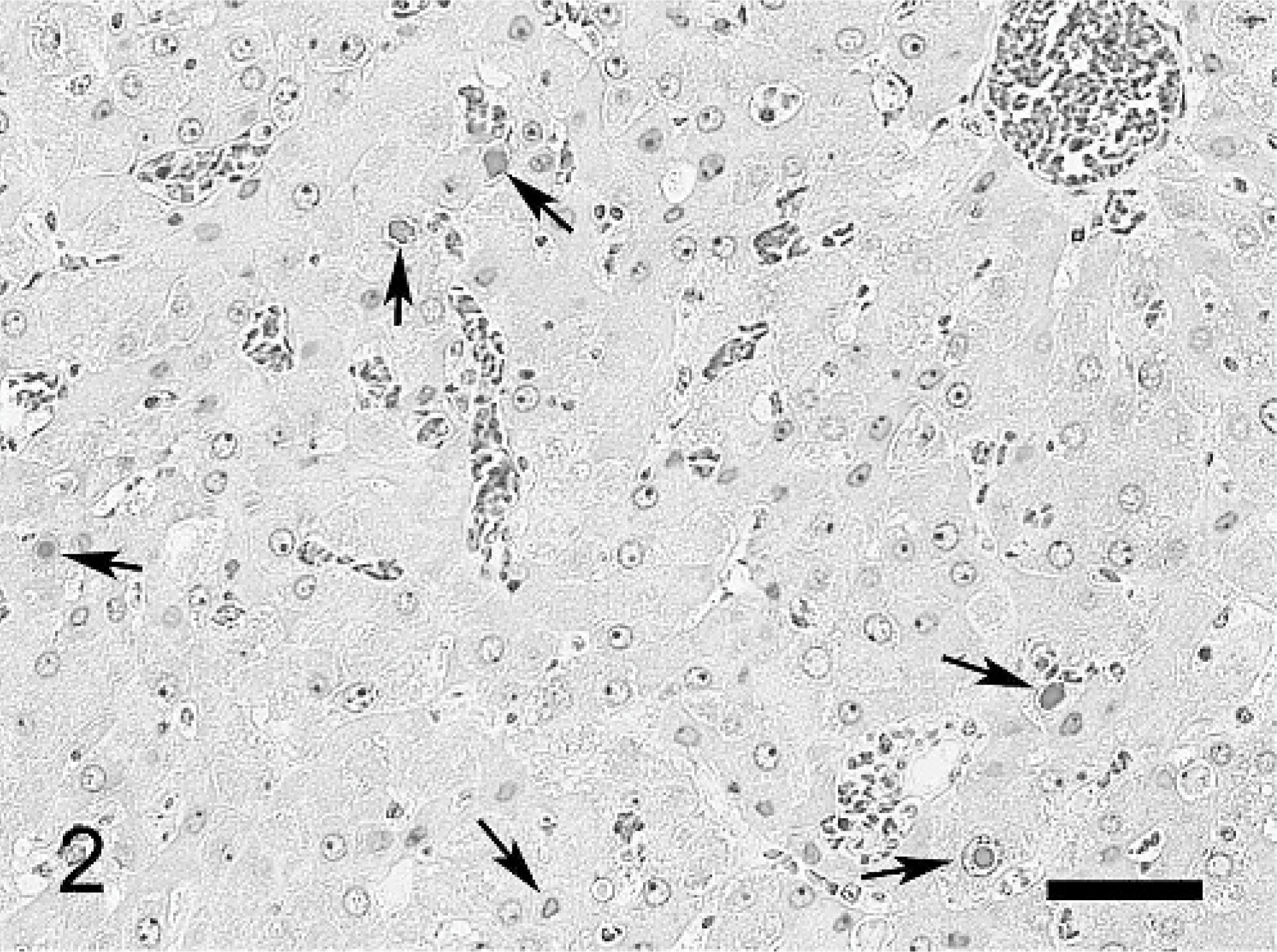
Liver; otter. Congestion of the liver has necrosis of the hepatocytes. Individual hepatocytes contain intranuclear inclusions (arrows). HE. Bar = 50 μm.
Formalin-fixed liver was examined by transmission electron microscopy. Samples were transferred to 2% paraformaldehyde, 2% glutaraldehyde, and 0.2% picric acid in 0.1-M cacodylate-HCl buffer (pH 7.2), rinsed, postfixed in 1% osmium tetroxide, and embedded in an epon mixture. One-micron-thick sections were prepared and stained with toluidine blue, and intranuclear inclusion bodies in hepatocytes were examined by optical microscopy. An 80-nm-thick ultrathin section was made using an LKB-V ultramicrotome with a diamond knife. The section was stained with uranyl acetate and lead citrate and examined with the JEM 100CX electron microscopy (JEOL, Tokyo, Japan). Transmission electron microscopy of these areas revealed characteristic hexagonal virus particles that measured approximately 70 nm in diameter in the nucleus of the hepatocytes, resembling adenoviral particles (Figs. 3, 4).
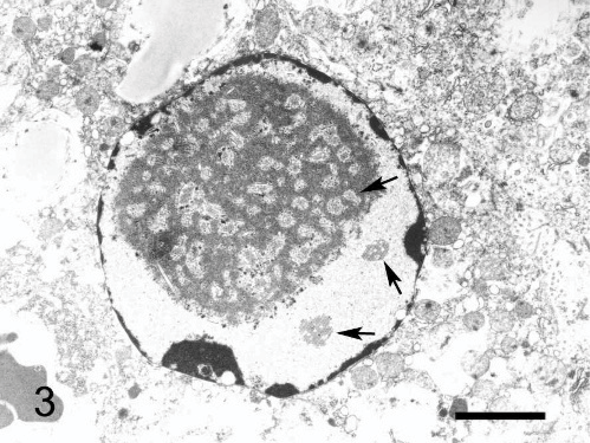
Liver; otter. Adenovirus particles (arrows) are seen in the nucleus of a hepatocyte. Uranyl and lead stain. Bar = 1 μm.
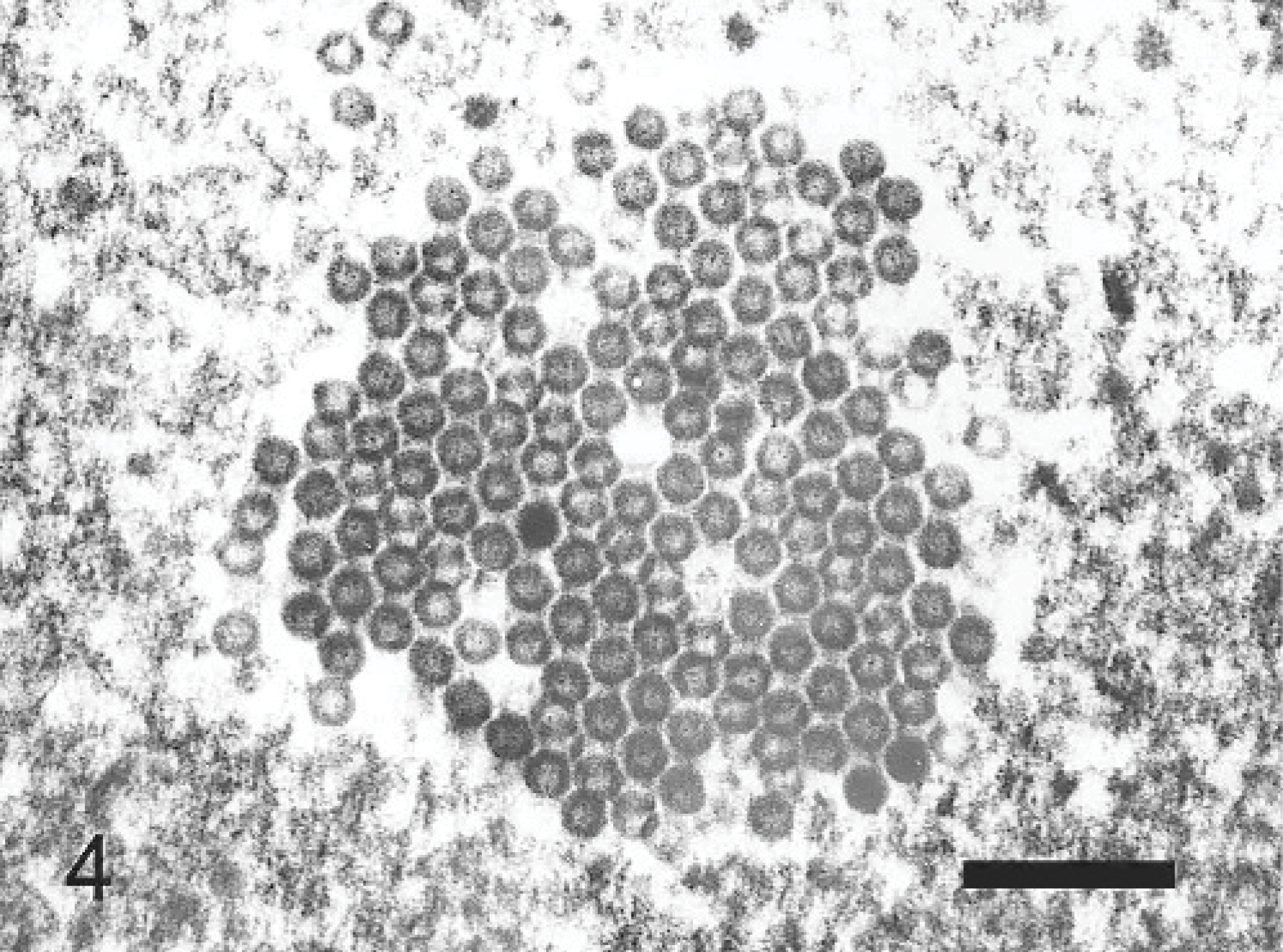
Liver; otter. Numerous pseudocrystalline arrays of isometric adenovirus particles are seen in the nucleus of a hepatocyte. Uranyl and lead stain. Bar = 300 nm.
Polymerase chain reaction (PCR) of formalin-fixed, paraffin-embedded liver sections was used to confirm whether the virus was CAV-1 or canine adenovirus type 2 (CAV-2) using a previously described method. 18 Briefly, the DNA was extracted from the formalin-fixed, paraffin-embedded liver sample using a DEXPAT kit (Takara Biomedicals, Shiga, Japan) according to the manufacturer's instructions. The CAV-1 DNA was extracted from a vaccine strain (Choongang Vaccine Lab, Daejeon, Korea) using the DNAzol (Molecular Research Center, Inc., Cincinnati, Ohio, USA) according to the manufacturer's protocol and used as the positive control. As a negative control, an extract of viral DNA from a CAV-2 strain (ATCC VR-800, American Type Culture Collection, Manassas, VA, USA) cultured in the canine kidney cells (Madin-Darby canine kidney [MDCK] cells; provided by Choongang Vaccine Lab) was used. The PCR conditions were as follows: initial denaturation at 95°C for 5 minutes, followed by 30 cycles of 95°C for 30 seconds, 56°C for 1 minute, 72°C for 1 minute, and a final extension step at 72°C for 10 minutes in a thermal cycler (Palm-Cycler, Corbett Research, Mortlake, Australia). The amplified products were resolved by 1% agarose gel electrophoresis and visualized by ethidium bromide staining. PCR indicated that the virus was CAV-1. The PCR products were purified using a GENECLEAN II kit (Bio 101 Systems, La Jolla, CA, USA) according to the manufacturer's instructions. DNA sequencing was performed using an ABI 3700 automated sequencer (Applied Biosystems, Foster City, CA, USA). The sequencing result of the PCR product was matched with the published GenBank sequence for CAV-1 (M60937). The PCR results of the liver sample were positive for CAV-1 (151 bp) (Fig 5).
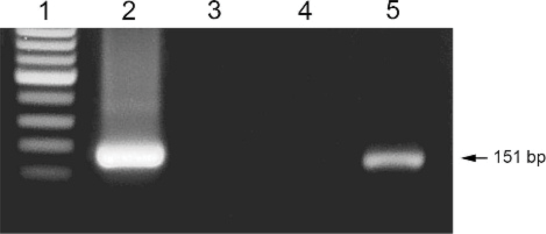
Polymerase chain reaction for the detection of canine adenovirus type 1 in the liver of an otter. Lane 1: 100 base-pair adder. Lane 2: negative control (no template). Lane 3: canine adenovirus type 2. Lane 5: liver sample.
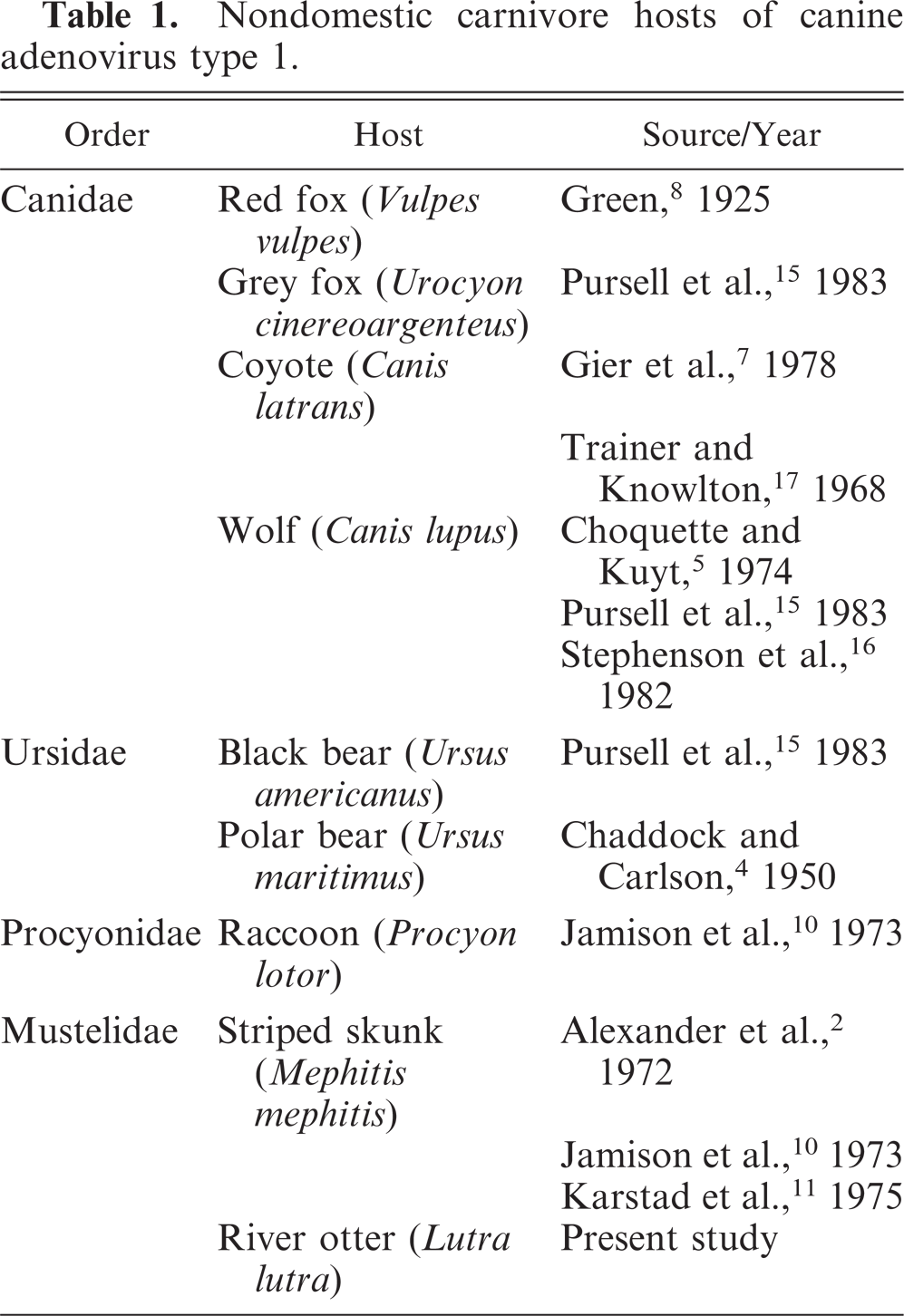
Viruses of domestic animals have been shown to infect wild animals. 3, 9, 19 Wild canids and mustelids including minks and skunks are most susceptible to CAV-1 (Table 1). 1, 14 Sea otters can also be infected with CAV-1. 6 Recently, a serologic survey was performed for selected viral agents in free-ranging North American river otters (Lontra Canadensis). 13 Among the 64 blood samples, none tested positive for antibody to CAV-1. CAV-1 causes acute hepatitis and fulminant hepatic necrosis accompanied by hemorrhages. In this case, severe viral hepatitis with necrosis and viral inclusions were also present. The infectious agent was confirmed to be CAV-1 by PCR and sequencing of the PCR products. The source of infection in this case might have been from other canids and mustelids in the zoo. These included the Himalayan wolf (Canis lupus chanco), coyote (Canis latrans), black-backed jackal (Canis mesomelas), red fox (Vulpes vulpes), African wild dog (Lycaon pictus), North American raccoon (Procyon lotor), raccoon dog (Nyctereutes procyonoides), timber wolf (Canis lupus), Siberian weasel (Mustela sibirica), least weasel (Mustela nivalis), yellow-throated marten (Martes flavigula), American mink (Mustela vision), and Eurasian badger (Meles meles) in the zoo. These animals were not in close proximity to the otter's outdoor cage. Captivity can be stressful, and susceptibility of the otter to CAV-1 may have been increased by being held in a zoo.
Nondomestic carnivore hosts of canine adenovirus type 1.
Species of otter were designated as follows by the International Union for the Conservation of Nature in 2000: 11 threatened species, 4 endangered, 3 vulnerable, 1 near-threatened, and 3 data-deficient. Only 1 species of Eurasian river otter lives in Korea, and it is designated as endangered. These otters tend to be thinly distributed in their ranges and are long-lived with a slow rate of reproduction. These attributes make them vulnerable to population decline. 6 Therefore, the Eurasian river otter has been designated a natural treasure in Korea. 6
To date, there is no reported case of CAV-1 infection in any zoo or wild animal from the Republic of Korea. Also, the other otters from same zoo did not exhibit any clinical signs or illness, and there were no deaths due to CAV-1 infection or by any other agent after this case.
This animal had not been vaccinated against CAV-1. This case suggests that CAV-1 is lethal to otters, and vaccination of otters with inactivated CAV-1 is recommended. In addition, proper hygiene is essential for preventing contact with other canine species.