
Abstract
Mast cell tumors are one of the most frequent skin tumors in dogs. Treatment decisions often depend on a wide range of clinical information and the main criteria for prognostic formulation are histological grade, mitotic count, Ki67 index, and KIT immunostaining pattern. NANOG is a pluripotency factor expressed by normal and cancer stem cells, which is a prognostic marker and a potential therapeutic target for several human tumors. In the present study, mast cell tumor samples from 41 dogs were evaluated for NANOG and Ki67 by immunohistochemistry. All samples were positive for NANOG but its expression was not correlated with Ki67 index and no significant differences were found with respect to histopathological grades, disease-related mortality, or survival. Our results suggest that, although related to pluripotency, NANOG expression does not correlate with proliferative activity, and is not a reliable prognostic factor for canine cutaneous mast cell tumors.
Keywords
Cutaneous mast cell tumors (MCTs) represent up to 25% of all skin tumors and 27% of all malignant neoplasms in dogs. 2 The macroscopic features may resemble several types of skin lesions, neoplastic or not, thus making biopsy of central importance for correct diagnosis. 2,13
In order to achieve the best response among treatments, histologic grading using the Patnaik and Kiupel systems, mitotic count, Ki67-labeling index, and lymph node status are the most important prognostic indicators for canine cutaneous MCT. 2,4,10,13 However, histologic grading is not enough to provide a reliable therapeutic decision. For this reason, other prognostic markers have been investigated in order to help predict MCT behavior including nuclear morphometry, 12 BAX expression, 1 and KIT labeling pattern. 5
NANOG is a key transcriptional factor that regulates self-renewal and pluripotency features in both embryonic and cancer stem cells. This homeobox, located at chromosome 27 of the canine genome, consists of 4 exons that encode a protein with 298 amino acids. 15,18 It is involved in several cellular functions that positively affect tumor development and progression, such as proliferation, motility, apoptosis, and crosstalk between cancer cells and stromal cells. 15,18,19
Even though NANOG transcription seems to be induced by OCT3/4 and SOX2 heterodimers, its expression has been demonstrated in OCT4-deficient embryos. 15,18,19 NANOG has unique properties that stand out from any other cell differentiation factor. Its overexpression, for instance, is capable to maintain embryonic stem cell pluripotency and self-renewal independently of the LIF-STAT3 pathway. 15,18 Moreover, NANOG expression has been identified on diverse types of human cancer, commonly being associated with tumorigenesis, metastasis, chemoresistance, and a poor prognosis. 11,18,19
The aim of this study was to compare the immunohistochemical expression of NANOG with histopathological grade, Ki67 index and postsurgical survival in canine cutaneous MCTs.
Samples of 41 canine cutaneous MCTs were selected from the Tumor Bank of the Laboratory of Comparative and Translational Oncology at Faculty of Animal Science and Food Engineering, University of São Paulo, Brazil (LOCT-FZEA-USP). Forty-eight tumors were obtained: 12 grade I, 26 grade II, and 10 grade III MCTs according to the Patnaik system, or 33 low-grade and 15 high-grade MCTs according to the Kiupel system. 4,10 Cases with multiple lesions were classified using the sample with higher histological grade, Ki67 and NANOG indices. The criteria for inclusion in the present study comprised complete clinical follow-up (minimum of 180 days) and wide surgical tumor excision, aiming for cure, with adequate amount of tissue margins, and without any other modality of treatment such as adjuvant radiotherapy or chemotherapy. Deaths not related to the disease and dogs that were still alive at the end of the study were censored.
The mean age of the dogs was 9 years and 19 were females (46%). Dogs of 10 different breeds composed the study population: mongrel dogs (12/41, 29%), Boxer (9/41, 22%), Labrador Retriever (5/41, 12%), Poodle (4/41, 10%), Dachshund, American Pitbull (3/41, 7% each), Brazilian Fila (2/41, 5%), Doberman Pinscher, Great Dane, and Miniature Pinscher (1/41, 2% each).
Thirty-five dogs presented 1 tumor, 5 dogs had 2 tumors, and 1 dog had 3 tumors (Supplemental Table S1). MCTs were found on the limbs (21/48, 44%), thorax (15/48, 31%), abdomen (5/48, 10%), inguinal region (4/48, 8%), and head and neck (2/48, 4%). In 1 case, it was not possible to determine the location of the lesion. Of the 41 dogs, 12 died due to the disease (29%), 11 died for other reasons (26%), and 18 were still alive by the end of the follow-up (44%).
Tumor samples were fixed in 10% buffered formalin for 48 hours and routinely processed for histopathology. Four-micrometer sections were placed onto silanized slides for immunohistochemistry. Antigen retrieval was achieved by microwave heating in citrate buffer for 15 min for anti-NANOG antibody, or in a pressure cooker for 2 min for anti-Ki67 antibody. Endogenous peroxidase was blocked by incubation with 1.75% hydrogen peroxide in methanol for 30 min and nonspecific interactions were blocked with 5% skimmed milk for 45 min. A primary rabbit polyclonal anti-NANOG antibody (code ab80892, Abcam, Cambridge, MA, USA, dilution 1:100) or a mouse monoclonal anti-Ki67 antibody (clone MIB-1, Dako Cytomation, Inc, Carpinteria, CA, USA, dilution 1:50) were applied overnight at 4°C. The slides were incubated with a secondary antibody (Advanced HRP, code K406889-2, Dako, Carpinteria, California, USA) for 25 min. Labeling was visualized with 3,30-diaminobenzidine and sections were counterstained with Harris’s hematoxylin. Negative control samples were processed the same way as the positive samples, with replacement of the primary antibody for normal rabbit IgG. Bone marrow slides were used as positive and negative controls. To validate the specificity of the antibody, a western blot of a canine cutaneous mast cell tumor was probed with the anti-NANOG antibody, revealing a single band of approximately 48 kDa (Supplemental Figure S1).
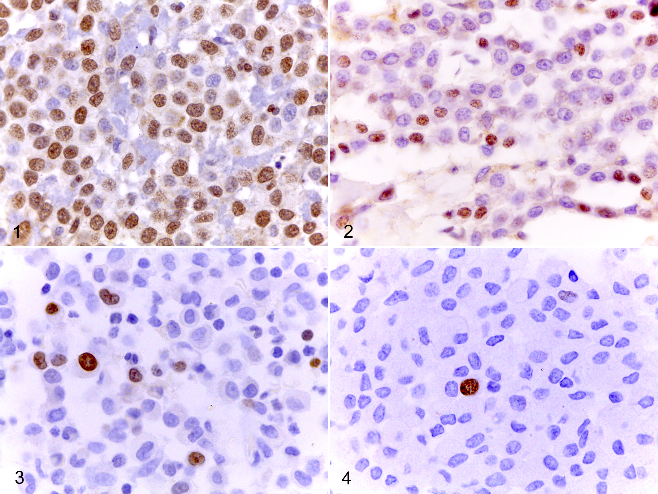
NANOG and Ki67 quantification were performed in five 400× microscopic fields considered to have the highest frequency of immunolabeling (“hot spots”). Images of the fields (0.08 mm2 each) were saved and positive and negative mast cells were counted using an image analysis software (ImageJ, NIH, USA) (Supplemental Table S1). All samples showed some degree of nuclear staining for NANOG (Figs. 1–4). Although the intensity of staining varied from weak to strong among samples, positivity was considered regardless of intensity for both markers in order to avoid any bias that could be added by setting a minimum threshold. The percentage of positive mast cells in each case (NANOG and Ki67 indices) were compared between groups using analysis of variance (ANOVA)/Kruskal-Wallis test followed by Dunn’s multiple comparisons test or Mann-Whitney test. Correlation analysis was performed using Pearson’s test and survival analysis using the Kaplan-Meier method followed by log-rank test. Associations with death due to disease were analyzed by Fisher’s exact test. Statistical analysis was performed using GraphPad Prism (version 6.0c for MacOS-X, GraphPad Software, GraphPad Software Inc) and Bioestat (version 5.0, Universidade Federal do Pará, PA, Brazil) and significance level was set as 5%.
Mast cell tumor, skin, dog.
According to Patnaik system, 68 ± 13% (mean ± SD) of the neoplastic mast cells were positive in grade I MCTs, and 57 ± 20% and 60 ± 14% in grade II and III, respectively (P = .36). 10 In the 2-tier system, 59 ± 19% of the cells were positive for NANOG in low-grade tumors and 64 ± 14% in high-grade tumors (P = .74). 4 NANOG positivity was 65 ± 13% for dogs that died due to the MCT and 58 ± 19% for censored cases (P = .37). NANOG and Ki67 indices were not correlated (P = .51, r = .10) (Fig. 5).
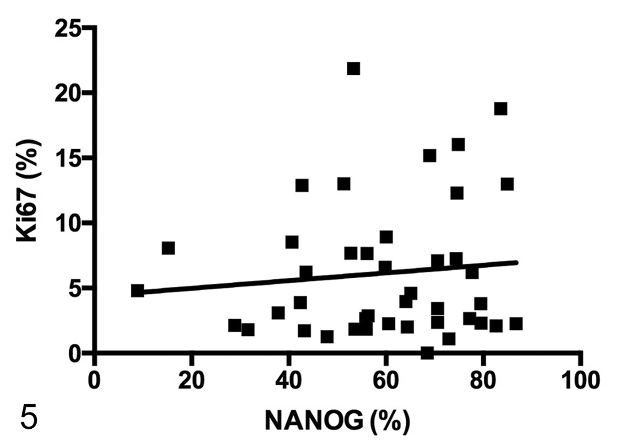
The percentage of neoplastic cells with immunolabeling for NANOG compared to that for Ki67 in canine cutaneous mast cell tumors. P > .05.
Thereafter, we determined a cutoff value in order to separate MCT cases into 2 groups based on the percentage of positive mast cells for NANOG using a ROC curve (cutoff = 65.1%). There were no significant differences between groups regarding mortality due to the disease (P = .52) or survival (P = .67, χ2 = .18, median survival undefined for both groups) (Fig. 6).
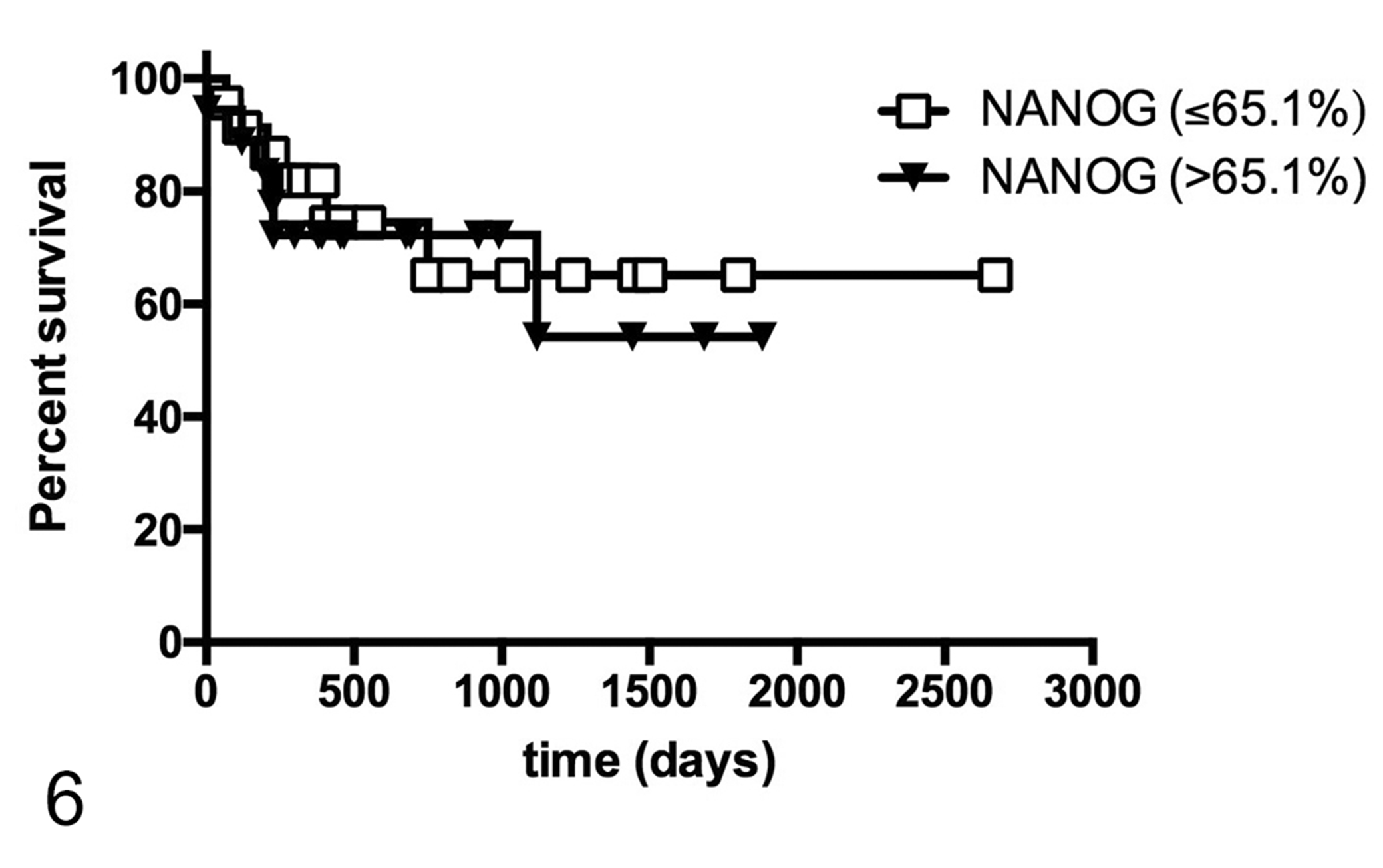
Overall survival curves for dogs with low (≤65.1%) and high (>65.1%) percentage of neoplastic mast cells with immunolabeling for NANOG . P > .05. Points indicate censored events.
We hypothesized that high NANOG expression could be related to cell differentiation and proliferation in canine cutaneous MCTs, thus implying a worse prognosis. Although we have demonstrated that NANOG transcriptional factor is expressed by neoplastic mast cells in dogs, its expression was not related to the degree of histological differentiation. Moreover, NANOG immunostaining was not a precise indicator for postsurgical survival or disease-related mortality in cases of canine cutaneous MCTs.
In a previous study, our research group obtained similar results for OCT4 pluripotency factor. 14 In that study, the immunolabeling varied among the 28 MCT samples, and was not related to survival, disease-related mortality and histological grades. Here we have also demonstrated that, although related to pluripotency, NANOG expression is not correlated with proliferative activity in canine cutaneous MCTs.
NANOG, OCT4, and SOX2 are key regulatory factors for the maintenance of pluripotency and proliferation of embryonic stem cells (ESCs). 15,18,19 Together, these molecules form a regulation loop, inducing their own transcription and the transcription of genes associated with signal transduction pathways, leading to the conservation of an undifferentiated state. NANOG, OCT4, and SOX2 are especially important during the early stages of embryonic development by the activation of TGF β and Wnt signaling pathways. 18,19
NANOG is expressed in several human cancers endowed with stem cell properties and commonly associated with carcinogenesis and poor prognosis. 7,17 –19 Knockdown of NANOG and OCT4 in human pancreatic cancer, for example, resulted in significant reduction of proliferation, migration, invasion, chemoresistance and tumorigenesis, both in vitro and in vivo. 6 Likewise, downregulation of NANOG gene in human gastric cancer cell lines resulted in reduction of both cell proliferation and metastatic ability followed by induction of cell cycle arrest and apoptosis. 3 NANOG is also found to be involved in tumorigenesis by enhancing clonogenic formation of cancer stem cells, stimulating the EMT process, and promoting cell proliferation, motility and invasion in colorectal cancer. 7
However, OCT4 and NANOG share several binding sites among genes that can induce or repress differentiation, including transcriptional regulators, growth factors and signaling molecules. 18 In spite of the fact that OCT4 deficient ESCs are still able to express NANOG, its expression alone is not enough for the maintenance of the undifferentiated state, suggesting that NANOG and OCT4 are functionally associated. 6,18
Although NANOG expression has already been described in some canine neoplasms, such as osteosarcoma and mammary tumors, to the best of our knowledge, the present study is the first to characterize NANOG expression in canine cutaneous MCTs, as well as to compare the results with histological grades, disease-related mortality and postsurgical survival. 8,9,16
Our results indicate that this pluripotency factor is not a satisfactory prognostic indicator for canine cutaneous MCTs. Similarly, previous results suggested that OCT4 is not a key malignancy factor for this type of tumor. 14 Further studies must be conducted in order to elucidate how NANOG, OCT4, and SOX2 pathways influence canine cutaneous MCTs development and progression, as well as the reason why NANOG expression does not correlate with malignancy despite its high immunohistochemical positivity.
Supplemental Material
Supplemental Material, DS1_VET_10.1177_0300985818789470 - Nanog Expression and Proliferation Indices in Canine Cutaneous Mast Cell Tumors
Supplemental Material, DS1_VET_10.1177_0300985818789470 for Nanog Expression and Proliferation Indices in Canine Cutaneous Mast Cell Tumors by Julia A. Joselevitch, Camila N. Barra, Thiago Henrique M. Vargas, Lidia H. Pulz, Adriana T. Nishiya, Silvia Regina Kleeb, José Guilherme Xavier, José Luiz Catão-Dias, and Ricardo F. Strefezzi in Veterinary Pathology
Footnotes
Acknowledgements
We thank the veterinarians from the Veterinary Hospitals at Universidade de São Paulo, Universidade Anhembi Morumbi and Universidade Metodista de São Paulo for submitting surgical specimens and Nilton Pedro dos Santos, Dr Juliano Rodrigues Sangalli, and Dr Juliano Coelho da Silveira for technical support.
Declaration of Conflicting Interests
The author(s) declared no potential conflicts of interest with respect to the research, authorship, and/or publication of this article.
Funding
This research was supported by Fundação de Amparo à Pesquisa do Estado de São Paulo (FAPESP, grants 2013/13252-8, 2014/20872-5, and 2016/03862-1).
Supplemental material for this article is available online.
References
Supplementary Material
Please find the following supplemental material available below.
For Open Access articles published under a Creative Commons License, all supplemental material carries the same license as the article it is associated with.
For non-Open Access articles published, all supplemental material carries a non-exclusive license, and permission requests for re-use of supplemental material or any part of supplemental material shall be sent directly to the copyright owner as specified in the copyright notice associated with the article.