
Abstract
Tissue microarray (TMA) is a time- and cost-saving technique allowing the simultaneous immunohistochemical evaluation of multiple tissue samples. The aim of this study was to assess the efficacy of TMA at classifying canine gastrointestinal spindle cell tumors as gastrointestinal stromal tumor (GIST), smooth muscle tumor (SMT), and non-GIST/non-SMT based on the expression of α-smooth muscle actin (α-SMA), desmin, and CD117. Thirty-four cases were investigated on TMAs, sampling 2 cores each. Immunohistochemistry was performed on TMAs and full sections, and the results were compared. Comparing full sections, TMA specificity and sensitivity were 100% and 93.8%, respectively, for α-SMA; 100% and 80.8% for desmin; and 100% and 100% for CD117. TMA allowed the identification of 6 of 6 GISTs, 25 of 26 SMTs, and 2 of 2 non-GIST/non-SMTs. One SMT was misdiagnosed as non-GIST/non-SMT. Based on these results, TMA-based immunohistochemistry is efficient at diagnosing canine gastrointestinal spindle cell tumors and might be applied on large caseloads in a research setting.
Keywords
Tissue microarray (TMA) consists of paraffin blocks containing multiple tissue samples deriving from original “donor” blocks. 6 One of the advantages of TMA is the simultaneous analysis of multiple samples using a single immunohistochemical stain (IHC), making it a time- and cost-saving technique. 6,8 Nevertheless, one major concern of TMA is its ability to represent the original sample, as the tissue samples are usually very small. For this reason, several studies have been conducted to validate TMA for IHC of specific lesions with specific panels of antibodies, including canine and feline mammary tumors, 7 canine tumors of the central nervous system, 10 canine islet cell tumor, 1 mast cell tumors, 3 and lymphoma in dog 2 and ferrets. 4
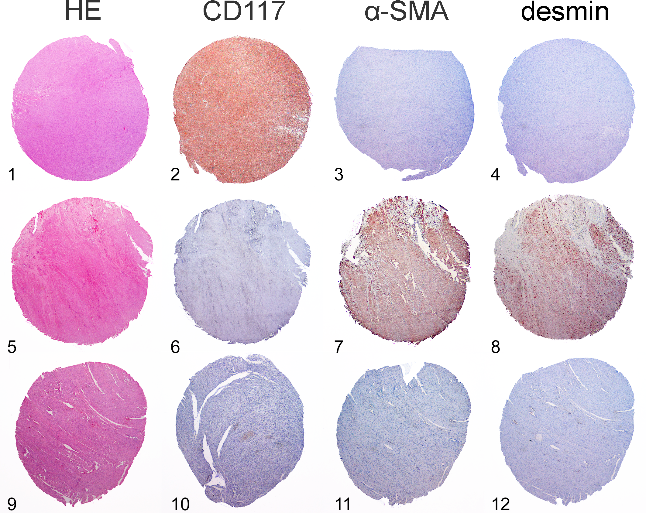
Canine spindle cell tumor of the gastrointestinal tract comprises a group of tumors including gastrointestinal stromal tumors (GISTs), smooth muscle tumors (SMTs), and non-GIST/non-SMTs. 5 As these neoplasms have a similar histologic appearance, usually characterized by interlacing bundles of spindle cells intermixed with scant collagen fibers, IHC is mandatory for their distinction. 5 The classification is based on the expression of α-smooth muscle actin (α-SMA), desmin, and CD117: GISTs express CD117, occasionally α-SMA, and are desmin-negative; SMTs are α-SMA– and/or desmin-positive and CD117-negative, while non-GIST/non-SMTs are α-SMA–, desmin- and CD117-negative. 5 This classification bears therapeutic implications, since anti-tyrosine kinase therapy in GIST is considered beneficial. 9
The aim of this study was to assess the efficacy of TMA for application of the α-SMA/desmin/CD117 panel to canine gastrointestinal spindle cell tumors to distinguish these 3 entities and to allow the application TMA to large caseloads in a research setting.
Thirty-four cases of canine gastrointestinal spindle cell tumor were selected from our database. Four consecutive full sections were prepared from each sample. One was stained with hematoxylin and eosin (HE) for histological reevaluation and for the selection of representative areas. The remaining 3 sections were used for IHC.
To construct the TMA, a sector map was drawn in Excel: each TMA contained 2 cores from each donor block and 1 core of normal hepatic or splenic tissue used as a landmark to orientate the sections.
The HE-stained section of each case was carefully observed to select 2 representative areas 3 mm in diameter characterized by neoplastic tissue without necrosis, inflammation, hemorrhage, or nonneoplastic tissue. Selected areas were marked on the slide and compared with the donor block to identify the exact sampling areas. A 3-mm skin punch was used to sample the donor blocks and to create the empty recipient blocks where samples were placed according to the sector map. From each TMA, an HE-stained section was evaluated to confirm the presence of representative tissue.
IHC on the full section of the donor block and on TMAs (Figures 1-12) was performed for desmin (mouse monoclonal, clone H76, dilution 1:200, Santa Cruz Biotech, Santa Cruz, CA), α-SMA (mouse monoclonal, clone 1A4, 1:450, Dako, Glostrup, Denmark), and CD117 (rabbit polyclonal, 1:200, Dako). Endogenous peroxidase was blocked by immersion in H2O2 0.3% in methanol for 30 minutes. Heat-induced antigen retrieval using pH 6.0 citrate buffer was performed for α-SMA and CD117, while incubation with pH 7.6, 0.05% trypsin at 37°C for 25 minutes was performed for desmin. Slides were incubated with the primary antibody overnight at 4°C, the reaction was revealed by a commercial streptavidin-biotin-peroxidase technique (ABC Kit Elite, Vector, Burlingame, CA) and visualized with 3-amino-9-ethylcarbazole (Dako). Slides were counterstained with Mayer’s hematoxylin. A positive control section of canine small intestine was used as positive control for α-SMA and desmin, while sections of canine mast cell tumor were used as positive control for CD117. Negative controls were obtained by omission of the primary antibody.
IHC was evaluated in a blinded manner, and results were compared between TMA and full sections and between the 2 cores of each case. The specificity and sensitivity of TMA (compared to evaluation of full sections) were calculated for α-SMA, desmin, and CD117. The diagnoses based on the reactivity on full sections and those based on the reactivity of TMAs were also compared.
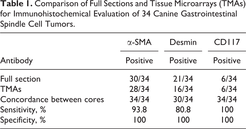
Thirty of 34 cases expressed α-SMA in full sections, 28 of which were α-SMA–positive also in TMA (Table 1). The 2 remaining cases were negative in TMA (false-negatives). No false-positive was identified. The reactivity of the 2 cores was concordant in all cases. Specificity of TMA was 100% and sensitivity was 93.8% for α-SMA. Twenty-one of 34 cases expressed desmin in full sections, often with multifocal distribution, 16 of which were also desmin-positive in TMA. The 5 remaining cases were negative in TMA (false-negatives). No false-positives were identified. The reactivity of the 2 cores was concordant in 30 of 34 cases. The specificity of TMA was 100% and sensitivity was 80.8% for desmin. Six of 34 cases expressed CD117 on full sections, and all were also CD117-positive in TMA. No false-positives or- negatives were identified. The reactivity of the 2 cores was concordant in all cases. Specificity and sensitivity of TMA were 100% for CD117.
Comparison of Full Sections and Tissue Microarrays (TMAs) for Immunohistochemical Evaluation of 34 Canine Gastrointestinal Spindle Cell Tumors.
Based on the IHC results on full sections, 6 cases were diagnosed as GISTs, 26 were SMTs, and 2 were non-GIST/non-SMT. TMA allowed the identification of all GISTs, 25 of 26 SMTs, and 2 of 2 non-GIST/non-SMTs. One SMT was misdiagnosed as non-GIST/non-SMT based on TMA results.
The IHC results revealed a high sensitivity and specificity of TMA for the diagnosis of canine spindle cell tumor of the gastrointestinal tract. Considering the 3 markers separately, CD117 was the most reliable on this type of TMA. As GIST diagnosis is based on CD117 expression, TMA identified all the GISTs. The reliability of α-SMA and desmin on this type of TMA was lower, resulting in a small number of false-negatives. Desmin false-negatives were probably secondary to the multifocal expression evidenced in some cases; α-SMA false-negatives were less common. As α-SMA expression is diffuse rather than multifocal, these false-negatives were more likely secondary to a procedural error since the immunohistochemistry was performed manually. Furthermore, the false-negative cores were located at the periphery of the TMAs, and this might have favored drying of the section or inadequate covering of the section with the reagents. We therefore repeated the staining for α-SMA, and the 2 cases gave a positive result, thus supporting the hypothesis of a procedural error. Despite 5 desmin and 2 α-SMA false-negatives, only 1 SMT was misdiagnosed as non-GIST/non-SMT since the simultaneous application of desmin and α-SMA identified the myoid differentiation in most cases. Furthermore, the use of 2 cores prevented false-negatives in the 4 cases in which desmin reactivity was discordant between cores.
Summarizing, the construction of 3-mm dual-core TMA allowed the classification of canine gastrointestinal spindle cell tumors in the large majority of cases and can therefore be used as an effective tool in the analysis of large caseloads in a research setting.
Footnotes
Acknowledgements
We thank Ms A. Collins for editing the English text.
Declaration of Conflicting Interests
The authors declared no potential conflicts of interest with respect to the research, authorship, and/or publication of this article.
Funding
The authors received no financial support for the research, authorship, and/or publication of this article.