
Abstract
The majority of primary intestinal lymphomas in dogs are T-cell lymphomas, with enteropathy-associated T-cell lymphoma (EATL) large cell type (type 1) being the most common. While most T-cell lymphomas express the T-cell marker CD3, there is increasing evidence that some human and canine T-cell lymphomas coexpress the B-cell marker CD20. We describe 3 cases of CD3+, CD20+, Pax5- EATL type 1 in dogs. All 3 cases had clonal rearrangement of T-cell receptor gamma. Initial clinical signs included weight loss, inappetence, diarrhea, and/or vomiting. The mean age was 9 years (range 3–12). Survival was highly variable ranging from 20 days to longer than 1.6 years. Considering the different chemotherapeutic response of T-cell versus B-cell lymphomas, accurate diagnosis of lymphomas coexpressing CD3 and CD20 as EATL type 1 based on histologic features and clonality results is important. Regardless, the clinical and/or prognostic significance of neoplastic T cells expressing CD20 is unclear.
Keywords
Historically, the vast majority of primary intestinal lymphomas in dogs have been reported as peripheral T-cell lymphomas, not otherwise specified. 3 While feline intestinal T-cell lymphomas have been well characterized and, similar to their human counterparts, have been subclassified according to cell morphology and pattern by the World Health Organization (WHO) as enteropathy-associated T-cell lymphoma (EATL) large cell (type 1) and small cell (EATL type 2); 7,8,13 this advance in classification has only recently been established in dogs. 2,8,13 In contrast to feline intestinal T-cell lymphomas, which are predominately EATL type 2 and characterized as a monomorphic population of small T lymphocytes often forming variably sized intraepithelial nests and plaques, canine intestinal T-cell lymphomas are most commonly reported as large cell type with or without accompanying inflammation and/or epitheliotropism or, according to human classification, EATL type 1. 2,8,13
In most intestinal T-cell lymphomas, neoplastic cells are expected to express the T-cell marker CD3 and are not immunoreactive for the B-cell marker CD20. However, there is an increasing body of literature of human peripheral T-cell lymphomas and fewer cases of cutaneous epitheliotropic T-cell lymphomas that coexpress CD20. 4,11 Recently, coexpression of CD3 and CD20 was reported in a cutaneous epitheliotropic T-cell lymphoma (mycosis fungoides) in a single dog. 1 While the clinical significance has yet to be determined in either species, recognition of such dual expression has important diagnostic implications that ultimately guide the therapeutic approach. To our knowledge, this is the first report of coexpression of CD3 and CD20 in primary intestinal T-cell lymphoma in dogs.
Three dogs were included in this case series. Tissues received included 3 and 4 small endoscopically biopsied sections of small intestinal mucosa from dogs 1 and 2, respectively, and 2 full thickness small intestinal biopsies from dog 3. Surgical biopsy specimens had been routinely fixed in formalin and embedded in paraffin. From each case, 5 µm serial sections were routinely stained with hematoxylin and eosin. CD3, CD20, and Pax5 immunohistochemistry, including dual labeling for CD3 and CD20 as well as dual labeling for CD3 and Pax5 were performed as previously described. 1,2 Polymerase chain reaction (PCR) for antigen receptor rearrangement (T-cell clonality) was performed on each case as previously described. 2
Dog 1 was a 12-year-old, male castrated Cocker Spaniel × Poodle that had weight loss, inappetence, and diarrhea for several months duration. Endoscopy showed a rough small intestinal mucosa, and small intestinal mucosal biopsies were taken. Dog 2 was a 12-year-old, female spayed Pembroke Welsh Corgi that had intermittent soft stool for 1 year duration and diarrhea with occasional hematochezia for approximately 4 months duration. The dog was treated with metronidazole and a change in diet to bland home cooked meals. The dog was then transitioned off metronidazole and onto tylosin powder. Endoscopy showed a diffusely hyperemic, friable, and granular small intestinal mucosa, and duodenal mucosal biopsies were taken. Dog 3 was a 3-year-old, male American Stafford Terrier that had vomiting and inappetence for 5-6 days duration, and an acute onset of lethargy, diarrhea, and inability to walk. The dog was febrile and had swollen joints, and abdominal exploratory surgery revealed a ruptured mass at the ileocecal junction, septic abdomen, and mesenteric lymphadenopathy. The mass and an enlarged cecal lymph node were resected.
On histopathologic evaluation, in mucosal tissue sections from dog 1, there were large lymphocytes moderately expanding the villar mucosal lamina propria and forming multifocal intraepithelial nests and plaques (Figs. 1–2). In mucosal tissue sections from dog 2, there was a monomorphic population of intermediate-sized lymphocytes with a similar distribution as noted in dog 1. In full thickness tissue sections from dog 3, there was a transmural infiltrate of monomorphic large lymphocytes arranged in sheets that extended into the adjacent mesenteric adipose tissue.
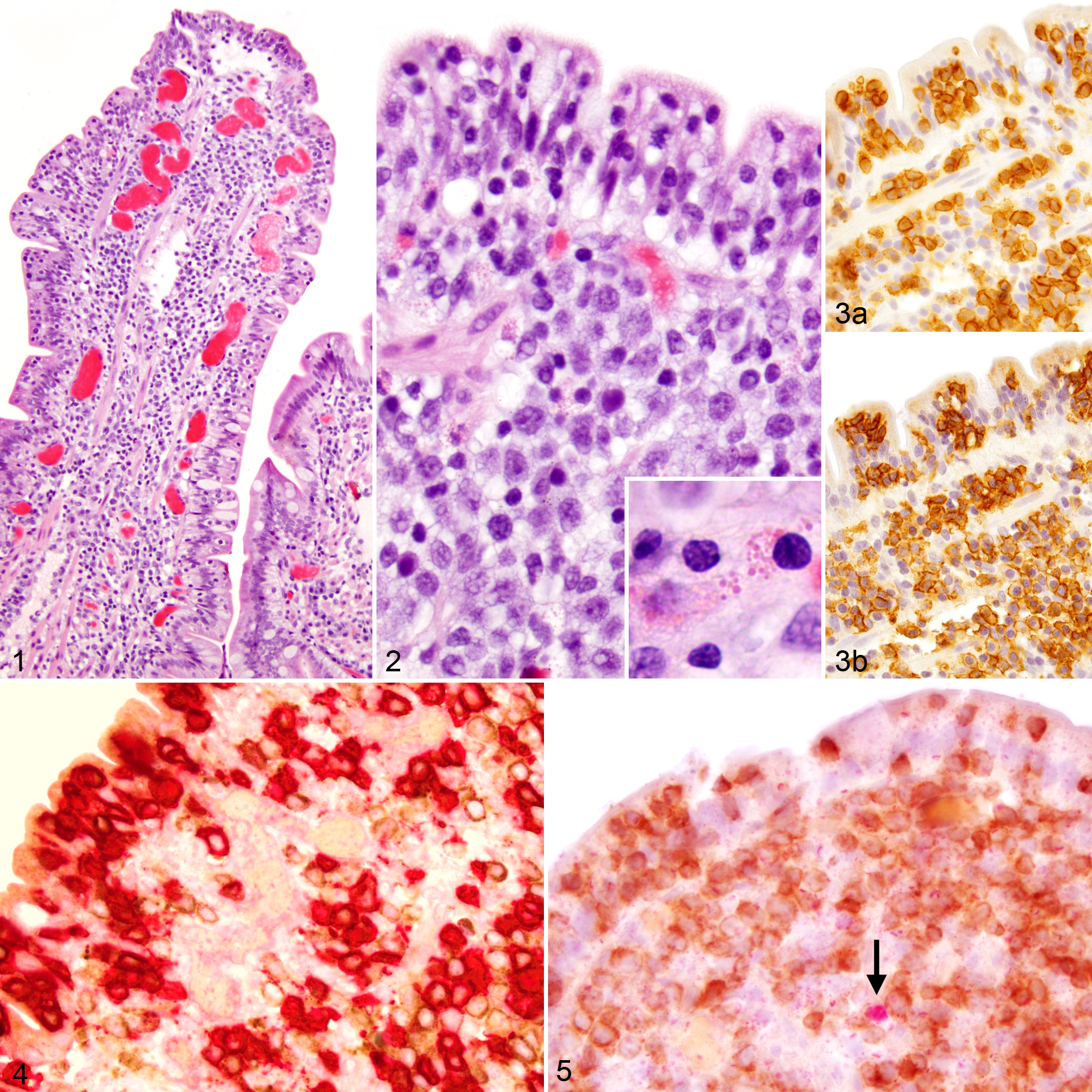
In all cases, the neoplastic lymphocytes had strong perimembranous labeling for CD3 (Fig. 3a). In addition, the majority of neoplastic lymphocytes, including those noted within the mucosal epithelium, had perimembranous labeling for CD20 of variable intensity (Figs. 3–4). These neoplastic lymphocytes were not immunoreactive for Pax5 (Fig. 5). PARR testing showed clonal rearrangement of the T-cell receptor gamma gene for all cases. Since the neoplastic lymphocytes in all 3 cases were negative for another B-cell marker, Pax5, and monoclonal rearrangement of the T-cell antigen receptor gene was confirmed for each case, a diagnosis of a CD3+, CD20+ enteropathy-associated T-cell lymphoma, large cell type (EATL type 1) was made for each dog.
For dog 1, prednisone therapy initially decreased clinical signs; however, at 20 days after the molecular confirmation of a lymphoma diagnosis, the animal developed weakness, profuse hemorrhagic diarrhea, vomiting, and inappropriate urination and subsequently developed increased respiratory effort and a tense painful abdomen. Due to a poor prognosis, euthanasia was elected at that time.
For dog 2, following the start of tylosin therapy and at 10 days after the molecular confirmation of a lymphoma diagnosis, the animal had not had any diarrhea. While the animal has not been examined since that time, the dog was reportedly alive at 1.6 years after the initial diagnosis.
For dog 3, ultrasound examination at 26 days postsurgery showed no signs of recurrence or progression of disease. At 96 days postsurgery, the dog had pain while defecating, and an ultrasound examination showed an 8.5 cm jejunal mass. The jejunal mass was debulked, and a modified T-cell multiagent chemotherapy protocol, including asparaginase, vincristine, lomustine, and prednisone, was started 8 days later. Chemotherapy was continued on a weekly basis in an alternating fashion. Progressive disease was noted on ultrasound examination at 159 days postdiagnosis. An abdominal exploratory surgery was performed at 181 days postdiagnosis, and the dog was euthanized due to complications and advanced stage disease throughout the abdomen.
CD20 is typically considered a marker of B-cell lineage. Unlike Pax5, which was negative in these cases and is expressed throughout the entirety of B-cell maturation, CD20 is normally expressed on the surface of B lymphocytes beginning at the pre-B cell stage before immunoglobulin µ heavy chains develop in the cytoplasm, and expression persists up to terminal plasma cell differentiation. 11 This transmembrane phosphoprotein has unconfirmed roles in both calcium transport and T-cell-independent antigen-induced activation of B cells. 11 However, in normal peripheral blood of humans, a subset of T lymphocytes with low expression of CD20 has been detected by flow cytometry, which are known as CD20dim T lymphocytes. 10 These CD20dim T cells are phenotypically different from CD20- T cells and are more likely to be CD8, CD45RO, and γ/δ T-cell antigen receptor positive; however, their function remains unknown. 5
It is unclear whether CD20 expression in this subset of canine T-cell lymphomas reflects aberrant expression of CD20 or whether these cells originated from a subpopulation of normal CD20dim T cells, and if either of these possibilities have any clinical relevance. 10 –12 A third theory involves false-positive labeling of the neoplastic T cells due to cross-reactivity of the anti-CD20 antibody. 12 This theory is less likely because the observed CD20 labeling in T cells was limited to neoplastic T cells only and not inflammatory T cells at greater distance from the lesion, and because such dual expression is rare in our routine biopsy service.
Similar CD20 expression has been recognized in a small percentage of human T-cell lymphomas and in a single case of a canine cutaneous epitheliotropic lymphoma. 1,4,11 In humans, reports include 2 descriptions of enteropathy-associated T-cell lymphoma with CD20 expression, one of which was initially misdiagnosed as B-cell lymphoma. 6,11 The prognostic and therapeutic implications of such CD20 expression is unclear. Follow-up information in the human cases was limited, but 1 of the 2 reports indicated a poor prognosis. 11 Regardless, a misdiagnosis of a T-cell lymphoma as a B-cell lymphoma can have serious clinical implications because T-cell lymphoma is typically more aggressive than B-cell lymphoma, resulting in a different prognosis and therapeutic strategy. In limited follow-up information for this case series in dogs, the biologic behavior was aggressive in 2 dogs and 1 dog was still alive after 1.6 years without further clinical information. Monoclonal therapy against CD20 has at least been shown to deplete CD20dim T lymphocytes in humans with multiple sclerosis, which raises the question whether targeted therapy against CD20 may be of value for T-cell lymphomas that have CD20 expression. 9
EATL type 1 lymphomas are often associated with transmural infiltration. 7,8,13 In 1 case (dog 3), there was peritonitis at the time of diagnosis due to rupture of the intestinal wall secondary to full thickness invasion of the neoplasm; however, the full depth of invasion could not be evaluated in the other 2 cases (dogs 1 and 2) due to submission of endoscopic biopsies. The aforementioned human case of a CD20-expressing EATL also had intestinal perforation. 10 No neoplastic cells were reported in the cecal lymph node in dog 3, and the oral and aboral margins of the resected intestinal mass were reported free of neoplastic cells at the time of diagnosis.
This case series demonstrates the existence of CD3+, CD20+ enteropathy-associated T-cell lymphoma in dogs and highlights the importance of combining histomorphology, clinical presentation, immunophenotyping, and PARR testing to properly diagnose these neoplasms. Further, given the potential prognostic and therapeutic implications, immunophenotyping should routinely be considered for not only diagnostic purposes. Recognition of this particular phenotype of canine EATL type 1 will allow collection of additional cases and investigation of their biological behavior to ultimately determine the clinical significance and prognostic value of CD20 expression in canine enteric T-cell lymphomas.
Footnotes
Acknowledgements
We thank Madison Operacz at the Michigan State University Veterinary Diagnostic Laboratory (MSU VDL) for performing the clonality testing, and the histology department at MSU VDL, particularly Tom Wood, for IHC. Gratitude to Dr James Walberg, VetPath Services, Stone Ridge, NY; Dr Kate Vickery, Hope Vet Specialists, Malvern, PA; Dr Julie Stanton, Cornell University Hospital for Animals, Ithaca, MI; and Animal Emergency and Referral Associates, Fairfield, NJ for submission of the cases and/or providing follow-up data.
Declaration of Conflicting Interests
The author(s) declared no potential conflicts of interest with respect to the research, authorship, and/or publication of this article.
Funding
The author(s) received no financial support for the research, authorship, and/or publication of this article.