
Abstract
Although cytology is a rapid diagnostic procedure in dogs, the cytologic criteria of endoscopic biopsies for chronic enteritis and intestinal lymphoma are not well defined. An immediate diagnosis using cytology would benefit patients by enabling prompt initiation of therapy. The objective of this study was to investigate the correlation between the results of endoscopic cytology and histopathology. In this study, 167 dogs with clinical signs of chronic gastrointestinal disease were included. On the basis of histopathology, the following diagnoses were determined: lymphocytic-plasmacytic enteritis in 93 dogs; eosinophilic enteritis in 5 dogs; small cell intestinal lymphoma in 45 dogs; and large cell intestinal lymphoma in 24 dogs. Two clinical pathologists retrospectively evaluated the endoscopic cytology of squash-smear preparations. The cytologic diagnoses of inflammation, small cell lymphoma, and large cell lymphoma were based on the severity of lymphocyte infiltration, the size of infiltrated lymphocytes, and eosinophil/mast cell infiltration. The clinical severity score was significantly increased along with the degree of lymphocyte infiltration evaluated by cytology. The cytologic diagnosis was in complete agreement with the histopathologic diagnosis in 136 of 167 (81.4%) cases. For the differentiation between enteritis and lymphoma, endoscopic cytology had a sensitivity of 98.6%, a specificity of 73.5%, a positive predictive value of 72.3%, and a negative predictive value of 98.6%. The log-rank test and Cox regression analysis showed that the results of cytology predicted the prognosis. These results suggest that endoscopic cytology is a useful technique to aid diagnosis of intestinal inflammation and lymphoma in dogs.
Keywords
Chronic enteritis and intestinal lymphoma are common intestinal diseases in dogs, causing persistent or relapsing clinical signs such as vomiting, diarrhea, anorexia, and weight loss. 1,29 Previous studies have shown that the prognosis of intestinal lymphoma, especially the immature large cell type, is very poor compared with that of chronic enteritis. 14,18,24 Therefore, early diagnosis and appropriate treatment are important for prolonged survival of dogs with intestinal lymphoma. As the clinical presentations of chronic enteritis and intestinal lymphoma are similar, it is often difficult to distinguish between these 2 diseases on the basis of laboratory tests such as blood tests, fecal examination, urinalysis, and imaging. Histopathologic assessment of the gastrointestinal tract via endoscopic or surgical biopsies is the most conclusive diagnostic tool for the differentiation between chronic enteritis and intestinal lymphoma. 4,29 Lymphocytic-plasmacytic enteritis (LPE) and eosinophilic enteritis (EE) are the predominant histopathologic diagnoses of chronic enteritis. 11 Unfortunately, it may be difficult to distinguish LPE from intestinal lymphoma, particularly when well-differentiated, small neoplastic cells are present. 4,5,29,30 Immunohistochemistry (IHC) aids in the diagnosis of intestinal lymphoma. 4,17 However, histopathology and IHC require, at least, several days to prepare the specimen slides. Cytological assessment may allow an earlier diagnosis of intestinal lymphoma, which may be important in those cases that have rapid progression.
Polymerase chain reaction for antigen receptor gene rearrangement (PARR) may be helpful in the early differentiation of inflammation and lymphoma. A stepwise diagnostic approach has been recommended, using histopathology as the first step followed by IHC and PARR. 4 Nonetheless, the early diagnosis and accurate differentiation of enteritis and intestinal lymphoma remain difficult, due to the delay in laboratory results and the low sensitivity of PARR analysis, respectively. The sensitivity of PARR in canine intestinal lymphoma was only 66.7%–76%. 4,9,14,24 In addition, clonal rearrangement was detected in approximately 30% of dogs with chronic enteritis, 14,24,26 suggesting that the specificity of PARR was also low for diagnosis of canine intestinal lymphoma. These restrictions emphasize the need to develop diagnostic tools for differentiating chronic enteritis and intestinal lymphoma.
Cytology is commonly used as a rapid diagnostic method in veterinary medicine, and cytologic findings correlate well with the histopathologic diagnoses for numerous tumor types. The diagnosis of multicentric lymphoma can often be determined via fine-needle aspiration cytology of the enlarged lymph nodes. 8,23 Additionally, fine-needle aspiration cytology of intestinal masses or enlarged mesenteric lymph nodes is helpful for the diagnosis of intestinal lymphomas. 13 However, the occurrence of secondary inflammation may hinder a definitive diagnosis, and intestinal masses or enlarged mesenteric nodes are not always observed.
Cytologic examination of intestinal mucosa obtained via endoscopic biopsy is a useful technique for the detection of intestinal diseases in humans. 3,7,25 To the authors’ knowledge, only a few published studies have compared the cytologic and histopathologic results of endoscopically obtained specimens in dogs and cats. 15,16,22 In these studies, there is controversy over the diagnostic value of endoscopic cytology. Jergens et al 15,16 suggested that the results of endoscopic cytology correlate highly with the histopathologic observations in dogs and cats. In contrast, Mangelsdorf et al 22 demonstrated that the sensitivity of endoscopic cytology is low in cats. Several factors may be involved in this discrepancy, including the differences in sample size, disease type, and criteria for cytologic evaluation. Case survival was not reported in previous investigations. The aim of this study was to develop a cytologic grading scheme for lymphocyte infiltration in the intestinal mucosa and a diagnostic algorithm for distinguishing enteritis from lymphoma.
Materials and Methods
Cases
This study included 167 dogs, with clinical signs of chronic gastrointestinal disease, that underwent endoscopic examination between March 2011 and July 2015 at the Veterinary Medical Center of the University of Tokyo. Informed consent was obtained from the owners, and the study protocol was approved by the animal care committee of the Veterinary Medical Center of the University of Tokyo.
The dogs included in this study comprised 17 intact females, 60 spayed females, 34 intact males, and 56 neutered males. The median age was 107 months (range, 13–187 months) and the median body weight was 5.5 kg (range, 1.2–33 kg). The following breeds with more than 2 individuals were included: Miniature Dachshund (n = 21), Toy Poodle (n = 14), Shiba Inu (n = 11), mongrel (n = 10), Yorkshire Terrier (n = 9), Chihuahua (n = 8), French Bulldog (n = 7), Pug (n = 7), Papillon (n = 6), American Cocker Spaniel (n = 5), Pembroke Welsh Corgi (n = 5), Shih Tzu (n = 5), Jack Russell Terrier (n = 5), Pomeranian (n = 5), Maltese (n = 5), Boston Terrier (n = 5), Beagle (n = 4), Miniature Pinscher (n = 3), Shetland Sheepdog (n = 3), Kaninchen Dachshund (n = 3), and Bernese Mountain Dog (n = 3). The remaining 23 dogs were of other breeds. The cases were scored for severity according to the canine chronic enteropathy clinical activity index (CCECAI). 1
Sample Collection
Gastroduodenoscopy, ileocolonoscopy, or both were performed under general anesthesia. In preparation for endoscopy, dogs were fasted for 12–18 hours. Multiple mucosal biopsies (6 in each area) were collected from the stomach, duodenum, ileum, and colon for histopathology. At least 1 biopsy specimen from the small intestine (duodenum and/or ileum) was used for cytologic evaluation.
Histopathology
The mucosal biopsy specimens from each site were fixed in 10% neutral buffered formalin for 48 hours, embedded in paraffin, and subsequently stained with hematoxylin and eosin (HE). The histopathologic sections were evaluated by 2 pathologists with Japanese College of Veterinary Pathologists board certification (J.K.C., K.U.). A histopathologic diagnosis of gastrointestinal inflammation was determined according to the World Small Animal Veterinary Association criteria. 6,19,21 A histopathologic diagnosis of intestinal lymphoma was determined from HE-stained sections. We further performed IHC for CD3 and CD20 expression on 34 of 167 cases that were difficult to diagnose by histopathologic examination only. 18,20 Lymphocyte epitheliotropism, heterogeneity, and nuclear size were evaluated to differentiate between intestinal lymphoma and inflammation. 4,17 Intestinal T-cell lymphomas have been recently classified into enteropathy-associated T-cell lymphoma (EATL) type 1 (small cell lymphoma) and type 2 (large cell lymphoma) in humans and dogs. 4 In this study, the lymphomas were classified into the following 2 categories—small and large cell lymphoma—based on the morphological characteristics of the proliferating lymphocytes. 18
According to the results of histopathology and IHC, the following diagnoses were determined: LPE in 93 dogs; EE in 5 dogs; small cell intestinal lymphoma in 45 dogs; and large cell intestinal lymphoma in 24 dogs (Figs. 1–6). All lymphoma cases were immunophenotyped for the expression of CD3 and CD20 by IHC.
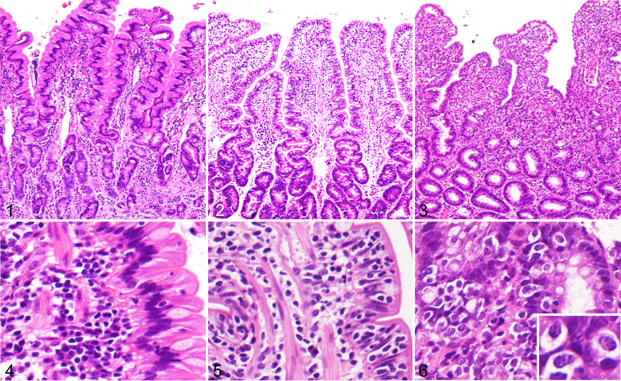
Lymphocytic-plasmacytic enteritis, duodenum, dog. Moderate increase in lamina propria lymphocytes with mild dilation of lacteals. Hematoxylin and eosin (HE).
Endoscopic Cytology
The cytologic specimens of endoscopic biopsies were prepared using the squash-smear technique. This technique involved placing a mucosal sample between 2 glass slides, applying gentle pressure to squash the samples, and subsequently smearing the above slide (Suppl. Video 1). Following the smear preparation, the slides were air-dried and stained with Wright-Giemsa (Suppl. Fig. 1).
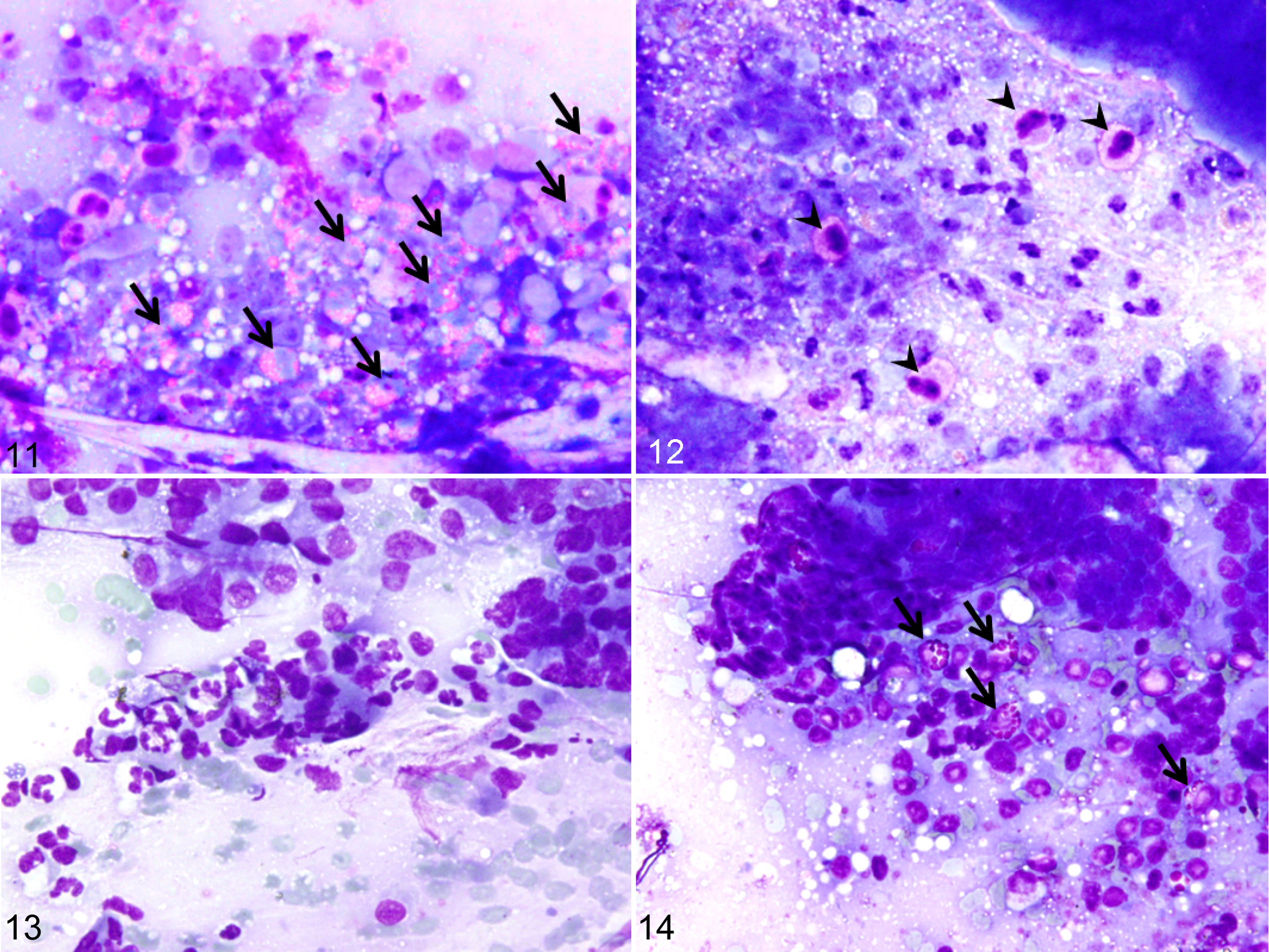
The cytologic specimens were reviewed in collaboration by 2 clinical pathologists (S.M., M.T.) who were blinded to the histopathologic results and clinical information. A grading system for the degree of lymphocyte infiltration consisted of the following categories (evaluation at 400× magnification): mild, infiltration of a few lymphocytes (0–4 cells per 400× field) on the edge of the epithelial cells (Fig. 7); moderate, presence of linearly aligned lymphocytes (≥5 and <20 cells per 400× field) or nests of lymphocytes (≥5 and <20 clustered cells per 400× field) on the edge of the epithelial cells (Fig. 8); and severe, characterized by the diffuse presence of small lymphocytes (≥20 cells per 400× field, Fig. 9). In addition to the degree of lymphocyte infiltration, lymphocyte size was assessed as follows: large cell infiltration, in which large cells comprise greater than 30% of the lymphocyte population (Fig. 10). Large and small cells were defined by their cellular size (larger or smaller than a neutrophil). 23 The cytologic grading in each case was determined by the most severely affected areas. The infiltration of eosinophils (≥5 cells per 400× field, Fig. 11) and mast cells (≥5 cells per 400× field, Fig. 12) was assessed. In addition, the infiltration of neutrophils (≥5 cells per 400× field, Fig. 13) and globule leukocytes 2 (presence or absence per 400× field, Fig. 14) was assessed for the analysis of prognostic factors. Degenerative changes of epithelial cells were not evaluated in cytologic specimens because artificial changes might be caused by squash-smear method.
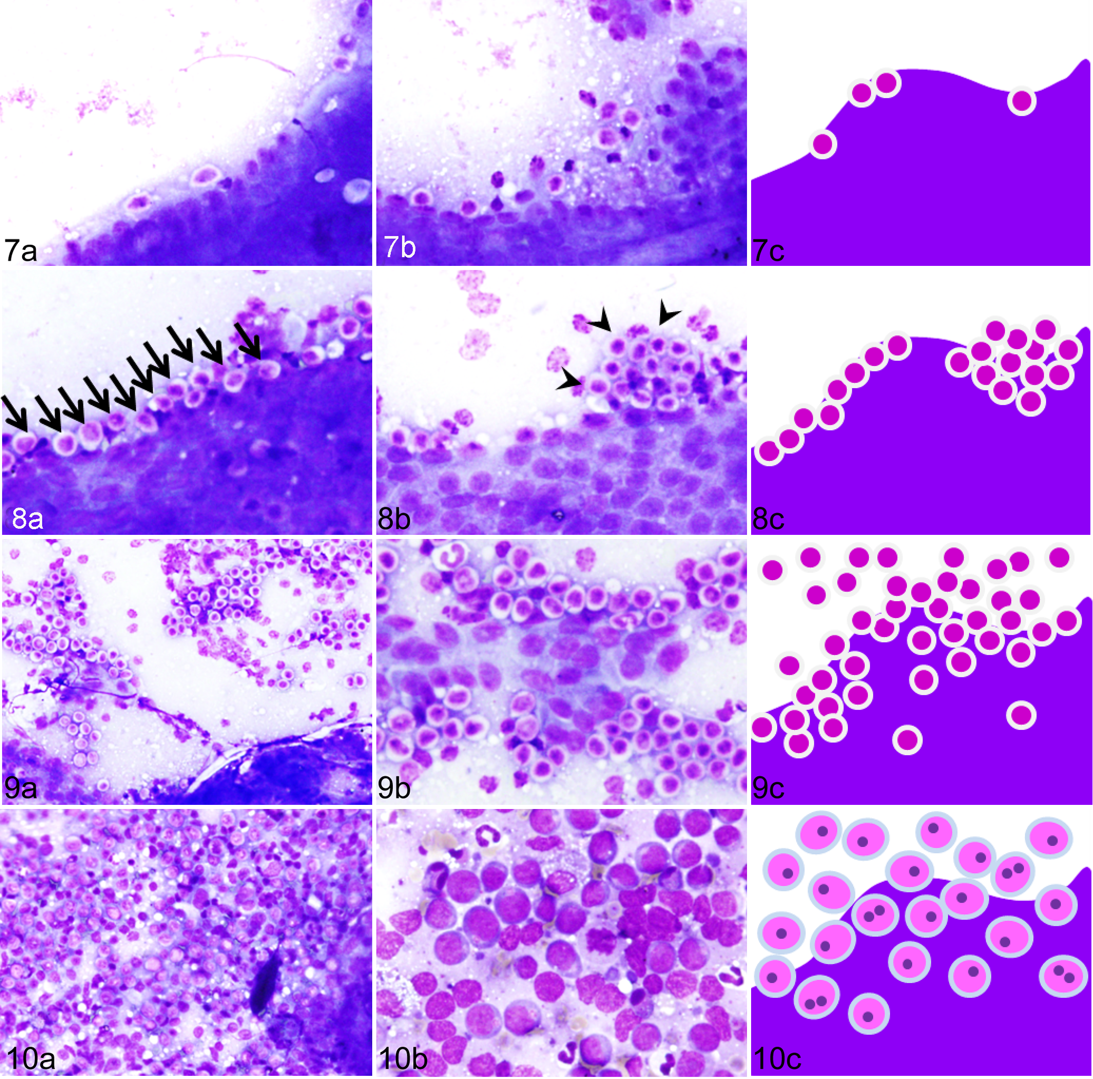
Lymphocytic-plasmacytic enteritis, duodenum, dog, graded as mild. Wright-Giemsa (WG) stain. (a, b) A few lymphocytes are seen at the periphery of the epithelial cells. (c) Schematic drawing of mild lymphocyte infiltration (0–4 cells per 400× field).
Eosinophilic enteritis, duodenum, dog. Wright-Giemsa (WG) stain.
Cytologic diagnosis was made by the following algorithm developed in this study (Fig. 15). A diagnosis of LPE was indicated by the observation of mild lymphocyte infiltration and absence of infiltration of eosinophils and mast cells; EE by mild lymphocyte infiltration and the presence of infiltration of eosinophils and mast cells; small cell intestinal lymphoma by the presence of moderate to severe lymphocyte infiltration and the prevalence of large cells comprising less than 30% of the lymphocyte population; and large cell intestinal lymphoma by the presence of moderate to severe lymphocyte infiltration and the prevalence of large cells comprising greater than 30% of the lymphocyte population.
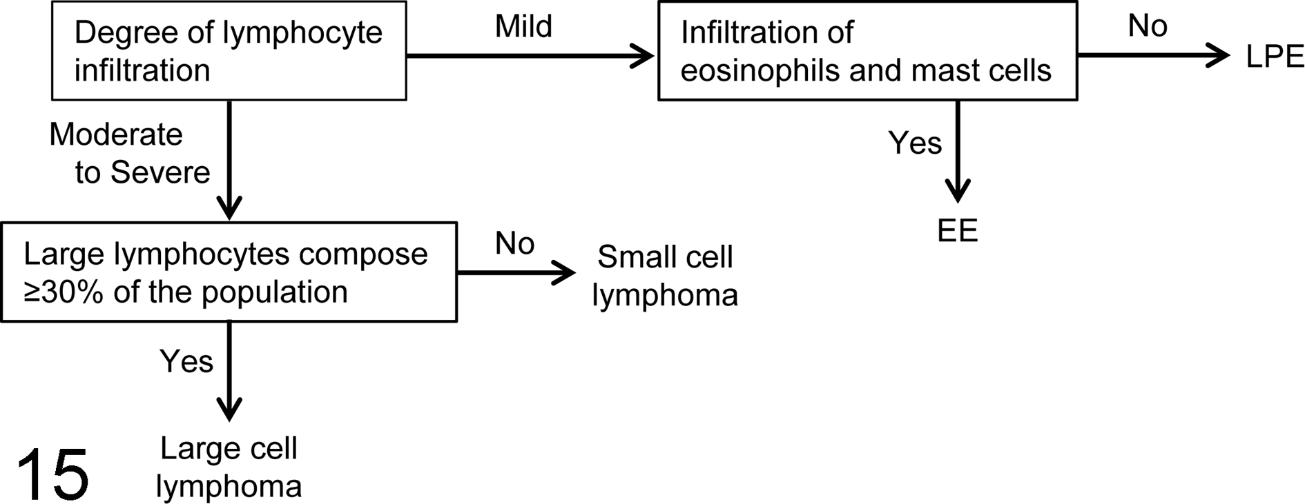
The diagnostic algorithm that was used to distinguish canine intestinal lymphoma from chronic enteritis, including lymphocytic-plasmacytic enteritis (LPE) and eosinophilic enteritis (EE), based on endoscopic cytology.
Case Outcomes
The survival time and current status (alive, deceased, or lost) were obtained for each case based on the medical records or a fax interview with the referring veterinarians. The overall survival was defined as the time from the endoscopic biopsy until the established cause of death of the animal at the end of the study (April 11, 2016).
Statistical Analysis
Statistical analyses were performed using JMP version 9 (SAS Institute, Cary, NC). The Kruskal-Wallis test was used to test for the overall differences in CCECAI score. The Steel-Dwass test was used to analyze the between-group differences. Survival curves in both histological and cytological diagnoses were generated with the Kaplan-Meier method, and overall survival was compared using the log-rank test. The Cox proportional hazard regression model was used to examine the prognostic value of each variable (age, CCECAI, neutrophil infiltration, globule leukocyte infiltration, and cytologic diagnosis). Statistical significance was defined as P < .05.
Results
Clinical Evaluation
The characteristics of each case are shown in Suppl. Table 1. Based on histopathology, 93 (55.7%) dogs were diagnosed with LPE. There were 12 intact females, 38 spayed females, 17 intact males, and 26 neutered males. The median age was 110 months (range, 13–187 months) and median CCECAI was 5 (range, 1–15). Five (3.0%) dogs were histopathologically diagnosed with EE. There were 2 spayed females and 3 neutered males. The median age was 45 months (range, 20–74 months) and median CCECAI was 5 (range, 4–6). All dogs with chronic enteritis (LPE and EE) received prednisolone (0.5–2 mg/kg) after endoscopy.
Cytologic and Histologic Diagnoses for Endoscopic Biopsies From 167 Dogs With Chronic Gastrointestinal Diseases.
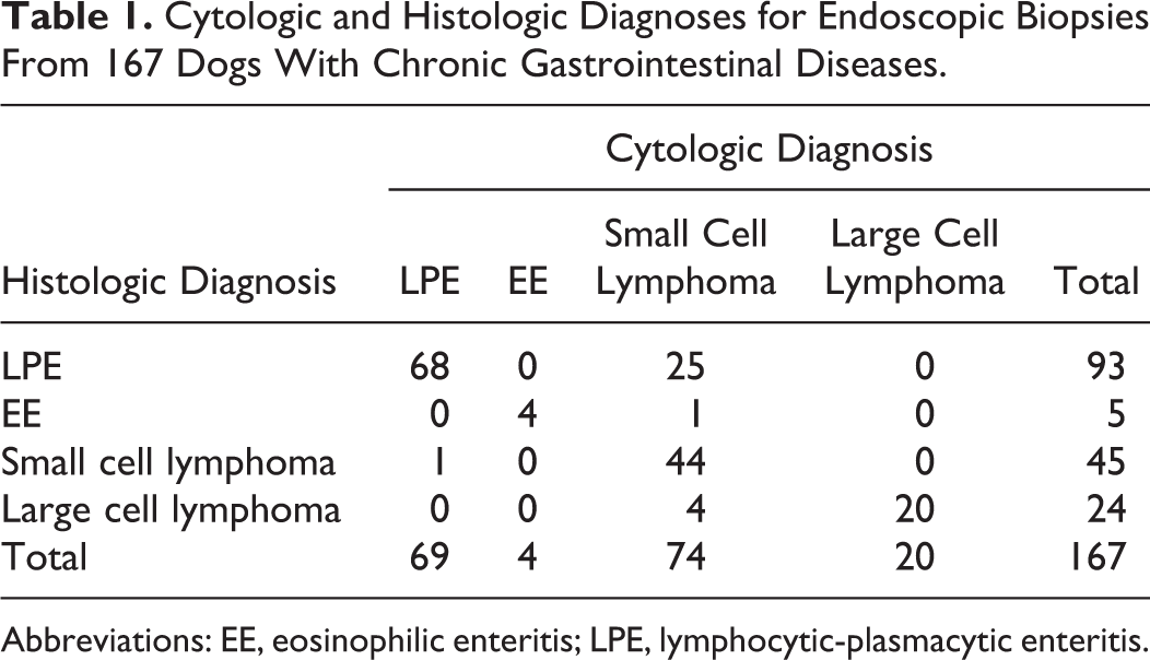
Abbreviations: EE, eosinophilic enteritis; LPE, lymphocytic-plasmacytic enteritis.
Forty-five (26.9%) dogs were histopathologically diagnosed with small cell intestinal lymphoma. All neoplasms were classified as T-cell lymphoma by IHC. There were 4 intact females, 16 spayed females, 11 intact males, and 14 neutered males. The median age was 100 months (range, 24–177 months) and median CCECAI was 7 (range, 1–16). All dogs received prednisolone (1–2 mg/kg), and 28 were treated with chlorambucil (2 mg/m2) in combination with prednisolone.
Twenty-four (14.4%) dogs were histopathologically diagnosed with large cell intestinal lymphoma. Of these, 20 (83.3%) neoplasms were classified as T-cell lymphoma, 3 (12.5%) as B-cell lymphoma, and 1 (4.2%) as T- and B-cell lymphoma. There were 1 intact female, 4 spayed females, 6 intact males, and 13 neutered males. The median age was 125 months (range, 71–186 months) and median CCECAI was 11 (range, 3–19). Seven dogs with large cell intestinal lymphoma received prednisolone (1–2 mg/kg), and 17 were treated with a 6-month modified version of the Wisconsin-Madison chemotherapy protocol (UW-25). 10 Therapeutic decisions were made with knowledge of the cytologic and histopathologic diagnosis.
Cytologic Evaluation
A total of 306 cytologic slides from 167 dogs were included. The details of the cytologic examination in each case are provided in Suppl. Table 1. Adequate cytologic specimens were obtained from 99.7% of the biopsies that were prepared via the squash-smear technique. As the infiltrated inflammatory cells primarily consisted of lymphocytes, the cytologic grading was determined according to the degree of lymphocyte infiltration (Figs. 7–10). Of the 167 cases, 73 (43.7%) were classified as mild, 50 (29.9%) as moderate, and 24 (14.4%) as severe, and 20 (12.0%) showed evidence of large cell infiltration. The CCECAI increased significantly along with the cytologic grading; however, a significant difference was not evident between the cytologic grades of moderate and severe (Fig. 16). Infiltrations of eosinophils, mast cells, neutrophils, and globule leukocytes were observed in 6 (3.6%), 15 (9.0%), 18 (10.8%), and 10 (6.0%) cases, respectively.
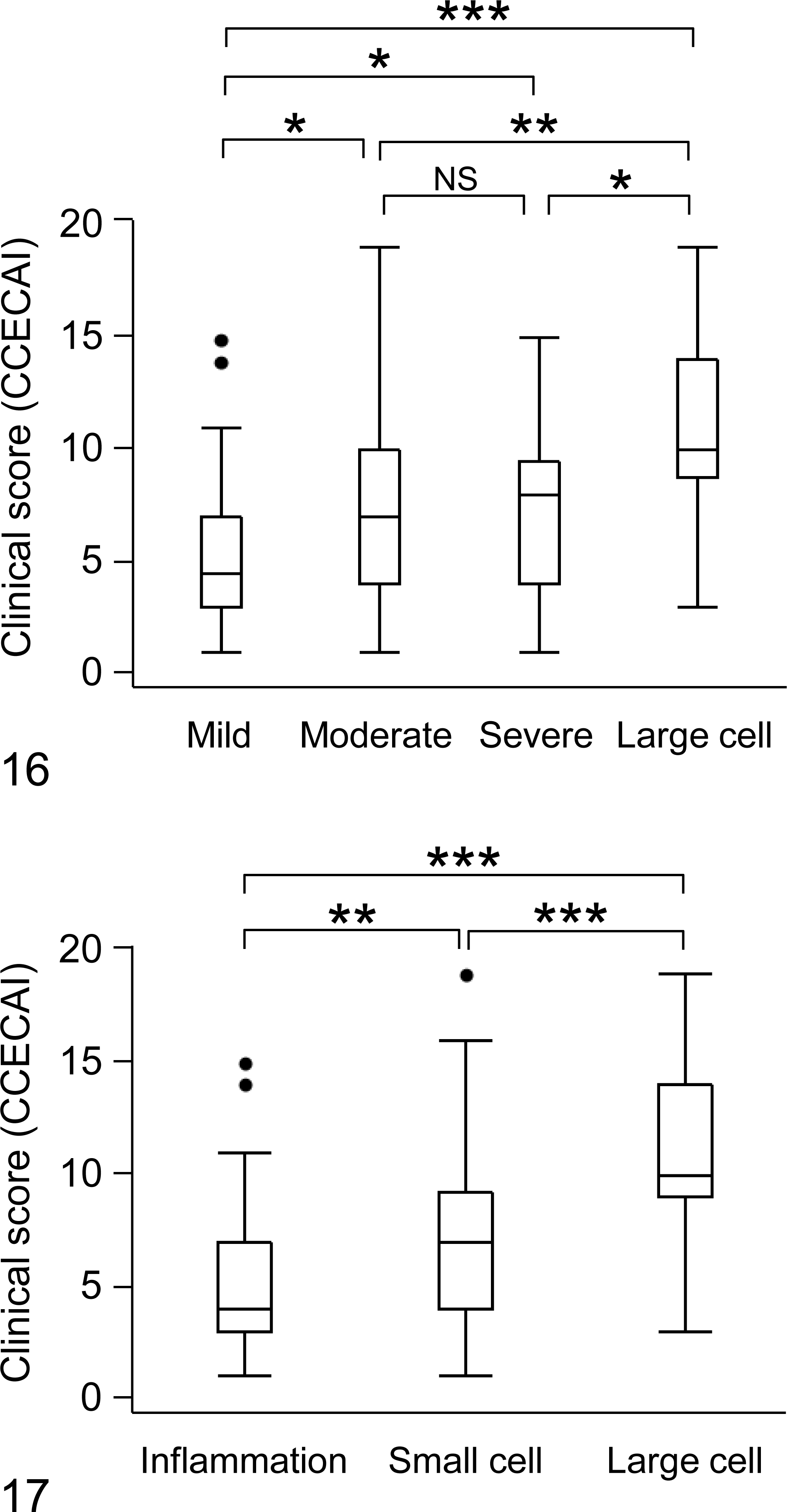
The clinical severity score (CCECAI) in each cytology grade is presented (mild, n = 73; moderate, n = 50; severe, n = 24; and large cell, n = 20). Box plots show the 25th percentile (lower edge of the box), median (solid line in the box), 75th percentile (upper edge of the box), and 90th percentile (whisker). The circles represent outliers. *P < .05, **P < .01, ***P < .001.
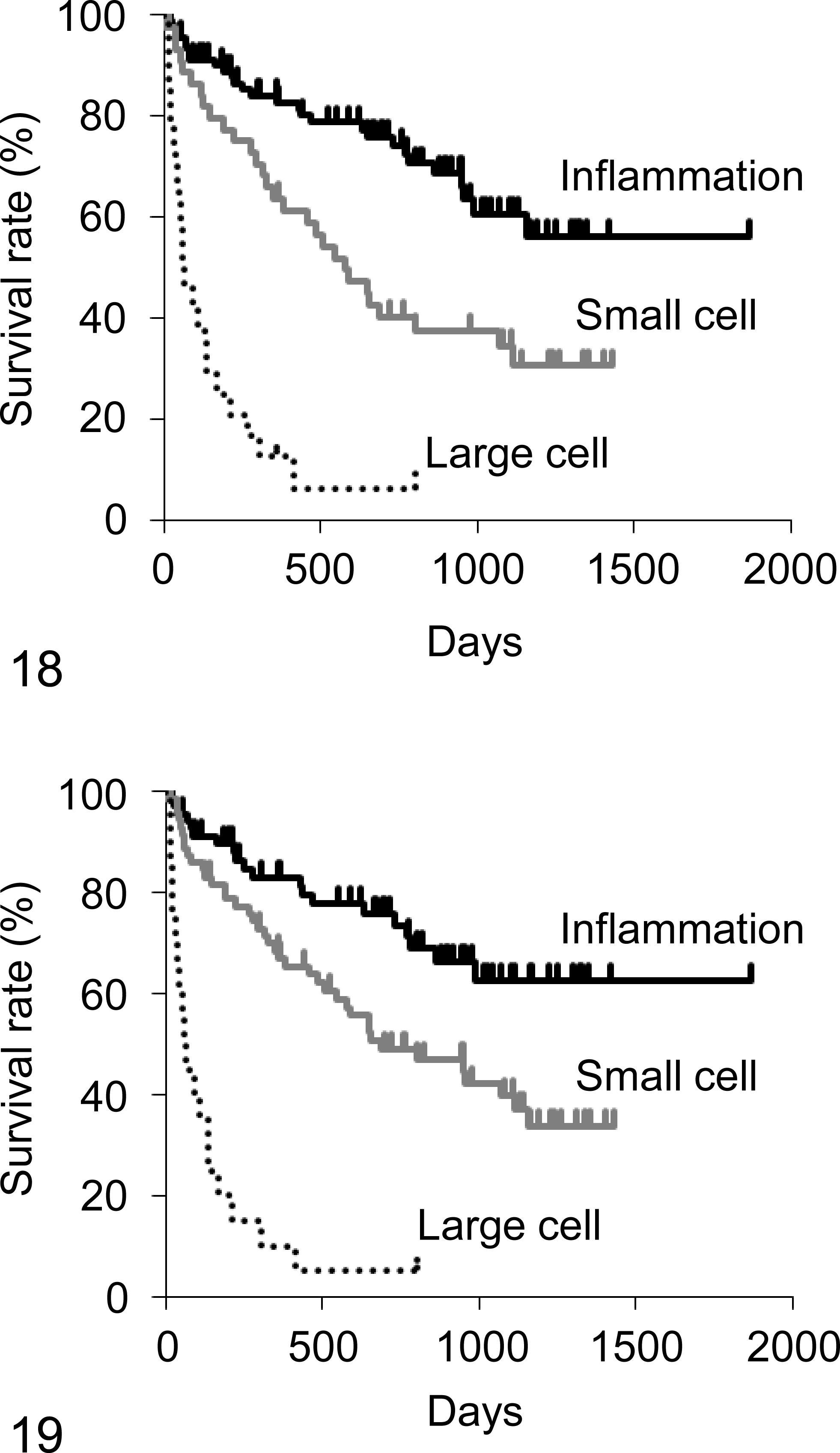
The Kaplan-Meier analysis of overall survival according to the histopathologic diagnosis. Histopathology was used to diagnose the cases of chronic enteritis (inflammation; n = 98), small cell intestinal lymphoma (small cell; n = 45), and large cell intestinal lymphoma (large cell; n = 24). Significant differences were found in the overall survival between cases of inflammation and small cell intestinal lymphoma (P = .0029), between cases of inflammation and large cell intestinal lymphoma (P < .0001), and between cases of small cell and large cell intestinal lymphoma (P = .0001).
Based on endoscopic cytology, LPE was diagnosed in 69 (41.3%) dogs; EE was diagnosed in 4 (2.4%) dogs; small cell intestinal lymphoma was diagnosed in 74 (44.3%) dogs; and large cell intestinal lymphoma was diagnosed in 20 (12.0%) dogs. The CCECAI was significantly higher in the order of large cell lymphoma, small cell lymphoma, and inflammation (Fig. 17).
Comparison of Cytologic and Histopathologic Diagnoses
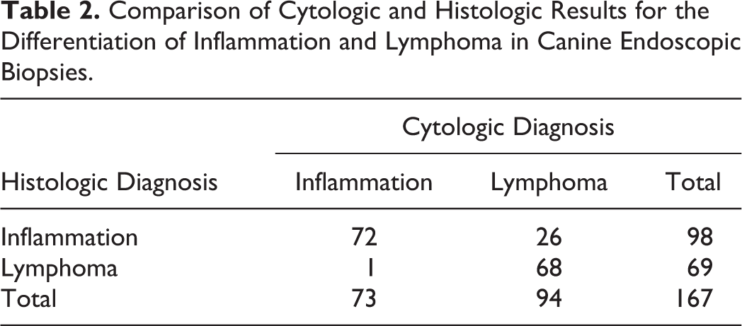
A summary of the cytologic and histopathologic diagnoses is shown in Table 1. Overall, the cytologic diagnoses were in complete agreement with the histopathologic diagnoses in 136 of 167 (81.4%) cases. The 2 × 2 agreement table, based on the classification of inflammation (LPE and EE) or lymphoma (small cell and large cell), is presented in Table 2. The sensitivity and specificity of endoscopic cytology for the diagnosis of lymphoma were 98.6% and 73.5%, respectively. False-positive results were obtained in 26 cases, and a false-negative result was obtained in 1 case. The positive and negative predictive values of endoscopic cytology were 72.3% and 98.6%, respectively.
Comparison of Cytologic and Histologic Results for the Differentiation of Inflammation and Lymphoma in Canine Endoscopic Biopsies.
Survival Analysis
The overall median follow-up period was 523 days (range, 1–1867 days). During the study period, 79 of 167 dogs died, cases of which 71 were considered to be due to the progression of intestinal disease. No dogs were euthanatized. Eighteen dogs were lost prior to the follow-up, and 70 dogs remained alive by the end of the study period.
The median overall survival of dogs with chronic enteritis, small cell intestinal lymphoma, and large cell intestinal lymphoma, according to histopathology, was 680 (range, 1–1867), 545 (range, 4–1433), and 63 (range, 13–800) days, respectively. The log-rank test showed that there were significant differences in the overall survival among the 3 histopathological groups (inflammation vs small cell lymphoma, P = .0029; inflammation vs large cell lymphoma, P < .0001; small cell lymphoma vs large cell lymphoma, P = .0001; Fig. 18). The median overall survival of dogs with chronic enteritis, small cell intestinal lymphoma, and large cell intestinal lymphoma, according to cytology, was 691 (range, 1–1867), 561 (range, 4–1433), and 63 (range, 13–800) days, respectively. In similarity with the histopathologic results, the log-rank test also showed significant differences in the overall survival among the 3 cytological groups (inflammation vs small cell lymphoma, P = .0178; inflammation vs large cell lymphoma, P < .0001; small cell lymphoma vs large cell lymphoma, P < .0001; Fig. 19).
Further analysis was performed to evaluate the correlations between overall survival and the 9 variables, including other cytologic findings (Table 3). Log-rank tests revealed that age (P = .0241), CCECAI (P < .0001), neutrophil infiltration (P = .0253), globule leukocyte infiltration (P = .0022), cytologic grading (P < .0001), cytologic diagnosis (P < .0001), and histopathologic diagnosis (P < .0001) were significantly associated with overall survival (Table 3). The Cox proportional hazard model was performed to screen variables with prognostic value. Among the candidate covariates, age, CCECAI, neutrophil infiltration, and globule leukocyte infiltration were included in the Cox regression model. The results of the Cox regression analysis are shown in Table 4. The variables that were individually associated with a shortened overall survival included CCECAI (hazard ratio, 2.0; 95% confidence interval, 1.2–3.2; P = .0073), a cytologic diagnosis of small cell intestinal lymphoma (hazard ratio, 1.8; 95% confidence interval, 1.0–3.1; P = .0437), and a cytologic diagnosis of large cell intestinal lymphoma (hazard ratio, 7.5; 95% confidence interval, 3.6–15.5; P < .0001).
Variables Associated With Shortened Survival Time of Dogs With Chronic Intestinal Diseases, Based on the Log-Rank Test.

Abbreviation: CCECAI, canine chronic enteropathy clinical activity index.
aMultiple testing was adjusted by Bonferroni correction.
Variables Associated With Shortened Survival Time of Dogs With Chronic Intestinal Diseases, Based on the Cox Proportional Hazard Model.
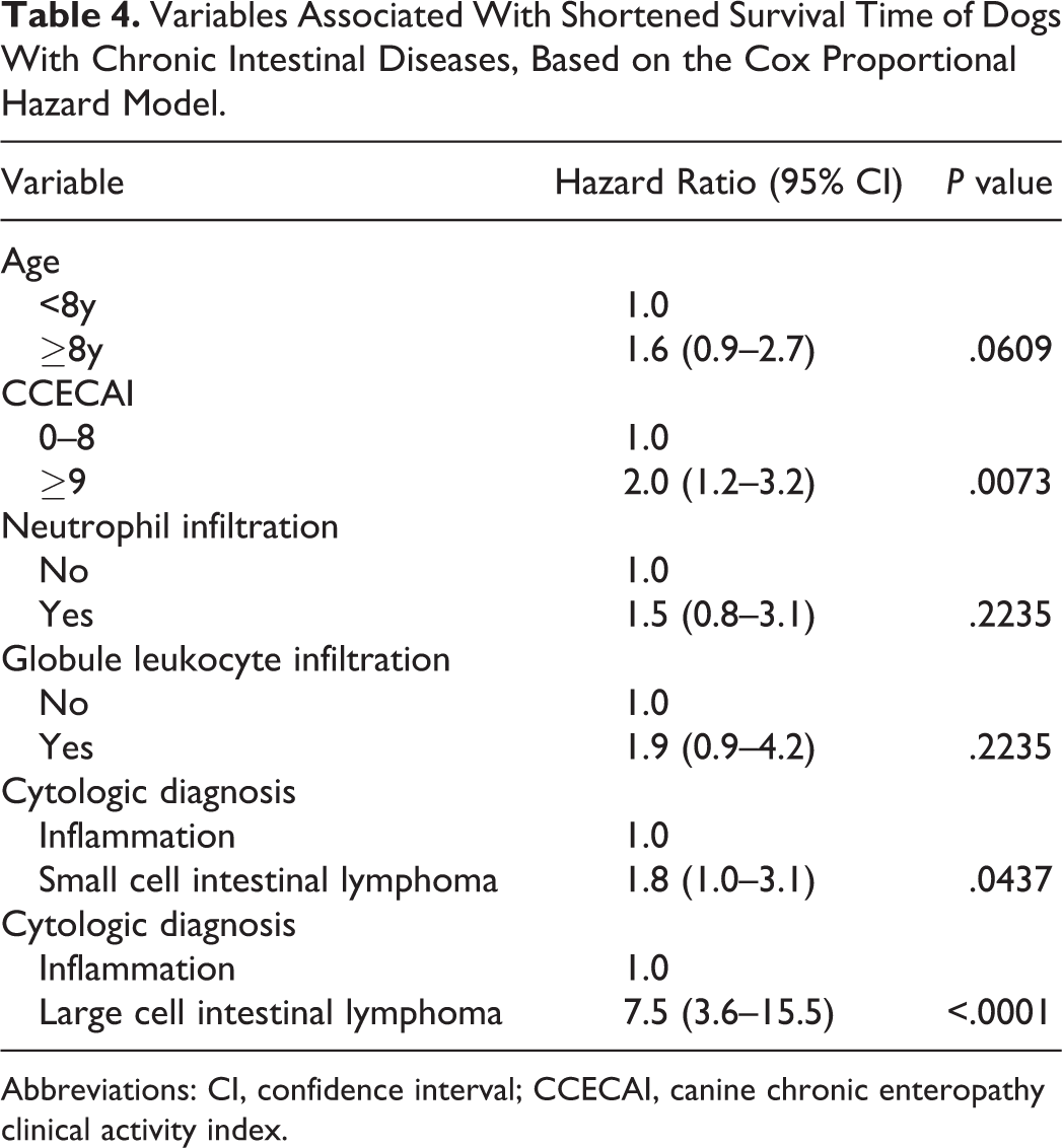
Abbreviations: CI, confidence interval; CCECAI, canine chronic enteropathy clinical activity index.
Discussion
This study showed that the findings of endoscopic cytology were concordant with histopathology, associated with disease severity, and predictive of prognosis for dogs with chronic enteritis and intestinal lymphoma. Although concurrent histopathologic evaluation is necessary, endoscopic cytology is a rapid and convenient diagnostic tool to aid differentiation of intestinal lymphoma from chronic enteritis, as the preparation of specimens is easy and the evaluation can be performed in the clinic.
Overall, the correlation between the cytologic and histopathologic results was good, which was shown as greater than 80% complete agreement for the determination of a specific diagnosis. Endoscopic cytology had a high (>98%) sensitivity for the diagnosis of intestinal lymphoma, whereas the specificity was relatively low (73%). Jergens et al 16 reviewed the endoscopic cytology of the small intestine in 122 dogs and 35 cats and reported results similar to ours (sensitivity, 92%; specificity, 80%). That study evaluated 11 categories (neutrophils, lymphocytes, plasma cells, eosinophils, macrophages, atypical cells, epithelial cells, bacterial flora, hemorrhage, debris/ingesta, and mucus), and the grades ranged from 0 to 7; 16 this may be too complicated to evaluate in clinical practice. Moreover, the study lacked data about the diagnostic value for small cell intestinal lymphoma. In the current study, we developed more simplified criteria for the differentiation between inflammation and intestinal lymphoma. Although the present and previous studies did not evaluate the consistency among the clinical pathologists, the prevalence of interobserver error may be reduced using our simpler criteria in comparison to that of the previous, more complicated system. 15,16 A large multicenter study is necessary to confirm the consistency between observers.
Small cell intestinal lymphoma was most commonly diagnosed by endoscopic cytology in this study. Of the 74 cases, 30 (40.5%) were in disagreement with the histopathological diagnoses. In many cases, we misdiagnosed LPE as small cell intestinal lymphoma. A possible explanation accounting for the prevalence of misdiagnoses is that the cases involving moderate lymphocyte infiltration are inclusive of both LPE and small cell lymphoma. The differentiation of LPE from small cell intestinal lymphoma in dogs and cats is significantly challenging, even with the use of histopathological examination. According to the cytologic criteria that we defined, dogs with moderate and severe lymphocyte infiltration were classified as cases of small cell intestinal lymphoma owing to the lack of significant difference in the CCECAI score between the 2 groups. The overall survival according to cytology and histopathology was significantly shorter in the following order: large cell lymphoma, small cell lymphoma, and chronic enteritis. Furthermore, the multivariate analysis revealed that the cytologic diagnosis of intestinal lymphoma was an individual, negative prognostic factor. Thus, we believe that the use of our cytologic grading criteria provided a valid diagnosis of intestinal lymphoma.
A previous study has demonstrated that a high CCECAI score was a risk factor for a negative outcome in dogs with chronic enteritis. 1 In the present study, dogs with a high CCECAI score exhibited a poor prognosis according to the log-rank test and Cox regression analysis. This indicated that CCECAI was also a prognostic factor for intestinal lymphoma. These findings suggest that an aggressive therapeutic intervention should be undertaken prior to the availability of the histopathologic results in dogs with a high CCECAI score and/or a cytologic diagnosis of intestinal lymphoma.
Of the 24 cases that were histopathologically diagnosed with large cell intestinal lymphoma, 4 (16.7%) were diagnosed by cytology as small cell lymphoma. These mismatches may be due to the presence of a multifocal distribution of neoplastic cells in intestinal lymphoma. 29 An accurate cytologic diagnosis is dependent on the mucosal site selected for biopsy and the number of tissue samples. For endoscopic cytology, it is essential to collect multiple mucosal biopsies from each site.
The presence of a significant number of neutrophils, or globule leukocytes, was observed by cytology in several cases. The log-rank test showed that the infiltration of neutrophils and globule leukocytes correlated with a reduced overall survival. However, the Cox regression analysis failed to reveal a significant correlation between this factor and the prognosis. The difference was most likely because the sample size was too small to detect a significant correlation in the multivariate analysis (neutrophils, 18 cases; globule leukocytes, 10 cases). More extensive studies will be necessary to determine whether there is an association between infiltration of these cells and prognosis.
This study has several limitations. First, not all information was available for each case due to the retrospective design of the study. Medications varied among dogs, which might have influenced the overall survival. Second, this study did not evaluate gastric and colonic specimens; the majority of the lesions in chronic enteritis and intestinal lymphoma were located in the small intestine. 28 The incidence of lymphocytes in the stomach and the colon was rare according to endoscopic cytology (data not shown). Third, the specimens were obtained by endoscopy in all cases; therefore, submucosal lesions were not examined in this study. Fourth, PARR analyses were not examined because this study aimed to compare the results of endoscopic cytology and histopathology. Recently, a stepwise diagnostic approach (histopathology followed by IHC and PARR) has been recommended for distinguishing chronic enteritis from intestinal lymphoma. 4,17 Further investigations are needed to evaluate the correlation among the above diagnostic methods and endoscopic cytology.
Generally, a major limitation of cytology is the sample quality. Inadequate cytologic specimens are often obtained, particularly during fine-needle aspiration, due to the insufficient number of intact cells, excessive contamination with blood, or smearing artifacts. 12,27 In this study, more than 99% of the specimens were adequate for cytologic evaluation. Only 1 slide was considered inadequate because of a failed Wright-Giemsa staining. In accordance with a previous report, 22 the squash-smear technique is an easy and reproducible method to obtain good quality samples in endoscopic cytology.
Conclusions
We established a simple cytologic grading system based on the degree of lymphocyte infiltration and size of infiltrating lymphocytes within the small intestines that correlated well with the histopathologic results. These cytologic grading criteria reflected the severity of the disease and, thereby, the prognosis of dogs with chronic enteritis and intestinal lymphoma. Our findings suggest that endoscopic cytology is useful as an adjunct to the diagnosis of canine chronic intestinal diseases.
Footnotes
Declaration of Conflicting Interests
The author(s) declared no potential conflicts of interest with respect to the research, authorship, and/or publication of this article.
Funding
The author(s) received no financial support for the research, authorship, and/or publication of this article.
References
Supplementary Material
Please find the following supplemental material available below.
For Open Access articles published under a Creative Commons License, all supplemental material carries the same license as the article it is associated with.
For non-Open Access articles published, all supplemental material carries a non-exclusive license, and permission requests for re-use of supplemental material or any part of supplemental material shall be sent directly to the copyright owner as specified in the copyright notice associated with the article.