
Abstract
Clouded leopards in North American zoological institutions have a high frequency of pheochromocytomas and were identified in 32 of 70 (45%) animals necropsied. Archival sections of adrenal gland from 20 adult clouded leopards with unilateral or bilateral pheochromocytomas collected between 1984 and 2011 were examined by light microscopy and immunohistochemistry, and case demographics were reviewed. Affected leopards were older than 10 years of age (mean, 16 years; range, 11–19 years), and males were overrepresented (12 males, 8 females). Pedigree analysis yielded no evidence for heritability. Five clouded leopards had bilateral neoplasms. Pheochromocytoma was the cause of death due to invasion of the caudal vena cava and fatal hemorrhage in 4 cases. Most pheochromocytomas were well-demarcated, nodular, and expansile masses composed of cords and packets of neoplastic polygonal cells. Five pheochromocytomas had vascular invasion, of which 4 resulted in hemorrhage that was the cause of death. One of the latter pheochromocytomas also had pulmonary metastasis. Ultrastructurally, neoplastic cells had cytoplasmic structures consistent with both norepinephrine- and epinephrine-containing granules. In all cases, neoplasms were immunohistochemically positive for chromogranin A, protein gene product 9.5, and synaptophysin. A subset of neoplasms evaluated by tissue microarray were positive for met-enkephalin and β-endorphin and negative for melan-A. Histologically, 7 of 20 (35%) clouded leopards with pheochromocytomas had retinal detachment, retinal degeneration, or intramyocardial muscular arteriosclerosis, suggestive of hypertension. Pheochromocytomas can cause mortality and may be a source of clinically significant hypertension in clouded leopards. These neoplasms share similar histologic, immunohistochemical, and ultrastructural characteristics with those of other species.
Clouded leopards (Neofelis nebulosa) are a medium-sized endangered felid from Southeast Asia and are protected internationally by Appendix I of the Convention on International Trade in Endangered Species. 35 In nondomestic felids, pheochromocytomas are uncommon and previously have been described in 1 clouded leopard and 1 jaguar at zoological institutions in Thailand and the United States, respectively. 4,24 In contrast, we have found that pheochromocytomas are common in clouded leopards from North American zoological institutions. A survey of adrenal neoplasms was undertaken by analyzing necropsy records of clouded leopards from North American zoological institutions. In this retrospective study, pheochromocytoma was the only adrenal neoplasm reported and was found in 32 of 70 (45%) clouded leopards.
Pheochromocytomas are neoplasms arising from the catecholamine-producing, neural crest–derived chromaffin cells of the adrenal medulla. 22 Similar neoplasms can also arise from extramedullary chromaffin cells and are referred to as paragangliomas. 1 Pheochromocytomas are uncommon in domestic felids, and little is known about their pathogenesis. 12 In animals, pheochromocytomas are commonly reported in adult ruminants, canids, and New World primates. 10,14,21 Pheochromocytomas are rare in other wildlife species, with few individual reports in the current literature. 6,9,30,32 Pheochromocytomas are also rare in humans, and the most common clinical presentation is secondary hypertension due to catecholamine secretion. 22 Domestic dogs and cats can also present in hypertensive crisis due to catecholamine-secreting pheochromocytomas, often resulting in lesions within the retina, brain, coronary vessels, and heart. 8,10,12,28
In humans, most pheochromocytomas are of sporadic occurrence, though a small percentage of these neoplasms are due to inherited germline mutations. 22 Multiple familial syndromes have been described, including multiple endocrine neoplasia (MEN) type 2A and MEN-2B, neurofibromatosis, von Hippel-Lindau syndrome and the succinate dehydrogenase gene family syndromes. 22 Multiple endocrine neoplasia-like syndromes have also been reported in animals, commonly in cattle, which develop thyroid C cell neoplasms along with pheochromocytomas. 36 Multiple endocrine neoplasia-like syndromes have also been reported in other domestic species, such as dogs, cats, ferrets, and horses. 7,11,17,27
In this study, we undertook validation of a series of antibodies that may be useful in demonstrating the adrenal medulla as the origin of neoplasms in the clouded leopard. Antibodies used in both humans and animals include chromogranin A, synaptophysin, PGP 9.5, inhibin, met-enkephalin, and β-endorphin, which are expressed in medullary epithelial cells, and melan-A, which is expressed in cortical epithelial cells. 15,16,20,22 In addition, p53, B-cell lymphoma 2 (Bcl-2), and Ki-67 expression, among others, have been evaluated to elucidate the mechanisms of carcinogenesis and to predict malignant behavior in humans but with variable results. 5,13,18,23,31,33 Expression patterns of these markers in clouded leopard pheochromocytomas have not yet been evaluated. Because of the apparent high prevalence and potential impact on medical management of clouded leopards, this study was undertaken to describe and characterize pheochromocytomas in this endangered felid via retrospective clinical history analysis, documentation of possible secondary hypertensive lesions, pedigree analysis, histology, immunohistochemistry, and electron microscopy.
Materials and Methods
Necropsy records from the Association of Zoos and Aquariums clouded leopard Species Survival Plan (SSP) were evaluated for the presence of adrenal neoplasia. Pheochromocytoma was the only adrenal neoplasm recorded. To confirm the diagnosis of these neoplasms, a subset of 20 cases was chosen based on availability of formalin-fixed, paraffin-embedded (FFPE) tissues from both adrenal glands. In cases with unilateral neoplasms, normal adrenal glands from the same animals were also evaluated. Animals had been housed in 5 of 23 North American zoological institutions represented in the clouded leopard pathology database. Available clinical histories, gross necropsy reports, and histopathology reports were reviewed. FFPE tissues were sectioned at 5 μm and stained with hematoxylin and eosin (HE). All neoplasms were examined histologically by 2 pathologists (S.C. and K.T.), the diagnosis of pheochromocytoma was confirmed, and all were evaluated for criteria of malignancy based on established parameters in the veterinary and human literature, including the World Health Organization (WHO) classification of tumors of the endocrine system of domestic animals. 16,20 Other organs available for examination in each case were also evaluated.
Pedigree analysis was performed using the Single Population Analysis and Records Keeping System (SPARKS) software from the International Species Information System (ISIS). The pedigree was evaluated subjectively for evidence of heritability.
Detailed immunohistochemical methods and information for each antibody are provided in Supplemental Methods and Supplemental Table S1. For PGP 9.5, chromogranin A, and synaptophysin, immunolabeling was evaluated in sections of domestic cat and clouded leopard adrenal gland and brain as well as additional clouded leopard tissues (heart, kidney, thyroid, intestine, stomach, skeletal muscle, lung, liver, lymph node, and spleen) to confirm site specificity and reactivity in this species. Negative controls for each antibody were prepared by replacing the primary antibody with phosphate-buffered saline (PBS).
A tissue microarray (TMA) was created from a subset of 7 clouded leopards (case Nos. 4, 5, 7, 14, 16, 17, and 18) based on block availability (Manual Tissue Arrayer, MTA1; Beecher Instruments, Sun Prarie, WI). Included in the TMA were canine and feline neoplasms with known expression levels and clouded leopard tissues, including the pheochromocytomas, normal adrenal glands, and control tissues as listed above. For each tissue, 5 cores were cut at a 0.6-mm diameter and placed into a recipient block. Histologic sections of the TMA were probed with 6 additional antibodies against β-endorphin, met-enkephalin, melan-A, p53, Bcl-2, and Ki-67. Negative controls for each antibody were prepared by replacing the primary antibody with PBS.
Immunoreactivity for all antibodies was assigned a grade of 0, 1, 2, or 3 based on the percentage of positive neoplastic cells within five 100× fields (0 = negative, 1 = <10% positive, 2 = 10%–50% positive, 3 = >50% positive). Pathologists were blinded to other case details when assigning a grade.
Transmission electron microscopy was used to further evaluate one of the pheochromocytomas (case No. 8). Formalin-fixed tissue was postfixed in 2% osmium tetroxide, prepared for electron microscopy, and examined with a Hitachi H600 transmission electron microscope (Hitachi America, Brisbane, CA).
Results
Case demographics and histologic lesions are summarized in Supplemental Table S2. Affected clouded leopards ranged from 11 to 19 years of age (mean, 16 years) and included 12 males and 8 females. Bilateral neoplasms were present in 5 of 20 animals (25%), of which 4 were female (80%). Most neoplasms were discovered as incidental findings at the time of necropsy. However, pheochromocytoma was the cause of death in 4 of 20 cases (20%) due to local invasion of the caudal vena cava and secondary fatal abdominal hemorrhage. One of these 4 fatal cases also had pulmonary metastasis. The remaining animals in the study died or were euthanized due to multiple causes, which in the majority of animals included degenerative musculoskeletal disease, chronic renal disease, or concurrent neoplasia (Suppl. Table S2).
Grossly, in most cases, the adrenal medulla was expanded by a well-demarcated, dark red, slightly soft, round neoplasm that compressed or partially replaced the adjacent cortex. The 4 malignant neoplasms that invaded the caudal vena cava were poorly demarcated, friable, necrotic, dark red, and hemorrhagic.
Histologically, most pheochromocytomas were small, well-demarcated, expansile, nonencapsulated masses that compressed or invaded the adjacent cortex (Fig. 1). Neoplasms were densely cellular, and cells were arranged in cords and packets within a fine reticular, well-vascularized stroma. Cells were polygonal or rarely spindloid, with an amphophilic to basophilic, granular cytoplasm. Anisocytosis and anisokaryosis were mild, with few mitoses. In 1 neoplasm that invaded the vena cava, neoplastic cells had marked cellular and nuclear pleomorphism, marked cytomegaly and karyomegaly, and numerous bizarre nuclei (Fig. 2). Histologic criteria for malignancy were present in 11 of 20 neoplasms (55%), including capsular and/or vascular invasion. Pheochromocytomas that invaded the vena cava also caused destruction of adrenal gland architecture, and immunohistochemistry was required to determine adrenal medullary origin. Pheochromocytomas from 2 cases also had admixed neoplastic cells from a second malignant neoplasm, lymphoma (case No. 8), and intestinal carcinoma (case No. 5).
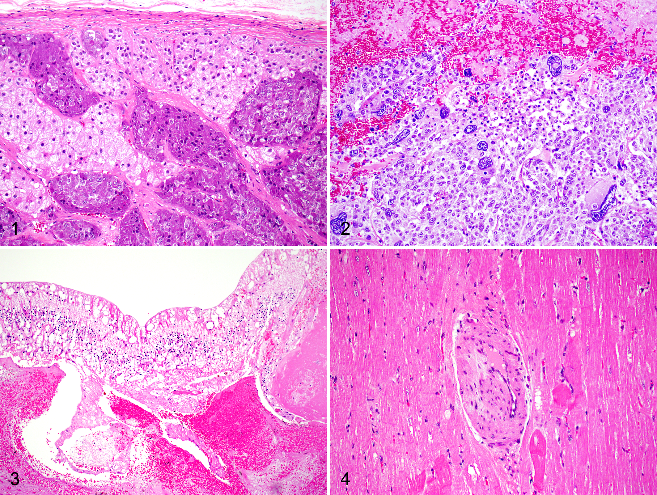
Histologic lesions of the eye or intramyocardial muscular arteries were present in 7 of 20 (35%) cases, which may be indicative of secondary hypertension (Figs. 3, 4). In 4 of 20 (20%) animals (case Nos. 7, 14, 16, and 18), ocular lesions consisted of partial or complete retinal detachment as well as retinal degeneration or atrophy. In the most severe case (case No. 18), histologic lesions included diffuse retinal degeneration, detachment, and hemorrhage, with loss of the retinal pigmented epithelium, photoreceptor layer, inner and outer nuclear and plexiform layers, and ganglion cell vacuolation. Retinal and choroidal vascular thrombosis was also present multifocally. Vascular medial hypertrophy or sclerosis was not observed in retinal or choroidal blood vessels. Three additional animals (case Nos. 4, 5, and 17) had arteriosclerotic changes within the intramyocardial muscular arteries. In these cases, multiple muscular arteries had a concentrically thickened tunica media with smooth muscle hyperplasia, characterized by increased numbers of haphazardly arranged and vacuolated smooth myocytes and an attenuated vascular lumen. Renal blood vessels in cases where kidney was available for evaluation were histologically normal.
Immunohistochemical labeling for chromogranin A, synaptophysin, and PGP 9.5 was positive in all clouded leopard pheochromocytomas (Table 1) and normal adrenal medulla and negative in the adrenal cortex. There was no unexpected labeling of the control clouded leopard tissues, confirming site-specific binding in this species. Positive cytoplasmic immunolabeling with chromogranin A was detected in all neoplasms (Fig. 5), with an immunoreactivity score of 3 in 19 of 20 (95%) cases. Positive synaptophysin immunolabeling was detected in all neoplasms (Fig. 6), although less than 10% of the neoplastic cells stained positive for synaptophysin in 7 of 20 (35%) cases. PGP 9.5 immunolabeling was detected in all neoplasms (Fig. 7), with strong cytoplasmic and some nuclear labeling. Most neoplasms had a PGP 9.5 immunoreactivity score of 3, while 4 of 20 (20%) had a score of 2.
Immunoreactivy Scores for Chromogranin A, Synaptophysin, and PGP 9.5 in 20 Pheochromocytomas From Clouded Leopards (Neofelis nebulosa).a
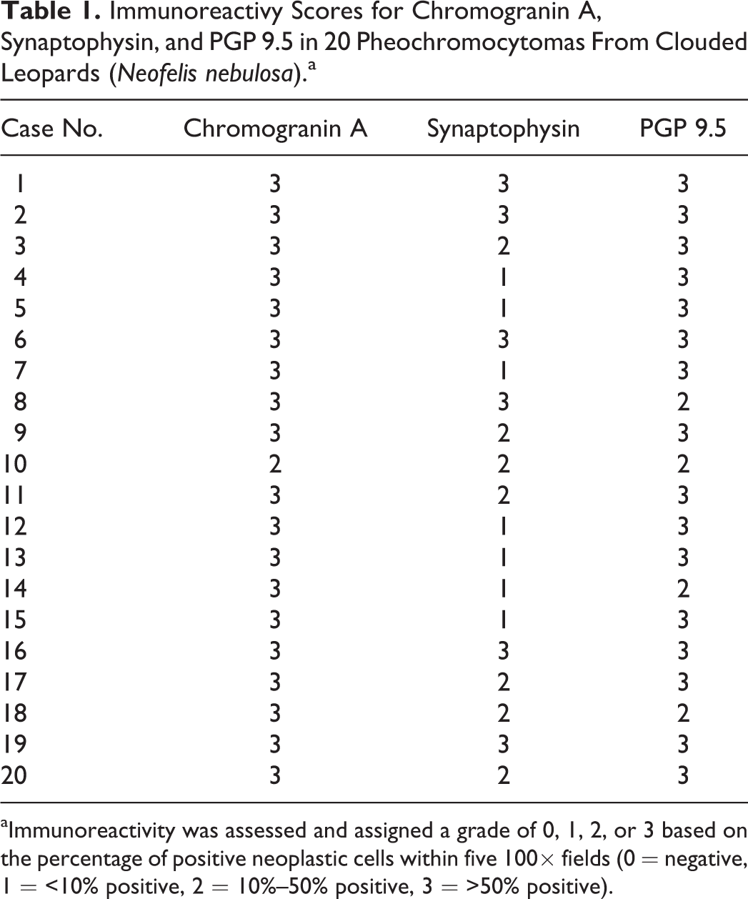
aImmunoreactivity was assessed and assigned a grade of 0, 1, 2, or 3 based on the percentage of positive neoplastic cells within five 100× fields (0 = negative, 1 = <10% positive, 2 = 10%–50% positive, 3 = >50% positive).
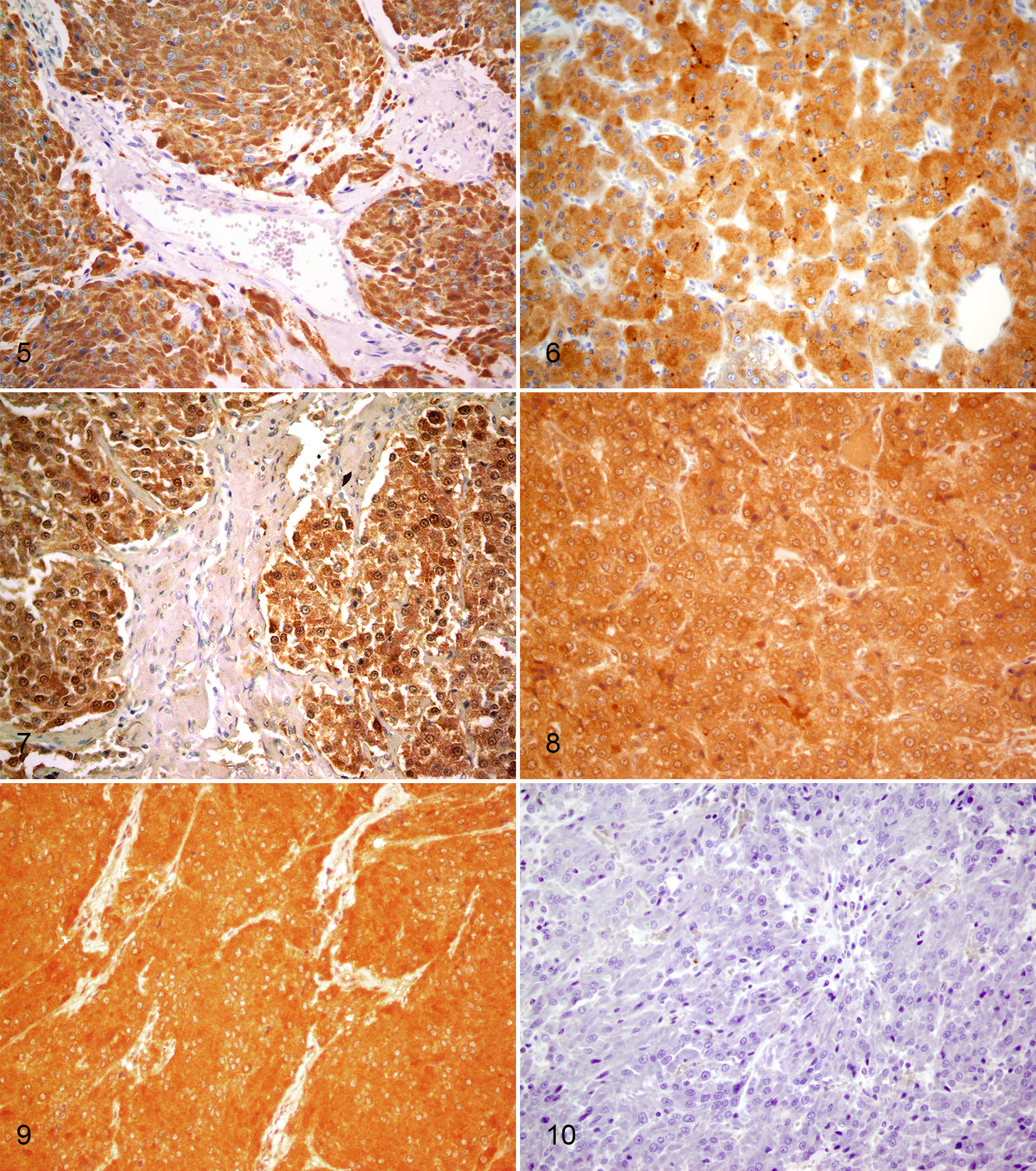
Histologic sections of the TMA were immunolabeled for β-endorphin, met-enkephalin, melan-A, p53, Bcl-2, and Ki-67 (Suppl. Table S3). There was no unexpected labeling of clouded leopard control tissues with antibodies to β-endorphin, met-enkephalin, or melan-A, confirming site-specific binding in this species. Positive cytoplasmic β-endorphin immunolabeling was detected in all 7 neoplasms tested by microarray (Fig. 8). All neoplasms had an immunoreactivity score of 3. Positive met-enkephalin immunolabeling was detected in all 7 neoplasms with cytoplasmic immunoreactivity (Fig. 9). Normal adrenal medullary chromaffin cells were also positive for β-endorphin and met-enkephalin. All 7 neoplasms had a complete absence of immunolabeling for melan-A, with an immunoreactivity score of 0 (Fig. 10), while the adrenal cortex of normal and neoplastic adrenal glands had positive cytoplasmic immunolabeling.
All neoplasms and normal adrenal glands had an absence of labeling for p53, Bcl-2, and Ki-67, with an immunoreactivity score of 0. All control clouded leopard tissues were negative for p53, in contrast to the positive control, a domestic feline fibrosarcoma with known p53 labeling. A clouded leopard intestinal carcinoma was immunolabeled, but there was also an absence of staining, and therefore we did not have a positive control for p53 in the clouded leopard. A clouded leopard lymph node had positive perinuclear Bcl-2 immunolabeling of scattered cortical and medullary lymphocytes. Several lymphocytes within a clouded leopard lymph node also had positive nuclear Ki-67 immunolabeling, consistent with immunoreactivity in this species, although the pheochromocytomas had no positive labeling for Ki-67.
Transmission electron microscopy was performed on case No. 8 (Figure 11). Neoplastic cells had numerous cytoplasmic, membrane-bound secretory granules consistent with both norepinephrine- and epinephrine-containing granules. Norepinephrine-containing granules had an eccentric, electron-dense core, with a prominent space between it and the surrounding membrane. Epinephrine-containing granules had a coarsely granular core with close apposition to the surrounding membrane. Both norepinephrine- and epinephrine-containing granules were approximately 150 to 250 nm in diameter, which is consistent with the granule diameter of both normal and neoplastic chromaffin cells. 3,22
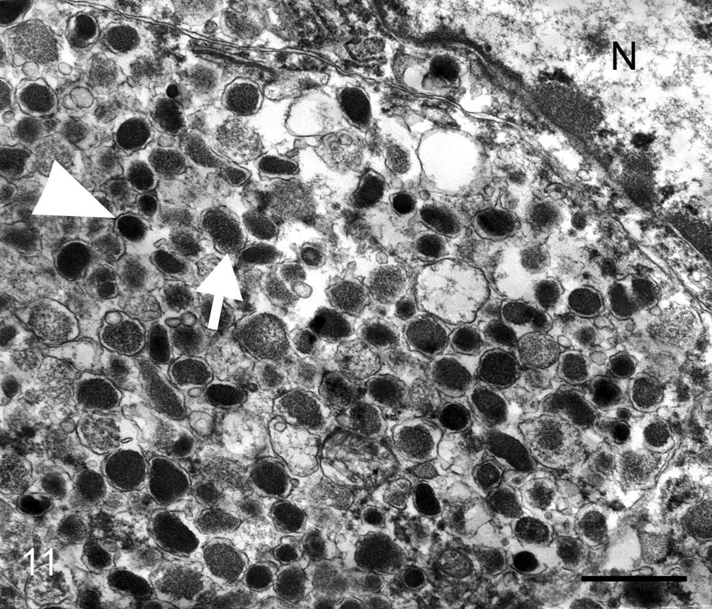
Pheochromocytoma, adrenal gland, clouded leopard. Neoplastic cells have both norepinephrine (arrowhead) and epinephrine (arrow) intracytoplasmic secretory granules. N, nucleus. Transmission electron microscopy. Bar = 0.5 μm.
Pedigree analysis was performed comparing the 20 animals in this study with the entire North American clouded leopard studbook. Clouded leopards with pheochromocytoma were scattered among 10 generations with no evident pattern of inheritance.
Discussion
Pheochromocytomas of the adrenal gland are common in captive clouded leopards, affecting one or both adrenal glands of primarily aged (>10 years) animals. There was no pattern of inheritance based on the pedigree analysis; however, the high prevalence within this population suggests that an inherited genetic component may play a role in the carcinogenesis of these neoplasms. There was no gross or histologic evidence of an inherited multiple endocrine neoplasia syndrome in this species.
In this study, most neoplasms were considered incidental findings, although many had histologically malignant characteristics. Differentiation between benign and malignant neoplasms based on histologic characteristics alone is difficult in both humans and animals, and a definite diagnosis of malignant behavior is often based on evidence of metastasis or vascular invasion. 16,20 In this subset, 4 of 20 (20%) animals had pheochromocytomas that invaded the caudal vena cava with subsequent fatal hemorrhage. Due to invasiveness and destruction of adrenal gland architecture, immunohistochemistry was required to determine that neoplastic cells arose from the adrenal medulla in these cases. Histologic criteria of malignancy were present in 11 of 20 (55%) cases, including capsular and/or vascular invasion. Of the bilateral neoplasms, 3 of 5 (60%) lacked malignant characteristics, and these cases did not have additional cardiac or retinal lesions. The presence of bilateral neoplasms does not appear to predict malignant behavior or lesions of secondary hypertension.
Antemortem diagnosis of hypertension in nondomestic felids is hindered by an inability to monitor blood pressure without anesthesia in many cases. None of the clouded leopards in this study had a documented history of hypertension, to our knowledge, and catecholamine levels were not assessed in this retrospective study. In humans, approximately 90% of pheochromocytomas are associated with secondary hypertension, often manifesting clinically as paroxysmal episodes. 1 Arteriosclerosis is observed in both humans and animals with hypertension but can also be common and clinically insignificant in aged domestic animals. 29 Arteriosclerosis has been observed in other animals with concurrent pheochromocytomas, such as nonhuman primates and dogs. 10,14,21 In clouded leopards, cardiac lesions were limited to the intramyocardial muscular arteries. No additional lesions were observed in the myocardium. Retinal lesions were consistent with those found in hypertensive retinopathy, such as hemorrhage, detachment, degeneration, and vascular thrombosis, although vascular medial hypertrophy or sclerosis was not observed in the clouded leopards. 34 Renal blood vessels were within normal limits in those cases with kidney available for histologic evaluation. The clinical significance of the retinal and arteriosclerotic lesions and their relationship to pheochromocytoma-induced hypertension in these clouded leopards is unknown. Of those cases with secondary lesions, 6 of 7 (86%) of the neoplasms were classified as malignant based on histologic characteristics.
Immunohistochemical techniques used to identify pheochromocytomas in domestic species were found to be applicable to the clouded leopard. Immunoreactivity to chromogranin A, PGP 9.5, β-endorphin, met-enkephalin, and, to a lesser extent, synaptophysin, in the absence of immunoreactivity to melan-A, can be used to help confirm a diagnosis of pheochromocytoma in clouded leopards. Inhibin, another marker for adrenal cortex, is highly species specific in our experience and was excluded from this evaluation.
Chromogranin A and synaptophysin are commonly used to diagnose pheochromocytomas in both humans and animals. 1,16 However, both of these proteins are present in secretory vesicles of neurons as well as a variety of neuroendocrine cells and are not specific for adrenal medullary neoplasms. 20,22 In this study, pheochromocytomas had consistent, strong immunoreactivity for chromogranin A but variable labeling for synaptophysin. This variable labeling may have been due to differences in epitope masking from fixation or due to variable expression. Of those neoplasms with a synaptophysin immunoreactivity score of 1, 6 of 7 (86%) were characterized as malignant. Synaptophysin may not be a useful marker in clouded leopard pheochromocytomas that have malignant histologic characteristics.
PGP 9.5 is a member of the ubiquitin hydrolase family of proteins that is expressed in neurons and neuroendocrine cells, as well as a number of other cells and various neoplasms in both animals and humans, and is not considered a specific marker for neuroendocrine tissues. 2,25,26 In clouded leopards, PGP 9.5 immunolabeling was present within neurons and neuroendocrine cells, including the adrenal medulla, but was not present in adrenal cortical cells and could be used to differentiate among cortical and medullary neoplasms in the clouded leopard tissues. β-Endorphin is an endogenous opioid peptide neurotransmitter present in neurons of both the central and peripheral nervous systems as well as chromaffin cells. 22 Met-enkephalin is also an endogenous opioid peptide that is co-released with catecholamines in the adrenal medulla. 22 β-Endorphin and met-enkephalin had strong immunolabeling of the clouded leopard adrenal medulla and pheochromocytomas but not the adrenal cortex. As expected, no pheochromocytomas had immunoreactivity for melan-A. Melan-A is encoded by the MART-1 gene in humans and is expressed in melanocytes and steroid hormone-producing cells, including adrenal cortical epithelial cells, and can be used to differentiate cortical from medullary adrenal neoplasms. 37
Bcl-2 is an antiapoptotic protein that inhibits steps in the intrinsic pathway of apoptosis. 19 In 1 study, Bcl-2 was significantly increased in malignant compared with benign pheochromocytomas of humans and might therefore be used as a prognostic indicator, yet other studies had conflicting results. 5,33,37 Bcl-2 has weakly positive immunolabeling of the normal adrenal cortex in the domestic cat but has not been evaluated in neoplastic adrenal glands of felids, to our knowledge. 19 Bcl-2 immunolabeling was absent in all clouded leopard pheochromocytomas examined by TMA. Control sections of normal lymph node from a clouded leopard had positive perinuclear immunolabeling of many cortical and medullary lymphocytes, consistent with antibody cross-reactivity with Bcl-2 of clouded leopards. Thus, the lack of immunolabeling in pheochromocytomas in this study suggests that Bcl-2 may not play a role in the pathogenesis of pheochromocytomas in clouded leopards.
Ki-67 is a marker for cell proliferation that is expressed in the nucleus between the G1 and S phase of the cell cycle. 5 As for Bcl-2, the antibody used appeared to cross-react with Ki-67 of clouded leopards, but the lack of immunolabeling of the neoplastic cells again suggests that Ki-67 is not a useful marker to predict malignant behavior in clouded leopard pheochromocytomas. Mitoses were observed histologically in some neoplasms, and Ki-67 immunolabeling would be expected in these regions. Therefore, it is unclear why there was labeling in the clouded leopard lymph node and not in the neoplasms with histologic evidence of proliferation. We must take into account that Ki-67 was examined on a tissue microarray, which is representative of some but not all of the tissue in section, and not all cases were available for evaluation of Ki-67.
None of the clouded leopard pheochromocytomas, intestinal carcinoma, normal adrenal glands, or other control tissues had immunolabeling for p53. It is possible that the p53 antibody used in this study may not cross-react with p53 of clouded leopards.
Adrenal pheochromocytomas are common necropsy findings in clouded leopards and a potential cause of sudden death in aged males and females of this species in North American zoological institutions. Because these neoplasms are a potential cause of death from caval invasion or metastasis, screening of adrenal glands in captive clouded leopards via ultrasonography during routine physical examinations may be warranted. These neoplasms are histologically similar to pheochromocytomas in humans and other species. Pheochromocytomas may be associated with secondary hypertension in this species, based on histologic evidence of retinal detachment and degeneration and of intramyocardial muscular arteriosclerosis in some cases. In 1 pheochromocytoma evaluated by electron microscopy, intracytoplasmic norepinephrine- and epinephrine-containing granules were present within neoplastic cells. Immunohistochemical markers, including chromogranin A, synaptophysin, PGP 9.5, met-enkephalin, β-endorphin, and melan-A, were validated for use in clouded leopards and can be used to differentiate adrenal medullary from cortical neoplasms. As in other species, a diagnosis of malignant or benign pheochromocytoma based on histology alone is difficult unless metastases or vascular invasion are present. Based on preliminary immunohistochemical results, Ki-67 and Bcl-2 do not appear to play a role in the pathogenesis of pheochromocytomas in clouded leopards. The reason for the high prevalence of pheochromocytomas in captive clouded leopards in North America remains unknown. The apparent high prevalence in the North American population may be due to consistent necropsy and disease surveillance, which are not routinely performed in free-ranging clouded leopards.
Footnotes
Acknowledgements
We thank Bonnie Breitbeil and Laurie Bingaman Lackey for their help with pedigree analysis and all participating clouded leopard SSP institutions. We also thank the histology laboratories at University of Illinois Veterinary Diagnostic Laboratory and Michigan State University Diagnostic Center for Population and Animal Health. Finally, we thank the following zoological institutions: Chicago Zoological Society’s Brookfield Zoo, Brookfield, IL; Cincinnati Zoo & Botanical Garden, Cincinnati, OH; Memphis Zoo, Memphis, TN; Omaha’s Henry Doorly Zoo & Aquarium, Omaha, NE; Smithsonian National Zoological Park, Washington, DC; and Staten Island Zoo, Staten Island, NY.
Declaration of Conflicting Interests
The author(s) declared no potential conflicts of interest with respect to the research, authorship, and/or publication of this article.
Funding
The author(s) received no financial support for the research, authorship, and/or publication of this article.
References
Supplementary Material
Please find the following supplemental material available below.
For Open Access articles published under a Creative Commons License, all supplemental material carries the same license as the article it is associated with.
For non-Open Access articles published, all supplemental material carries a non-exclusive license, and permission requests for re-use of supplemental material or any part of supplemental material shall be sent directly to the copyright owner as specified in the copyright notice associated with the article.