
Abstract
Forensic entomology can be useful to the veterinary professional in cases of animal cruelty. A main application of forensic entomology is to determine the minimum postmortem interval by estimating the time of insect colonization, based on knowledge of the rate of development of pioneer colonizers and on insect species succession during decomposition of animal remains. Since insect development is temperature dependent, these estimates require documentation of the environmental conditions, including ambient temperature. It can also aid in the detection and recognition of wounds, as well as estimate the timing of periods of neglect. Knowledge of the geographic distribution of insects that colonize animal remains may suggest that there has been movement or concealment of the carcass or can create associations between a suspect, a victim, and a crime scene. In some instances, it can aid in the detection of drugs or toxins within decomposed or skeletonized remains. During animal cruelty investigations, it may become the responsibility of the veterinary professional to document and collect entomological evidence from live animals or during the necropsy. The applications of forensic entomology are discussed. A protocol is described for documenting and collecting entomological evidence at the scene and during the necropsy, with additional emphasis on recording geographic location, meteorological data, and collection and preservation of insect specimens.
Forensic entomology, or the application of arthropod science to legal matters, has steadily increased its prominence within the forensic sciences. 5,28,41,46,77 While this definition allows for a broad interpretation of the scope of this science, forensic entomology is most commonly associated with use in death investigations.
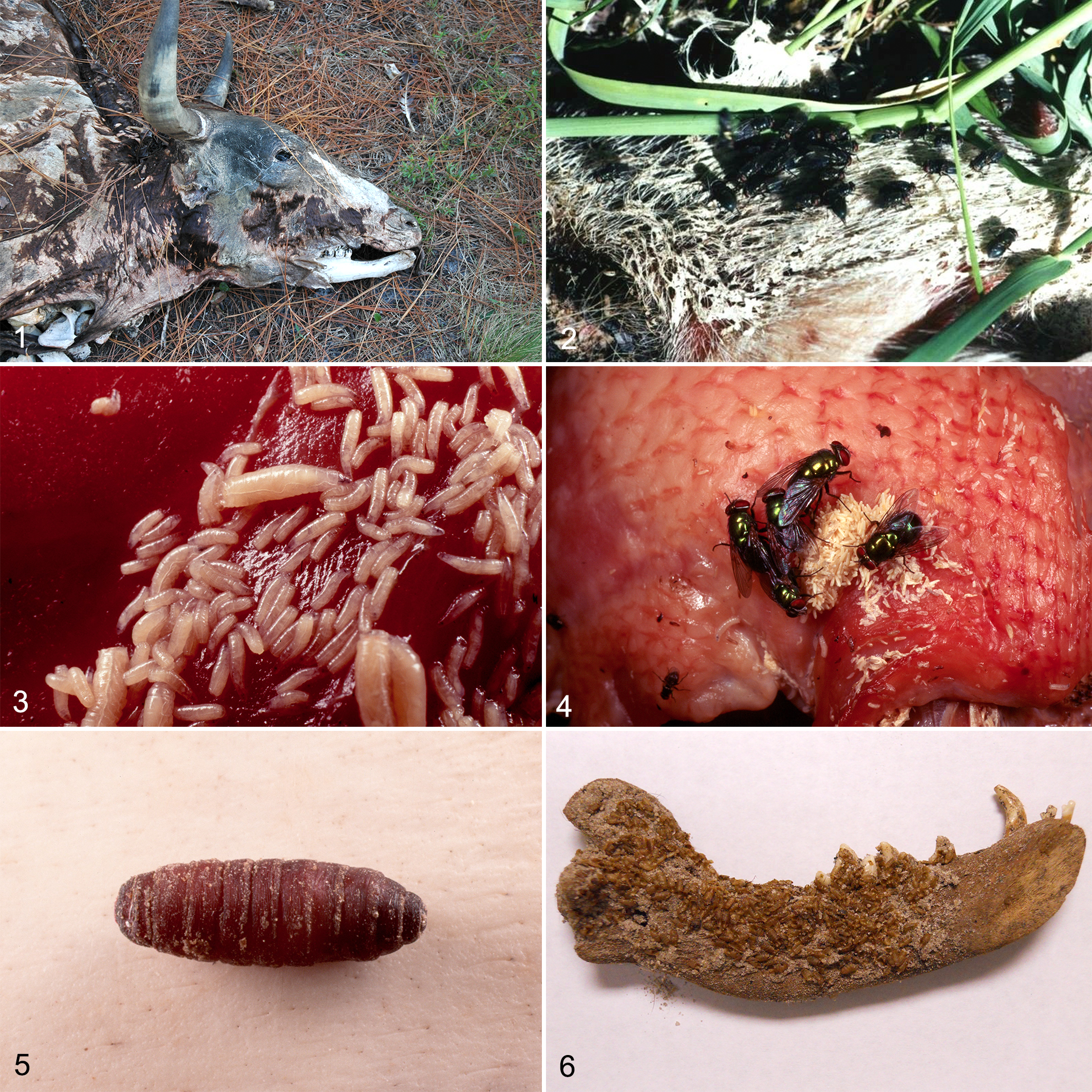
Although entomology has been used at crime scenes for centuries, research in recent decades has resulted in a significant advance of forensic entomological knowledge. 8,26,150 As a result, it is now commonly known that insect and arthropod evidence can assist in estimation of the postmortem interval (PMI) by the time of insect colonization on remains. 4,9,40,41,46,53 It can also aid in the detection and recognition of wounds, serve as indicators of perimortem and postmortem treatment of remains, and demonstrate neglect in both humans and animals 7,26,27 (Fig. 1). Arthropods may also be used to create associations between a suspect, a victim, and a crime scene. In some instances, it can aid in the detection of drugs or toxins within decomposed or skeletonized remains. 43,73 Recent research has even focused on the analysis of insect gut contents to determine the species on which fly larvae had fed. 43,87 These uses have made forensic entomology an invaluable tool for the death investigator.
While the bulk of forensic entomological applications has been in human death investigations, recent years have seen an increase in forensic entomology cases involving animals. 11,72,121 The application of forensic entomology in cases involving animals is relatively straightforward since the bulk of forensic entomological research has been carried out on animal models (Suppl. Table S1). This yields a large amount of data to use when associating insect species and development time with animal remains. It is possible to answer questions about the circumstances of an animal’s death by looking at decomposition and insect succession studies on the same animal species. This leads to a direct application of forensic entomological research to this particular discipline.
The evaluation of entomological evidence at a scene has the potential to give investigators valuable information about the circumstances of animal death or neglect. The major area of emphasis, however, is the analysis of insect species identification and succession to determine the time of colonization of either a living or dead animal that is infested with fly larvae and perhaps the geographical location of that infestation. 41 Many police agencies, veterinary pathologists, and animal welfare agencies throughout the United States are now requesting entomologists—knowledgeable in the behavior and biology of carrion insects—to assist in answering critical questions pertaining to the neglect and/or death of an animal.
Time of Colonization: General Considerations
The most commonly requested analysis by a forensic entomologist is estimation of the PMI, which is the time from death of the animal until the discovery of that animal by investigators. 68 During the PMI, various processes eventually lead to complete decomposition of animal remains, including the effects of bacteria, disarticulation by carnivores, weathering, and insect colonization. Each of these contributing factors has stereotypical timing that governs the arrival and duration of the factor, along with its overall effect on the animal remains. This timing is somewhat consistent but can be altered due to biotic and abiotic factors around the remains. Therefore, using any one of these processes of decomposition to estimate the PMI may be imprecise due to the uncertainty of the timing and mitigating factors of the analyzed process. 68
As a factor in the decomposition process, insects are known to arrive at and colonize decomposing remains very quickly after death if the remains are accessible. These decomposers feed upon animal tissue and have been known to remove all soft tissue within a matter of days. The most common member of this decomposer guild are the blowflies, which colonize in the larval or maggot stage, and is a well-known inhabitant of carrion. Female flies use olfactory and visual cues to search for remains on which to lay their eggs, and the resulting larvae use those remains as a food source. The cues used to find these remains are complex and not fully understood; they include volatile compounds related to both bacteria and the decomposing tissue but may also include other factors not yet identified. 128
This unknown quality explains why, although flies are often able to find and colonize remains with minutes of death, sometimes flies colonize an animal before death when wounds are present (ie, myiasis) or sometimes hours or days after death despite an otherwise normal process of decomposition. 6,39,44,45,80 This variation in colonization time presents a problem when attempting to use insects to determine PMI: the complex nature of their searching and colonizing mechanisms means they may or may not lay eggs on animal remains minutes after the animal dies. Entomologists therefore create a “time of colonization estimation” when analyzing insects on remains. 4 This estimate, which calculates how long insect larvae have been feeding upon the tissue of a living or dead animal, is determined by using known developmental rates for insect species that are attracted to fetid hair coats, wounds, or decomposing remains. The time of colonization can be estimated through the knowledge of insect assemblages on animals at different times during the decomposition process. 21,41,128 It allows an entomologist to account for female flies being attracted to a festering wound in an animal before death or being repelled from an animal after death. Often times, the time of colonization estimation equals the PMI, but unless an entomologist knows for sure the insects did not colonize wounds antemortem or that there was no delay in colonizing tissue after death, it is impossible to call the time maggots spent on decomposing remains the official PMI. Stating this time as the time of colonization allows for interpretation of the insect evidence in light of other circumstances such as presence of wounds or a placement of a covering on the remains.
The use of time of colonization for PMI estimation relies on the assumption that fly larvae colonizing the animal remains arrive after death. As most practicing veterinarians can attest, however, maggots may be present in the wounds of living animals; this is called myiasis. 17 In these cases, adult flies will lay their eggs on the open wounds of animals, and the maggots will hatch from those eggs to feed on the dead or living flesh of those wounds. Forensic entomologists may use the same techniques on living animals infested with maggots as used on animal remains infested with maggots. These techniques, outlined below, will give investigators a time of colonization estimation, not a PMI estimation, and the difference reflects the period of neglect or the period of time when wounds were present. 11,66 This situation is useful when an animal is still alive but may cause confusion during the postmortem investigation if antemortem myiasis is assumed to be postmortem colonization. If myiasis is used to determine the postmortem interval, then the calculated PMI would be significantly longer than the true PMI. This is why entomologists determine time of colonization rather than PMI. The analysis of insect larvae gives information about the insect development only, not if that insect developed on a living or dead animal or if those insects arrived at the moment of death, before death, or significantly after death.
The most useful group of insects associated with the decomposition of animal carrion and with myiasis is the family Calliphoridae, the blowflies and bottle flies. 41 These flies are usually the first insects to appear on carrion and are attracted to fecal matter encrusted in a hair coat or to open wounds on living or deceased animals. A few species of Calliphoridae, such as Cochliomyia hominivorax and Chrysomya bezziana, are obligate parasites and result in myiasis. 74 However, many species of Calliphoridae may infest the wounds of living animals, feeding on the necrotic tissue. 37,55,64 The adults can locate and colonize attractive sites within minutes and are therefore the most active and abundant insects found in the early stages of decomposition 128 (Fig. 2). The larvae of the Calliphoridae are responsible for consuming most of the soft tissue on remains, in just a few days. 47
Calliphoridae and other flies go through a basic life cycle, one that is studied in detail by forensic entomologists. Female blowflies search for oviposition sites through chemical and visual cues. 69 The chosen oviposition sites are often associated with decomposing animal tissue and the scents that accompany that decomposition. 153 The eggs are laid in protected areas, such as inside natural bodily openings, in wounds, within skin folds, and around wrappings such as sheets, blankets, and garbage bags. The eggs hatch into the first of 3 larval stages, known as instars. Each larval instar lasts a particular amount of time, dependent on ambient temperature. Higher ambient temperature yields decreased larval developmental time, while lower ambient temperature yields increased larval developmental time. Each larval instar feeds on the animal tissue, using the proteins and energy to facilitate growth. After the end of the third instar, the maggots leave the feeding sites on the animal to find protected places in which to pupate. The pupal stage of fly development is a nonfeeding stage; the third instar larva crawls under an object or burrows into the soil and hardens the outer surface of its body to form a protective casing. Within this casing, the larva uses an enzymatic and hormonal cascade to break down the larval body and reform it into that of the adult fly. This process can take weeks to months, depending on ambient temperature. When the adult fly is ready to emerge, it breaks open the anterior end of the pupal casing and crawls out into the air. 104 This life cycle is common among all flies, and each stage is present (from egg to larval instars to pupa) regardless of the fly species. The commonality among different species of flies allows entomologists to study those species that arrive on animal wounds and carcasses, as well as determine the time necessary to reach the observed developmental stage. 41,47,128
Entomology is not limited to just identifying the time of colonization of insects on remains or within wounds. The forensic entomologist’s knowledge of an insect’s geographical distribution may help an investigating agency determine where a death or neglect occurred and associate a suspect with a particular animal. The ability to determine where initial colonization occurred is possible due to the limited geographic distribution of particular insect species. If an insect species colonizing an animal is associated with a particular habitat, then that species can link the animal to that habitat. 1,19,38,61 For example, Calliphora alaskensis, a species of blue bottle fly, is found in cold northern regions throughout Alaska, Canada, Wyoming, and Colorado. In its southern range, it is only found at high elevations. In contrast, Lucilia mexicana, a species of green bottle fly, is found in warmer southern regions, from Texas south through Brazil. 149 Similarly, Lucilia cuprina is most commonly found in urban habitats during its peak seasons, while Compsomyiops callipes is found primarily in rural habitats. 38 The presence of C. alaskensis larvae on remains found in warm areas or the presence of C. callipes on remains found in urban habitats would indicate the movement of those remains after colonization and give investigators a clue as to where the animal died. Just as important would be the absence of insects where they should be present, such as in instances where animal remains are moved to a secondary location after the process of decomposition and insect colonization has begun. 41
Methods to Determine the Time of Colonization
There are many methods that a forensic entomologist may employ to estimate the time of colonization. 70 However, 2 methods are commonly used in casework. 41,47,128 The first uses the rate of development of pioneer colonizers, those insects first to arrive on an animal. One of the pillars of knowledge in the field of forensic entomology is the application of the insect’s temperature-dependent development to the time of colonization estimation.
The basic premise of temperature-dependent rates of development is that insects develop faster as temperatures increase until they reach a species thermal maximum, and they develop slower as temperatures decrease until they reach the species thermal minimum or minimal developmental threshold. 36 This temperature response can be illustrated by an S-shaped growth curve. 114 Countless organisms exhibit this type of development, and it is often used as a predictive indicator by scientists from botanists to bacteriologists to zoologists. 16,48,54,99,151 The process of decomposition itself is highly dependent on degree day accumulation. 97 One method to apply this temperature dependency is to calculate the accumulated degree hours (or accumulated degree days) necessary for insects to reach the observed developmental stage. This method allows forensic entomologists to account for fluctuating ambient temperatures and individual species thresholds while estimating developmental time. To accomplish this, the insects colonizing an animal must be properly identified, and historical meteorological data close to the scene must be collected. 128 It is important to note that entomological reports can never be more accurate than the accuracy of the temperature data. 52
The second method of time of colonization estimation uses insect succession by analyzing the presence or absence of particular insect species. 83,128 It is based on the premise that different insect species are attracted to different stages of decomposition, and each wave of colonizers feeds upon the resource for a generation. The act of feeding fundamentally changes the resource, thereby rendering it unusable to species within the current wave yet attractive to other species in subsequent waves. The blending waves of insects can span weeks or months, making this method most useful for time of colonization estimations that span many weeks or months. 41
While the decomposition process is a continuous process, researchers have categorized stages within this process in an effort to better understand the rate and mechanisms involved. 6,23,44,66,112 Flies, especially the Calliphoridae, tend to arrive during the fresh stage of decomposition and become most abundant during bloat and early decay. Their numbers dwindle during advanced decay, and they are no longer attracted to the remains once they reach the dry stage. House flies (family Muscidae) and flesh flies (family Sarcophagidae) tend to arrive at remains during the bloated stage and continue feeding on those remains until the end of advanced decay. Beetles that feed directly on remains tend to arrive much later in the decompositional process, arriving during early or advanced decay and remaining during dry decay. 128 Much effort has been spent attempting to quantify the exact timing of insect arrival and duration of insect feeding during decomposition, but the continuous nature of the decompositional process makes this difficult. In short, flies arrive at remains first and leave when those remains dry out, while beetles arrive later and continue feeding during dry decay.
The drawback of using succession in forensic cases is that it requires a tremendous amount of empirical research, and its application is limited to cases from geographic areas similar to those in which the research was conducted. 41,52 For instance, a forensic entomologist could not easily apply succession data obtained from northern regions (eg, Canada) to southern habitats (eg, Texas) and expect accurate estimates. The extreme differences in climate and habitat render similar successional patterns highly unlikely. Before a case can be properly analyzed, research must be conducted within the same geographical habitat as the case to determine the extent that climate and species diversity have on the pattern of insect succession. In fact, succession data may be even more limited. For example, succession data obtained from the eastern shore of Virginia may not be valid in the mountainous areas of the eastern portions of the state. Therefore, depending on terrain, the forensic entomologist may need multiple data sets from within a relatively small geographic region. Thus, overall applicability of using succession data as an estimation of the PMI during the initial few weeks of death or neglect can be quite limited due to this lack of data.
The Entomologist at the Scene, and Collection Procedures
It is rare that an entomologist will be on hand at the scene where insect evidence must be collected. Luckily, entomological collection procedures are straightforward and require little in terms of specialized equipment. A basic knowledge of forensically important insects along with some basic preservation materials and environmental data are all most investigators need to adequately collect entomological evidence. Collection of this type of evidence may result in the unavoidable disturbance of the remains or the scene itself, but some insects and other arthropods may be collected later in the investigation process. A plan for the collection of the evidence should be detailed in advance to ensure proper collection and preservation of arthropods so an entomologist may successfully analyze the evidence at a later date.
Entomological evidence collection can be broken down into the following 6 stages.
Stage 1: Preparation of Entomological Collection Equipment
Proper equipment allows for efficient collection of evidence at the scene. Entomological Evidence Scene Form (Suppl. Table S2). This form allows for accurate recording of scene conditions and should be filled out completely and shipped with the evidence. Insect net (student type with mesh bag is suggested). Collection vials. Screw-cap vials (4-dram size) with neoprene inserts may be ordered from biological supply companies. Other sealable screw-cap, water-tight containers may be used as necessary. Collection and preservation chemicals. Soft-bodied insects must be properly killed and preserved before shipping. Larvae may be placed in near-boiling water for 15 seconds (hot water killed; thermos) and then placed in 80% ethyl alcohol in collection vials. If hot water is not feasible, ethyl acetate or commercially available killing solutions may be used to kill the larvae before placing in 80% ethyl alcohol. Featherweight (or light touch) forceps. Forceps with a light tension are available from most biological supply companies and allow for the collection of soft-bodied insects, such as fly larvae, without damage. Typical forceps may be used if caution is exercised, but too much force will kill maggots intended for rearing. Paper towels or cotton balls. These may be used in kill jars and for cleaning utensils. Kill jars. Commercially available jars are designed to kill living adult, hard-bodied insects. The glass jars are filled with 1 cm of plaster to absorb killing fluid (ethyl acetate or acetone). The jars are “charged” by pouring approximately 10 ml of ethyl acetate onto the plaster and allowing it to absorb. Live adult insects are sealed into the jar, where the fumes from the ethyl acetate asphyxiate the organism. If commercial kill jars are not available, any wide-mouth jar will suffice. Soak several cotton balls or wadded paper towels in ethyl acetate (or acetone) and place into the jar. The sealed jar will kill adults in the same fashion as above. Larval insects, which are soft-bodied, should not be placed in the kill jar. Plastic sealable containers. Small plastic containers, such as disposable snack containers, will aid in collecting and/or shipping live larvae. Aluminum foil. Foil may be used to construct pouches to hold live larvae and a food source inside shipping containers during shipment. Vermiculite. Use vermiculite to fill the bottom of the plastic collection containers or the shipping containers containing live larvae. The vermiculite allows for larvae to migrate away from the food source when they are finished feeding and will absorb excess fluids. If vermiculate is not available, sand or dirt from the scene will suffice. Labels, adhesive and nonadhesive. Nonadhesive, heavy bond paper is ideal for creating labels to place inside collection containers (for both preserved and live specimen containers). Adhesive paper labels are necessary for the outside of collection containers. Precut adhesive “mailing” labels from an office supply store may be used. Graphite pencil. Pencil lead should be used to make labels, since preservative fluids will cause ink to smear and not adhere to the paper. Small hand trowel or garden spade. A trowel is necessary for soil samples and for digging for migrating larvae or pupae in outdoor scenes. Soil samples should be placed in plastic containers for shipping. Thermometer. A digital thermometer is preferred, but any type is suitable for ambient temperature collection. Camera. An SLR camera, lens, and flash are used for photo-documentation of the scene and entomological evidence found therein. A macro lens will be helpful in photographing the insects, while a standard lens should be used for general scene photos. Disposable latex, polyethylene, or nitrile gloves. Ruler or other measuring device to use as a scale in photographs. Shipping containers. Styrofoam containers with lids are the best shipping containers as they offer good insulation from temperature extremes. Corrugated cardboard boxes are inexpensive and readily available and may also be used. Camel hair brush (optional). A camel hair brush may assist with the collection of eggs or very young larvae without damage.
Stage 2: Visual Observations at the Death Scene
This stage is especially important if an entomologist is not present to collect the data, as environment, weather, state of decomposition, and surrounding vegetation may significantly affect the species composition and growth rate of insects on an animal. This stage begins by completing the “Entomological Evidence Scene Form,” which is designed to give the entomologist a complete picture of the scene from the investigator’s point of view.
Record the exact location of the site, including address (if known) and GPS coordinates if possible. If an address is unavailable, record the nearest cross streets and include the address of nearby landmarks (eg, 1 mile east of Route 41 and Cays Dr, Miami, FL, near the Everglades Fishing and Tours Co). Include photos of the area, along with photos of nearby landmarks to allow for easy location on a map. This step is essential to the entomologist, since this information allows for location of proper meteorological data.
Photograph the scene from a distance and include environmental photos. If the scene is indoors, include photos of windows, doors, and vents. Take several photos of the head, which is often where insect colonization begins. Be sure photos include detail of the eyes, mouth, nostrils, ears, and any sites of probable trauma. Several photos should be taken to indicate the overall stage of decomposition of the remains. Make sure to include close-up or macro photos of all observed insects and maggot masses.
Supplement the photographs with descriptions of the insects present at the scene. If you are unfamiliar with forensically important insects, simply describe any arthropod you see (eg, “White smooth maggots in a large mass,” “large black beetles with orange spots”). General descriptions along with photos and collected specimens are generally sufficient to give an entomologist an idea as to the species composition present at a scene. When in doubt, take close-up or macro photos of observed insects at the scene.
Note and sketch the position of the remains relative to compass points, windows, doors, sun, shade, or other significant environmental features. Make detailed notes of any trauma, dismemberment, burning, or wrapping of the remains. These situations may alter the insect colonization patterns and are important for an accurate analysis of evidence. Note the presence of insect activity in the immediate vicinity of the remains and look for migrating larvae and pupae under or in nearby objects. Check for the presence of dead adult insects on or near the body or on windowsills of indoor scenes.
Stage 3: Collection of Meteorological Data
Insects inhabiting remains grow at different rates depending on ambient temperature. Accurate collection of ambient temperatures at the scene and the identification of the closest National Weather Service (NWS) temperature stations with historical data allow for rigorous analysis of insect evidence. In the United States, the assisting veterinarian and local law enforcement should be able to identify the nearest National Weather Service recording station by contacting the nearest NWS field office.
For proper scene temperature data, record the ambient air temperature at a height of about 1.2 m in the shade. If the scene is indoors, record the ambient temperature and include photographs and information about heating or cooling systems, especially set thermostat temperatures. Additional temperature data should be recorded according to scene circumstance. The remains surface temperature (taken from the upper surface of the remains), the ground-remains interface temperature, the maggot mass temperatures, and soil temperatures from directly under the remains need to be noted as appropriate. Include an estimate of the duration of exposure to sun or shade at an outdoor scene or the position of the remains in relation to vents in heated or cooled indoor spaces. Relative humidity should also be recorded at the scene if possible. These readings can be made from standard dial-faced hygrometers and compared with NWS recordings for the local area.
Ideally, a series of ambient temperature recordings should be made at the scene for up to 5 days after body recovery. This allows for comparison of historical data to scene temperatures and to correlate the historical data with the temperature data collected during scene processing. Such correlation can be crucial evidence in later courtroom testimony. Generally, 4 temperature recordings are made spaced throughout each 24-hour period. However, if this is not possible, recordings should be made during the time of expected temperature minimums and maximums. Automated temperature and humidity recording units are available at scientific supply houses and may be left at a scene for a period of time to collect hourly data. This is ideal for temperature correlation.
Stage 4: Collection of Insects From the Scene
Insects and other arthropods should be collected from both the remains and the area directly beneath and around the remains. As appropriate, insects found under nearby objects or along windowsills in indoor scenes should be included. These insects are what the forensic entomologist will analyze and should be a representative sample of all organisms and stages observed at the scene. A representative sample of insects noted at the scene should be killed and preserved while at the scene to indicate stage of development when collected. Some may be collected live and shipped to the entomologist for rearing.
Using the insect net, sampling should start with collecting a representative sample of all adult insects present. For flies, at least 50 specimens of each species should be collected (if possible). Using either a gloved hand or forceps, 10 to 15 adult beetles of each species present should be collected (if possible). Different species should be preserved and labeled separately. Live adult specimens may be placed in a kill jar and then into another dry container or into alcohol once deceased. It is not necessary to attempt to keep the adult insects alive during shipment to the forensic entomologist. If collecting live insects, never mix beetles with flies, as beetles are often predators and will consume the other entomological evidence. Each collection container should be labeled with the case number, collector’s name, the date and time of collection, the location of the site, and the location on the remains from which the specimens were collected. Containers should be rigid to avoid crushing during transport.
Next, collect a representative sample of the fly larvae from the remains. This is the most important collection and should include 50 to 60 of each observed species. Focus on collecting representative samples of the sizes, body shapes, and colors of the insects observed (Fig. 3). Do not collect only the largest or only the smallest insects present. Because flies tend to lay their eggs in cracks and crevices, maggot masses will usually be found in the orifices of the body and in areas of trauma. The head and wounds are often colonized first and should be the focus of the collection, since they will usually contain the oldest and largest maggots. However, with some animals, almost any place on the hair coat may be the first area of colonization, depending on the length, thickness, and possibly fetid nature of the hair coat (Fig. 4). The layperson can distinguish some species differences based on body shape or coloration and separate accordingly. Different species should be packed separately, if possible.
It is important to preserve this representative sample of maggots at scene, so the forensic entomologist can determine the age of the maggots during collection. If larvae were collected during necropsy or after removal from the scene, the delay in collection and the temperature at which the remains were kept must be indicated in the report. Maggots may be preserved through immersion in near-boiling water for 15 seconds and then placed in alcohol, or through immersion in a mixture of kerosene, alcohol, and acetic acid (KAAD) and sand, then placed in alcohol. Placing larvae directly in alcohol without submersion in hot water or KAAD will result in larval decomposition and difficulty in subsequent identifications. All vials should be properly labeled on the inside and the outside. Use a pencil to fill out any labels dropped into alcohol to ensure retention of the writing.
Live larval samples may be important for evidence analysis. If this is the case, place a few representative samples of each species on a food source (beef liver, pork, or even wet cat food will suffice), and place the food source in an aluminum foil cup. Living maggot species should not be mixed, as some are predatory and will consume other maggots as a food source. Place the cup of vermiculite in a rigid container, and poke some holes in the top to allow for air circulation. Each container should be labeled with the case number, date and time, site location, and the location on the body from which the specimens were collected. When maggots have been collected from a mass, this should be indicated on the label. If live insects are included in the shipment to a forensic entomologist, make sure to indicate this during the initial contact with the entomologist so proper rearing facilities can be prepared.
Beetle larvae, or grubs, should be collected in the same manner as fly larvae. Collect a representative sample, approximately 10 to 20 of each species, from the remains. Adults should be placed in a charged kill jar then preserved in ethanol, while larvae may be placed directly into ethanol. Each container should be properly labeled, and both live and preserved samples should be obtained as appropriate. Again, never mix beetle larvae with maggots, as grubs will feed on maggots and destroy the insect evidence.
Fly pupae indicate a more advanced life stage, and thus possibly older samples, and a representative sample of 50 to 60 pupae from on or around the remains should be collected. Pupae are small, rigid, and brown to black casings in which maggots develop into adult flies (Fig. 5). They look similar to roach egg casings or mouse droppings. Maggots tend to wander in the prepupal stage just before pupation, so the area around the remains should be searched for larvae and pupae that may have moved away from the remains and become distributed in the environment. In outdoor settings, maggots tend to bury themselves in the first inch or so of soft topsoil or migrate under rocks and limbs. Indoors, maggots will migrate under baseboards, rugs, mats, and other nearby objects. Some fly species may pupate directly on the remains (Fig. 6). Fly pupae do not feed, so live samples only need to be placed into a sealed container with some vermiculite or similar substrate.
Soil samples should be collected from directly beneath the remains as soon as appropriate. A core sample (about 8 cm wide and 15 cm deep) should be collected from the ground directly beneath the major body areas (head, torso, upper legs). These samples should be properly labeled and placed into a cardboard container. These samples will contain insect specimens that burrowed into the ground for protection or pupation and insects that inhabit the soil beneath the remains rather than on the remains themselves. These species are not usually recovered unless soil samples are taken.
After returning from the scene, official weather data should be collected and should span the time period in question. In the United States, these data can be easily obtained by contacting the nearest National Weather Service Office or from the National Climatic Data Center website (http://www.ncdc.noaa.gov/). Find the weather station situated closest to the scene, and record its identification number and distance from where the remains were found. Maximum and minimum daily temperatures, humidity, and precipitation measurements should be requested. If hourly data are available, they should also be requested and added into the report. Depending on the case, additional data such as cloud cover, wind speed, river stages, tides, and soil moisture may be requested by the forensic entomologist.
Collection of Entomological Evidence During the Necropsy
The best practice is to collect entomological evidence at the crime scene. Failure to do so may result in more advanced insect life stages being recovered, as well as entire species going undocumented and not collected. This may occur because many Diptera species undergo a prepupal wandering phase in which they disperse away from the remains and into the environment. If the remains were removed from the scene before insect collection, or insect collection happened during necropsy, the standard procedure of live and preserved collections from each area of colonization should be followed as for the scene protocol. However, one important aspect of the collection at necropsy is to document the temperature at which the remains were kept and the time logged into and out of refrigeration. Duration in a freezer or refrigerator should be noted, as well as the ambient temperature of the laboratory where the larvae were collected. Larvae will continue to grow during transport and storage, and the forensic entomologist will need this information to fully account for insect age during evidence analysis.
If possible, collections should be made both before and during necropsy. At necropsy, insects may be present that were not readily visible on scene or were feeding deeply within the remains. These insects will also be chilled and therefore easier to collect. The necropsy allows for closer inspection of wrappings or other items associated with the remains. Insects found in wrappings may be different species from those observed directly from the remains or in a different developmental stage, so they are essential to collect as evidence.
Stage 5: Packaging and Shipment of Insect Evidence
Insect evidence should be shipped to a forensic entomologist as quickly as possible, especially if there are live samples included in the evidence. All evidence should be packaged in rigid containers (avoid plastic bags) to avoid crushing during shipment. Containers with live specimens should contain the insects on their food source, wrapped in aluminum foil, and sitting on vermiculite or other absorptive material. This will allow for migration and burrowing of living specimens and also allow for absorption of fluids that may leak during shipment.
The shipment should include all scene notes, the completed scene form, weather information, and photographs. Basic contact information should also be included so the entomologist may ask for additional information if necessary.
Ongoing Research
Currently, much research is being devoted to improving molecular techniques in forensic entomology, especially where species identification and gut contents analysis is concerned. 29,61,133,148,149 Advances are also being made toward establishing basic statistics on the reliability of the time of colonization estimations based on entomological evidence, and computer modeling techniques are being employed to reduce statistical error. 36,84,99 It is anticipated that computer applications being developed will aid law enforcement in capturing more data from the death scene that can be used to improve the accuracy and precision of information-intensive computer modeling programs.
With a variety of analytical techniques at their disposal, forensic entomologists are well prepared to assist those involved in the investigation of animal neglect or death when entomological evidence is recovered. However, investigators are advised to contact a forensic entomologist before services are actually needed. This established line of communication along with a small amount of prior training in insect collection and preservation can greatly improve the usefulness of entomological evidence.
Footnotes
Declaration of Conflicting Interests
The author(s) declared no potential conflicts of interest with respect to the research, authorship, and/or publication of this article.
Funding
The author(s) received no financial support for the research, authorship, and/or publication of this article.
References
Supplementary Material
Please find the following supplemental material available below.
For Open Access articles published under a Creative Commons License, all supplemental material carries the same license as the article it is associated with.
For non-Open Access articles published, all supplemental material carries a non-exclusive license, and permission requests for re-use of supplemental material or any part of supplemental material shall be sent directly to the copyright owner as specified in the copyright notice associated with the article.