
Abstract
Fifteen dogs were found dead in a house that was on fire. Several of these dogs were partially burned. Four dogs were submitted for postmortem examination, 2 of which were determined to have died prior to the fire. Of the 2 submitted fire fatalities, only 1 dog had burns on its body (dorsum and right side of body). Internally, both dogs had soot deposits mixed with mucus in the larynx, trachea, and primary bronchi. Microscopically, soot was identified within both airways and alveolar spaces. There were no macroscopic or microscopic indications of vital heat exposure. High levels of carboxyhemoglobin were detected in the 2 dogs tested. The findings in this case support the use of postmortem examination and toxicology testing to allow for determination of vital reaction to heat and fire fumes.
In cases where humans or animals are found dead and burned, there are major challenges to the forensic investigator to determine a cause of death. Forensic pathologists are required to determine if the subject was alive when exposed to the fire fumes and determine if there was vital heat exposure. In human forensic investigations, the most important signs of vitality for exposure to fire fumes are the identification of soot deposits within the respiratory tract, esophagus, and stomach, as well as elevation of carboxyhemoglobin (COHb) in the blood. 2 Signs of vital exposure to heat include edema and vesicular detachment of the mucosa of the pharynx, larynx, and/or upper esophagus. 2 However, there is a paucity of literature regarding the postmortem findings and vitality in dogs exposed to fumes and heat from a fire. 7,10 This case report describes the findings from the postmortem examination of dogs involved in a house fire.
The home of a suspected animal hoarder was reported to be on fire. Fifteen dogs were found dead at the scene, while 6 dogs were found alive and transported to a local veterinary hospital for further evaluation. Four of the 15 dead dogs were submitted for postmortem examination. Two of the animals were severely decomposed and in the stage of dry decay and were determined to have died prior to the fire. The other 2 dogs were more recently deceased with a postmortem interval of approximately 24 hours. External examination of 1 of the recently deceased dogs revealed severe burning on greater than 50% of the body (predominately on the dorsum and right body wall) with severe charring and splitting of the skin, and there were no singe marks or deposition of soot noted on the eyelids. No evidence of burns was identified on the other 3 dogs (including both severely decomposed dogs).
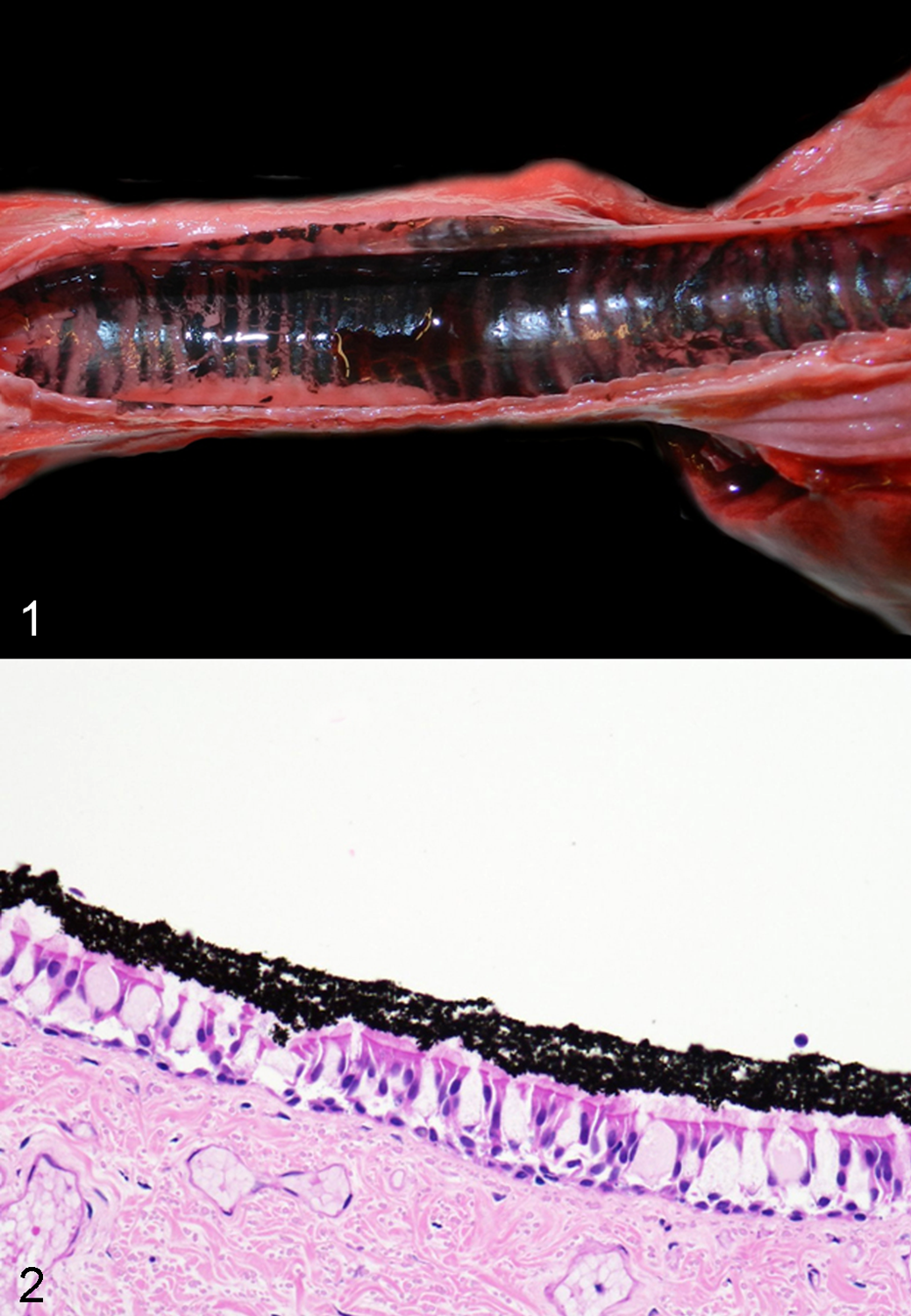
Internal examination of the 2 recently deceased dogs revealed deposition of soot (black pigmented material) mixed with mucus within the pharynx, larynx, trachea, and primary bronchi (Fig. 1). Oral mucous membranes and musculature were red. Microscopic findings were limited to the respiratory system in these 2 dogs. There was soot overlying the ciliated epithelium of the trachea and bronchi and the epithelium of bronchioles (Fig. 2). There was minimal soot within alveolar spaces. In the dogs that were severely decomposed, only the larynx and proximal trachea were available for examination, and no soot was identified in either animal, which would be consistent with death prior to the fire.
Samples of blood collected during the postmortem examination from the 2 recently deceased dogs were tested for carbon monoxide (CO), via COHb, and cyanide (CN–). Carboxyhemoglobin was determined using a sensitive and robust headspace method that used gas chromatography with a thermal conductivity detector (GC/TCD) for CO. 6 Results of the analysis revealed 65% and 56% COHb. The blood samples were found negative for CN–.
Based on the findings of soot within airways and elevated COHb, the cause of death was exposure to toxic fire fumes (CO) resulting in asphyxia (category: vitiated atmosphere). Vital exposure to heat was not observed within the animals examined, and it was determined that the burns on the 1 dog developed after death.
Discussion
Determination of vital exposure to both fire fumes and heat is of major importance in forensic fire investigations. In some instances, bodies are burned after death in an attempt to destroy evidence such as stabbing or gunshot wounds. 9
There are 2 main signs of vital exposure to fire fumes in burned bodies that are reported in the forensic literature. 2,9 The first is deposition of soot within the respiratory tract, including the larynx, trachea, and bronchi. Some cases also have soot deposits in the esophagus and stomach, but soot deposition in the respiratory tract is always present in such cases. 2
The second evidence of vitality is the elevation of COHb in the blood. In antemortem testing, the mean normal COHb levels were 6.1% (range, 5.6%–6.4%), and the COHb values in dogs that survived after exposure to a kennel fire ranged from 8.8% to 37%. 1 In the present case, COHb values were determined using headspace GC/TCD, rather than the more traditional UV/VIS techniques because GC/TCD was specifically designed to deal with the complexities involved in the analysis of deteriorated postmortem blood. 6
Toxic gases are produced in fires and include CO, carbon dioxide (CO2), and CN–. CO is produced by the incomplete combustion of carbon-containing molecules. 8 It readily crosses the respiratory epithelium and competitively and preferentially binds to hemoglobin, forming COHb. The CO binding reduces the oxygen (O2)–carrying capacity of hemoglobin, leading to hypoxemia and hypoxia. Hydrogen cyanide is formed from the combustion of carbon- and nitrogen-containing materials, such as plastics, wool, and silk.
Lethal levels of COHb have been studied in several species. In humans, CO poisoning begins at 20% COHb, and levels between 50% and 80% COHb may be fatal. 11 Levels of COHb of fire victims are typically >10% COHb. 2 In nonhuman primates, levels less than 30% are not considered life-threatening. 4 In dogs, atmospheric levels of 13% CO resulted in death within 1 hour after COHb levels reached 54% to 90%. 11 The COHb levels in the dogs of this case fall within the lethal range. Other toxic gases in the fire may have contributed to death, as synergistic effects have been shown for fire gases such as CO and CO2. 5 It deserves mention that suffocation due to lack of oxygen in a fire has been disproved since suffocation in humans occurs when the O2 concentration in air is less than 10 vol % as fires will smolder at 15 vol %. 2,3
Vital exposure to heat is of concern to the forensic pathologist. In 1 review, macroscopic evidence of exposure to heat included burn blisters on the skin; edema and vesicular detachment of the mucosa of the pharynx, larynx, and/or upper esophagus; absence of soot and/or burns in the corner of the eyes; incompletely singed eyelashes; and petechial hemorrhages in the conjunctiva. 2 Microscopic signs of vital exposure to heat include edema and vesicular detachment of the mucosa of the pharynx, larynx, and/or upper esophagus; increased secretion of mucus; and pseudo-goblet cell formation. 2
In this case, the 2 recently deceased dogs succumbed to exposure to toxic fire fumes (based on soot aspiration and elevated COHb), which resulted in asphyxia (category: vitiated atmosphere). In this report, we discussed the postmortem findings of fire victim dogs and the methods necessary to determine vitality in fire victims. The veterinary pathologist will inevitably be involved in arson investigations and the ability to accurately determine the cause of death will be of major importance to the arson investigators.
Footnotes
Declaration of Conflicting Interests
The author(s) declared no potential conflicts of interest with respect to the research, authorship, and/or publication of this article.
Funding
The author(s) received no financial support for the research, authorship, and/or publication of this article.