
Abstract
We provide here an overview of the state of applied techniques in the estimation of the early period of the postmortem interval (PMI). The biological methods included consist of body cooling, CSF potassium, body cooling combined with CSF potassium, and tissue autolysis. For each method, we present its application in human and veterinary medicine and provide current methodology, strengths, and weaknesses, as well as target areas for improvement. We examine current and future molecular methods as they pertain to DNA and primarily to messenger RNA degradation for the estimation of the PMI, as well as the use of RNA in aging wounds, aging blood stains, and the identification of body fluids. Various types of RNA have different lengths, structures, and functions in cells. These differences in RNAs determine various intrinsic properties, such as their half-lives in cells, and, hence, their decay rate as well as their unique use for specific forensic tests. Future applications and refinements of RNA-based techniques provide opportunities for the use of molecular methods in the estimation of PMI and other general forensic applications.
Keywords
The postmortem interval defined
The postmortem interval (PMI), also called the time since death, is the time during which a human or animal has been deceased and is the time lapse between death and the discovery of the body. After death, the bodies of humans and animals are subject to physical, chemical, and biological changes described as decomposition. Macroscopic changes observed in the early PMI are decreasing body temperature, rigor mortis (stiffening of limbs), and livor mortis or hypostasis (pooling of blood by gravity into the parts of the body closest to the ground).11,19,38 A microscopic change in the early PMI is cellular autolysis with loss of cell adhesions.51,120
Numerous other parameters change over time in the early PMI, such as morphologic changes in WBCs, 18 changes in blood glucose and electrolyte concentrations, and changes in enzyme activities.11,36 At later stages of the PMI, bodies will be subject to bacterial decomposition, referred to as putrefaction. Insects may also colonize the remains and assist in soft tissue decomposition.11,69 The mineral matrix of ossified bones is resistant to degradation after death, and a fully decomposed body remains “skeletonized” for >8–10 y. 19 In many cases, especially if foul play is suspected, estimating the PMI can be of central importance in establishing a chronology of events, including or excluding suspects and/or their alibis.
The link between animal abuse, domestic violence, and other deviant human behaviors is well known, and the legal system is evolving toward more systematic investigation and prosecution of animal cruelty cases. 67 The resulting increase in animal crime investigations has revealed gaps in veterinary forensic pathology, especially associated with estimating the PMI. Because animals are often harmed or slaughtered and left abandoned in the environment, reliable field-efficient methods are essential for the estimation of PMI.
Challenges in estimating the PMI in field cases
To date, no best method exists that accurately and reliably estimates the PMI. Extensive research has been published,1,38,60,69,97,112 but only a few of the techniques described in the literature are sufficiently field-efficient to be applied for forensic purposes in human death investigations, 123 and even fewer are applicable in animal cases. 11 A realistic PMI is indirect and based on the time between when a person or animal was last seen alive and when the body was discovered. In general, when estimating the PMI, ideal candidate parameters are those that change over time along a known trajectory. 37 In field situations, these parameters could be measured and the PMI could then be estimated from that known change over time. 37 A large number of potentially ideal candidates exist; however, the change in these parameters over the PMI is often influenced significantly by intrinsic or extrinsic factors. The best-known example is the decrease in body temperature after the death of a human, which is known to vary significantly depending on the clothing worn, body coverings such as blankets or sheets, body mass, the surface on which the body lays after death, ambient temperature, air movement, or if the surrounding environment is dry or wet.36,123 In animals, the decrease in body temperature is dependent mostly on body mass and type of haircoat, thus cooling differs across species as well as breed. 11
The methods described for the early PMI are considered by some researchers 36 as tasks to be executed by a trained and experienced forensic pathologist. However, the pathologist is rarely available to visit the scene, and therefore, in addition to the training of medical investigators in validated techniques, reliable techniques are essential to improve the accuracy and use of methods that estimate the PMI. Another issue in obtaining a reliable PMI is that the time of death may emerge only later during the investigation when the body has already been autopsied and the scene has been released. This can be corrected by standardizing the methods of investigation of any human corpse or animal carcass at a scene or during the autopsy.
To date, data on PMI estimation in animals is insufficient to estimate accurately the PMI in field cases, especially for use in legal investigations in which foul play is suspected. New molecular techniques have been considered using RNA types to assist in various fields of forensic sciences, including the estimation of the PMI.6,86,100,115,116
PMI as a tool in human and veterinary medicine
Domestic pets, such as dogs and cats, are often subject to abuse, neglect, and non-accidental and ritualistic killings.21,90 Many animals are also used for illegal blood sports, including primarily dogs and poultry.21,90 Cattle 74 and horses23,70 are often victims of cruelty, neglect, non-accidental killing, theft, and illegal slaughter, and establishing the timeline of events as well as the time of death of these animals, is a crucial element in investigations. Evidence gathered during these investigations should rely on rigorous scientific principles, which contribute essential information regarding the circumstances of abuse and death, the time of abuse, and the PMI.
The literature on estimating the PMI in domestic animals is limited and focuses on changes in body temperature, occurrence of rigor mortis in different limbs, eye, and CSF potassium (K+) increase, skin discoloration, as well as hypostatic congestion, internal changes of various organs, including morphologic changes at the cellular level, and immunologic changes based on the decreasing intensity of B- and T-cell staining.11,21,90 Most of these studies conclude that further research and data are needed before being validated and declared field efficient; none of these techniques is currently used in forensic investigations.69,120,123
Biological methods for estimation of the early PMI
Body cooling
Applied techniques in human medicine
Mathematical models have been established29,65,123 for the bi-exponential rectal cooling curve, 36 which take into consideration the initial plateau followed by an exponential drop of rectal temperature. After the plateau, the rectal cooling curve has a sigmoid shape that can be modeled. Nomograms are available for human bodies and allow the PMI to be estimated with a 95% CI for a body of a certain weight, from a single measurement of the rectal and environmental temperatures up to 23°C. The nomogram can estimate the PMI up to 80 h depending on the body weight with a 95% CI of ± 7 h if a correction factor is used. For shorter PMIs and lighter bodies, the 95% CI is ± 2.8 h, with or without correction factors. If, for example, a human body is found naked, on a dry and thermally inert surface, lying fully extended on the back, in still air, and in the absence of surrounding sources of radiant heat, no correction factor is needed for the PMI reading. In any other situation (e.g., moving air, body covered with one or more dry layers, wet coverings, wet body surface, in still or moving water), tables for some correction factors are available and can be used for estimation of the PMI. 36
Situations in which the nomogram method cannot be used include: 1) the presence of strong radiant heat near the body (e.g., radiators, heaters), 2) significant but unknown changes in cooling conditions (e.g., open windows and doors), 3) marked and recurrent changes in climatic conditions, 4) transportation of the body (e.g., the place where the body is found does not correspond to the place where the person died), and 5) cases of death as a result of hypothermia. Other limitations include situations in which the environmental temperature is >23°C, if/when the patient had a fever, or was in a prolonged agonal state.36,123
The nomogram method using body cooling is only useful up to 80 h postmortem, depending on body weight, and is based on measured, evaluated, or estimated parameters providing a single mean value. Each parameter has a potential source of error, and if an inappropriate correction factor is chosen, the estimated PMI could be highly inaccurate. The nomogram determines a time window of the PMI with a 95% CI, which indicates with 95% confidence that death occurred within that time. Thus, PMI determination is only an estimate. Unknown events occurring during the PMI can influence the cooling of the body; hence, the measured, evaluated, or estimated parameters will not represent the circumstances accurately, and the calculated PMI will be incorrect.36,123
Applied techniques in veterinary medicine
Decrease in body temperature in dogs has been reported twice.21,90 The ambient temperature in both studies varied significantly; therefore, the findings are not comparable. Nonetheless, rectal temperatures decreased along a parabolic curve in both studies. An extensive study performed in pigs examined the decrease in body temperature of the eyeball, orbit soft tissue, rectum, and muscle tissue. 46 A single-exponential model applied to eyeball cooling provided a reasonable estimate of the PMI up to 13 h after death; after 13 h, muscle and rectal temperatures were better estimates of PMI. Decreases in eye K+ were seen in dogs when measured 1.5 and 7 h after death. 90 Rigor mortis of hindlimbs persists in dogs up to 24 h, and elbow rigidity was lost between days 3 and 7. 21
CSF K+
Applied techniques in human medicine
The rise in K+ within the CSF is the result of the increased permeability of cell membranes that starts during early autolysis.36,60 The rise in cisternal K+ occurs at a constant rate and is related to body temperature. In the first 20 h of the PMI, the increase in cisternal K+ is not only correlated strongly with the PMI but is also independent of the environmental temperature. The deviation between the real and the extrapolated PMI is ± 1.5 h in the first 15 h postmortem. This deviation can be reduced by considering the body’s cooling temperature. The 95% CI limit is ± 1.4 h in the first 15 h of the PMI and ± 1.03 h in the first 10 h.
Applied techniques in veterinary medicine
In a study of various K+ electrolyte changes in blood, CSF, and vitreous humor in dogs, CSF K+ concentrations increased markedly over 48 h, but correlation with the PMI was not described. 97
Combined body cooling and K+
Applied techniques in human medicine
According to some researchers, the most precise available method to estimate early PMI in humans is an assessment of cisternal CSF K+ levels, taking into consideration the rectal temperature using the nomogram method.36,38 Some limitations of applying the combined analysis of cisternal K+ and rectal temperature are: 1) slowly progressing chronic diseases with electrolyte imbalance, 2) concurrent toxic or infectious processes, 3) intracranial or intracerebral hemorrhages with bleeding into the ventricles or cisterns, and 4) cases in which the patient died of hypothermia.
Applied techniques in veterinary medicine
A single study examined time and temperature effects on postmortem biochemical changes in canine CSF; CSF K+ did not change significantly after death and was found to therefore not have field-efficient diagnostic forensic value for estimation of the PMI. 98
Tissue autolysis
After death, the blood supply to tissues is lost, and cells undergo autolysis. Although autolysis is a process well-known to histopathologists, little research has been done on autolysis. 120 The process of autolysis is driven by phosphorus-rich enzymes, including alkaline and acid phosphatases, adenosine triphosphate, 5′-nucleotidases, and glucose-6-phosphatase.76,107 Microscopic characteristics of cell autolysis resemble those of necrosis, and the distinction is not always possible. 51 Necrosis is a type of cell death described in living tissues; its pathognomonic characteristic is the presence of an associated inflammatory reaction, except in cases of peracute ischemic or toxic necrosis. 51 Histologically, if present, inflammatory cell infiltrates and normal tissue surrounding areas of focal-to-extensive necrosis allow the distinction between necrosis and autolysis. 71 Autolysis is more evenly distributed across the entire organ because it usually progresses at the same rate throughout the tissue during decomposition.15,71,120
Autolysis affects different cell types at different rates. As in living tissue, some cells are much more resistant to hypoxia than others.12,51 Early degradation is observed in the mucosa of intestine, gall bladder, the parenchyma of pancreas, and cells in the adrenal medulla, followed by autolysis of neurons and last by connective tissue.12,71 As with most other changes observed after death, the activities of enzymes responsible for autolysis are greatly reduced by the refrigeration of bodies or tissues. 120
Applied techniques in human medicine
After brain death, autolysis of the brain is seen as reduced neuronal nuclear staining, reduced numbers of neuronal nuclei, and increased pallor of the neuropil without associated glial reaction.79,102 At 5–22 h postmortem, karyorrhexis and neuronal Nissl substance dissolution increase progressively. 99 Neuronal cytoplasmic vacuolation, basophilic cytoplasmic stain, and nuclear and cytoplasmic swelling are also described as autolytic changes. In a natural death, autolytic changes in the brain are first discernable as swelling of the neuronal nucleus and cytoplasm with increasing chromatolysis and liquefaction of the cytoplasm that may or may not involve the nucleus. 76 These initial changes appear at different times after death according to various sources cited and are observed as early as 30 min or as late as 3 h postmortem. 76
The autolytic changes of skeletal muscle tissue are less well documented, and studies use postmortem electrical stimulation as a surrogate. The changes observed are interruptions of individual muscle fiber continuity with loss of cross-striation, interruptions of fiber continuity, and segmental and discoid disintegration of fibers. To date, autolytic changes are not used by medical examiners to estimate PMI (Hamilton WF, pers. comm., 2021 Sept 29).
Applied techniques in veterinary medicine
In a study of the evolution of postmortem changes at 22°C and 8°C in equine brain, liver, and skeletal muscle tissue during the first 72 h of the PMI, brain autolysis was the least predictable. 120
Mammalian hepatocytes imbibe plasma during autolysis, causing the formation of eosinophilic non–membrane-bound cytoplasmic inclusions similar to those formed during sub-lethal injuries, such as hypoxia, intoxication, malnutrition, nutrient deficiency, and some viral infections. 15 The sequence and rate of autolytic changes of hepatocytes and bile ducts over 48 h were studied in guinea pig livers kept at 20°C after death. 104 The observed changes were hepatocyte cytoplasmic eosinophilia after 3 h, cytoplasmic vacuolation by 36 h, and lysed nuclei and hepatocyte individualization by 48 h. In portal areas, the separation of bile duct epithelium from the basement membrane and chromatin margination in nuclei was present in most bile ducts by 24 h. In rat livers, hepatocyte nuclear chromatin condensed and hepatocytes individualized over 6 h of postmortem autolysis. 107 Significantly contrasting results were observed in canine livers, in which the bile duct epithelium detached from the basement membrane after 3 d. Most hepatocyte nuclei were autolyzed by 7 d, and the most significant hepatocyte autolysis overall was observed after 3 wk 21 in the aforementioned equine study 120 ; liver autolysis occurred as early as 1 h after death and progressed over 72 h. The changes observed were hepatocyte individualization, separation of bile duct epithelium from the basement membrane, and bile duct epithelial pyknosis and cytoplasmic vacuolation.
Studies examining postmortem changes in muscle have been reported in horses and dogs. Equine skeletal muscle had significant postmortem disruption of myofiber continuity, hypereosinophilia, loss of striation, and sarcoplasmic floccular fragmentation at 22°C and only sarcoplasmic eosinophilia at 8°C. 120 The autolytic skeletal muscle changes in dogs were described in postmortem traumatized muscles exposed to seawater, and changes were similar to those described for electrically stimulated human muscles, namely rupture of fibers, and segmental and discoid disintegration of fibers. 105
Methods for estimating the late PMI
When the body enters the later stages of the PMI, the best method to estimate the time since death in humans is through forensic entomology. This is true for cases of early or advanced putrefaction, advanced stages of soft tissue decomposition, presence of saponification or mummification, and for skeletonized remains. This requires the presence of insects, or signs of previous insect activity (e.g., empty pupal cases), on the body or near the body (e.g., in soil below or near the body, in clothing, or under objects near the body). Forensic entomology is outside the field of expertise of most pathologists, and a forensic entomologist should be consulted to assist 115 with the estimate of PMI. 47
Given the limited number of forensic entomologists, it is unlikely that a qualified expert will be present at the scene. Therefore, proper documentation and collection techniques for entomologic evidence must be utilized to ensure complete and representative sampling of insect samples at the scene. Subsequent collections can be made during the autopsy to enhance the entomologic collection and documentation process; improper collection and documentation will limit the accuracy of an estimated PMI. 47
Molecular methods for estimating the early PMI
DNA
A comprehensive review has summarized the results of >40 studies of the degradation of DNA after death. 112 The reported results were mixed: many studies reported a linear correlation between DNA degradation and time since death; others observed a trend or no correlation. Most studies were done on animal tissues, fewer used human organs. There is insufficient knowledge of the influences on the DNA decay process, and extrapolation of animal data to human cases is highly discouraged. 112
RNA
New insights into the stability and quality of different RNA types make RNA a potentially useful candidate in forensic sciences.86,100,115,116 The 3 major classes of RNA in eukaryotes are messenger RNA (mRNA), ribosomal RNA (rRNA), and transfer RNA (tRNA). 54 Other classes of RNAs include small and other non-coding RNAs (ncRNA), 22 which play a role in controlling and fine-tuning gene expression. Various ncRNA-containing microRNAs (miRNAs), and PIWI-interacting RNAs (piRNAs) exist, as well as medium-length RNAs (snoRNAs) and longer transcripts (lcnRNAs).
Overview of cellular coding and non-coding RNA
The different RNA types can be grouped into coding RNAs and ncRNAs, which have different functions, structures, sizes, and stabilities, and are located in different parts of the cell (Table 1).
Summary of function, special structure, size/length, location in cells, half-lives, and special characteristics of coding (mRNA, tRNA, rRNA) and non-coding (miRNA, piRNA, snoRNA, lcnRNA, circular RNA) RNA.
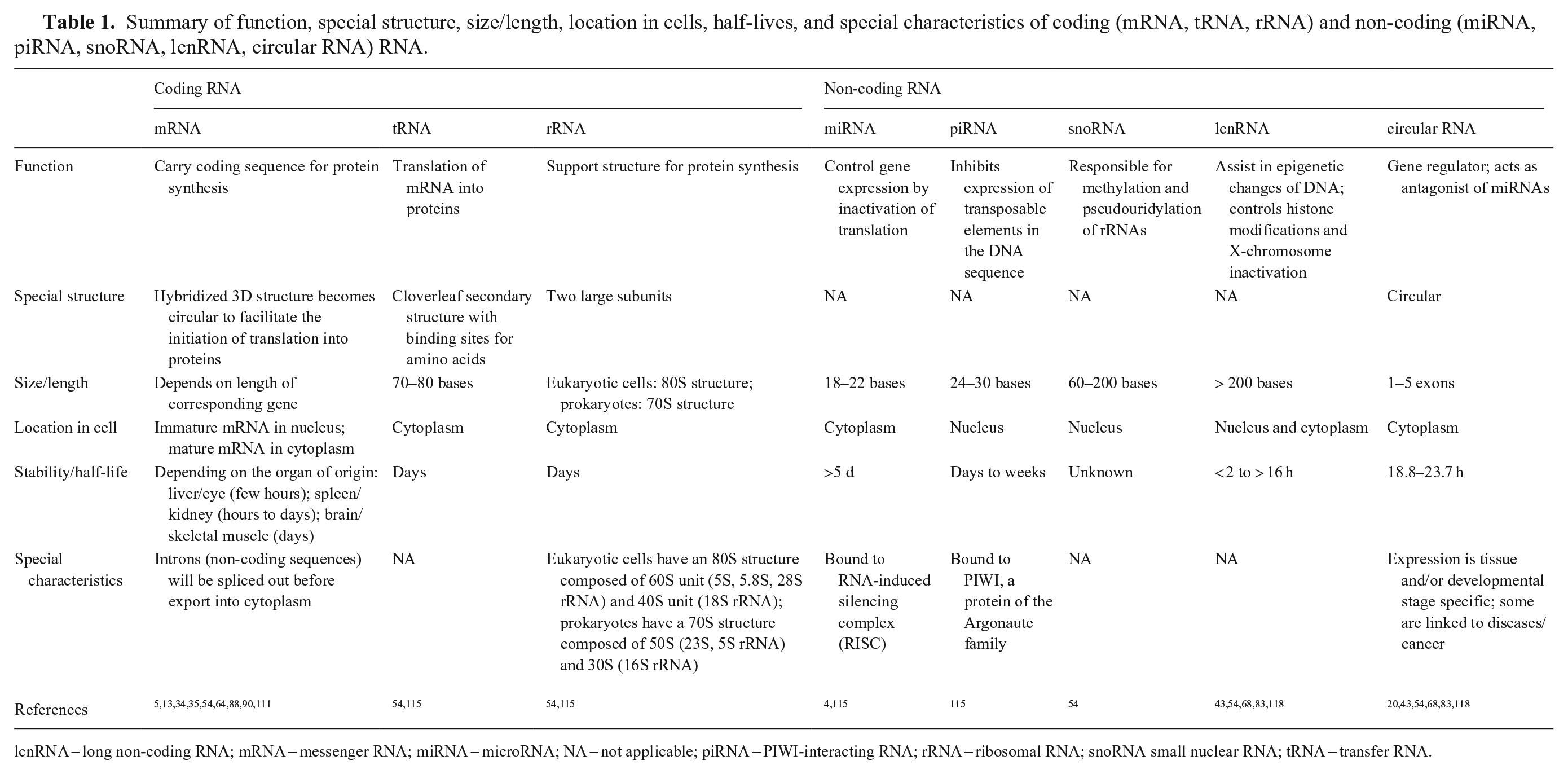
lcnRNA = long non-coding RNA; mRNA = messenger RNA; miRNA = microRNA; NA = not applicable; piRNA = PIWI-interacting RNA; rRNA = ribosomal RNA; snoRNA small nuclear RNA; tRNA = transfer RNA.
Use of RNAs in forensic sciences
RNAs reflect the dynamic status of the cell given that they appear in response to a specific stimulus. Certain RNA types are active molecules and participate in the control of gene expression and can also reflect the health or disease status of the cell or organ. Expressions of mRNAs and miRNAs have specifically been used as biomarkers in the detection of disease and/or cancer.68,85,93,94,111 RNA species are also known for their short half-life and sensitivity to environmental stress or strains. The miRNAs represent an exception given that they are very stable and have long half-lives. 115
RNA and the PMI
A major characteristic of total RNA is its prompt beginning of decay after death; the general belief among scientists is that this decay is too rapid to make RNA of any use for forensic science. However, researchers have found that RNA is not so unstable.86,100,115,116 This new insight into RNA stability could be used to possibly determine the PMI, in addition to having other forensic applications. 100 In contrast to DNA, which is primarily used for the identification of an individual, RNA gives information about the dynamic metabolic status of the cell and the functional status of an organ. RNA expression profiles vary constantly in cells for most genes and reflect responses to stimuli. 54 Some genes are considered more constitutive in their expression and are referred to as housekeeping genes. The stability of these housekeeping genes has been questioned in recent literature,68,85,93,94 and expression profiles have shown variation over time for these genes, mainly in response to the functional status of the cell. The advantage of RNA over proteins is that mRNA appears earlier than proteins in the cell and reflects the gene expression status more precisely than would proteins.
During gene transcription in a living organism, DNA is transcribed into mRNA in response to a cell stimulus in order to respond to the cell’s or organ’s tissue metabolic needs. 54 The mRNAs will then be translated into proteins that will accomplish a function in response to the initial cell stimulus. Once sufficient protein is synthesized, the mRNA is no longer needed, and various mechanisms are in place to degrade the now superfluous mRNAs. These mechanisms include enzymes (RNases) and small RNAs. This process is referred to as RNA turnover. The fate of mRNAs in various tissues after death is unknown and it was commonly thought that ubiquitous RNases would degrade mRNAs immediately after death.4,14,17,34,92,111,121,127 However, more recent studies have shown that isolation of intact RNA from postmortem tissue is possible for several days after death. 115 Environmental factors such as sunlight, humidity, and high temperatures will influence the mRNA decay rate and need to be taken into consideration at the time of sampling. This insight into mRNA postmortem stability has sparked various researchers’ interest. The literature now focuses on the potential use of total RNA or mRNAs of specific genes in the hope that information about the PMI can be obtained.24,57,58,100,109,113,115
Most studies focus on extracting sufficiently intact mRNA from postmortem tissues,27,32,106,125 and some studies compare the quality and integrity of extracted mRNA at different times after death. 95 The most popular tissues for such postmortem RNA studies are brain,5,13,34,35,88,89,110 bone,50,114 tooth pulp,87,124 gingiva, 24 eye, 64 heart,35,84 skeletal muscle, 52 lung, 16 and skin. 30 The stability of RNAs was analyzed as RNA integrity number (RIN), using conventional or reverse-transcription real-time PCR (RT-rtPCR), or by using microarray techniques. Most of the studies concluded that RNA is sufficiently stable in postmortem tissues, with slow degradation rates.27,32,106,125 Most studies report that, although RNA stability decreases after death, RNA data are still potentially useful for the estimation of PMI.56,57,95,100,109
Most studies have used rats and mice for mRNA decay rate studies; domestic animals are underrepresented in these types of studies.24,56,58,59,100,122 The postmortem stability of porcine skeletal muscle was examined over 48 h; a declining RIN was observed as well as a decrease in the quantitative assessments of various GAPDH transcripts. 26 The same research group evaluated the stability of porcine skeletal muscle over 24 h, using a microarray gene expression analysis, and concluded that the data obtained did not show any effect of postmortem time. 26 A semi-quantitative study evaluated the mRNA degradation profile of brain, lung, heart, and liver in a single rat. 41 The study evaluated the 28S rRNA band peak area over time and observed a rapid decrease in the liver, followed by the heart and lung, with brain showing the slowest rate of decrease over 7 d. RT-rtPCR technology was used to evaluate the increasing Ct values of 4 housekeeping genes (GADH, β-actin, HPRT, IL-1β) over time. Another study using rats used microarray and real-time fluorescent quantitative PCR to screen 217 mRNA markers and concluded that cell division cycle 25 homolog B (Cdc25b) had the best correlation with time within 24 h after death. 109 A study observed a time-dependent correlation between HIF-1α protein and its mRNA in rats. A high signal was observed in the stratum basale of the oral mucosa 1–3 d after death, a gradually decreasing signal was observed at 4–5 d, and no signal was seen at 8–9 d postmortem. 24 A decreasing tendency of the amplification products of GAPDH mRNA during 48 h PMI in the mouse liver was detected using 2-step fluorometric RT-rtPCR and a nucleic acid protein cryoscope. 122 The stability of isolated RNA from Atlantic salmon in postmortem brain, muscle, liver, and kidney was evaluated over 48 h postmortem. 101 Conventional PCR illustrated decreasing band intensity to total disappearance of 18S rRNA, 28S rRNA, β-actin, and thyroid hormone receptor β in the selected tissues over time. 101 Finally, a similar approach was used to follow the stability of RNA postmortem in bovine reproductive tissue in which total RNA yields remained stable up to 96 h. 25
Studies have used decay profiles of the more stable miRNAs to estimate the PMI using tissues from humans, rats, and mice. The overall results are similar to those for mRNA. MicroRNAs also decrease over time, and authors propose that miRNAs could also aid the estimation of PMI over somewhat longer times (up to 12 d) compared to mRNAs.55,56,59,63,66,72,100,113
How does mRNA decay?
Once mRNA has served its purpose, and enough protein has been synthesized, mRNA will degrade. This process is controlled by small RNAs, and the process of mRNA degradation follows a precise chronology of steps. The classical degradation pathway4,14,17,34,92,121,126 starts with a slow phase of deadenylation of the poly-A tail at the 3′-end with significant reduction in the length of the poly-A tail. Once the deadenylation is sufficiently advanced, the 3′-end is recognized by 3′-exonucleases that will degrade the mRNA in a 3′ to 5′ fashion. This is described as the fast phase. Occasionally, the 5′-cap is removed by a decapping enzyme and the 5′-end is then recognized by a 5′-exonuclease that degrades the mRNA in a 5′ to 3′ fashion. On rare occasions, mRNA is recognized by an endonuclease 111 that cleaves the mRNA within the coding sequence causing the newly created ends to be recognized by the corresponding exonuclease. The half-life of mRNAs varies from minutes to days depending on the organ. 115
RNA and estimation of the age of blood stains
Blood stains are among the most important types of investigative aids in crime scene analysis of humans, and are also of central importance in investigations of animal cruelty cases, especially in suspected dog- or cock-fighting cases. DNA profiles of the blood can identify the individuals involved in the event, and blood spatter analysis can help reconstruct the scene.2,3,7,10,48,91,127 Until recently, it was impossible to know when a blood stain had been deposited. Various laboratory techniques have been investigated and most are complementary to one another in the long-term as well as in the short-term age estimation of blood stains. 10 The age of a blood stain can be estimated by measuring the 18S rRNA:β-actin mRNA ratio using RT-rtPCR.2,3,8,10,91 This technique compares the relative degradation of β-actin mRNA to that of 18S rRNA over time. The 18S rRNA is significantly more stable than β-actin mRNA, and the relative amounts of the 2 RNAs change predictably over time. This technique can distinguish 6-d-old blood stains from 30-d-old stains and potentially a 90-d-old blood stain. 3
Two studies2,91 have determined that the 18S rRNA:β-actin mRNA ratio decreases in a linear fashion over 150-d2,91 and 28-d2,91 study periods, and that the ratio was consistently moderately higher in female subjects than in male subjects. Others 10 observed the same linearity over 150 d without a significant difference between males and females. One study 8 used semi-quantitative duplex PCR with 2 fragments of β-actin mRNA. The researchers based the technique on the assumption that the quantity of RT-rtPCR products resulting from sequences near the 3′-end of the RNA will be greater than the amplified products near the 5′-end in degraded samples. For competitive PCR, an RT-rtPCR target homologous to the mRNA of cyclophilin was used. This group of researchers 8 concluded that this method was reliable for the quantification of RNA degradation in dried bloodstains stored up to 15 y. However, the method can only be applied to samples with known storage conditions because environmental factors will influence RNA stability.
Identification of body fluids using mRNA and miRNA
Use of mRNA to identify body fluids
The 5 body fluids of most significant importance in human forensic sciences are vascular blood, menstrual blood, semen, vaginal secretions, and saliva.31,33,44,45,53,75,96,103,119 Previously, the tissue of origin, and therefore the type of body fluid, was determined using protein-based presumptive testing, 53 and it is only recently that mRNA profiling has emerged as an alternative strategy. Various researchers31,44,45,53 have used mRNA markers to identify different body fluids. A series of identified mRNA markers have been used for blood, saliva, semen, menstrual blood, and vaginal mucosa to accurately identify the fluids using multiplex endpoint RT-rtPCR and rtPCR. Body fluids can also be distinguished quantitatively based on the delta Ct (ΔCt) of 3 different genes using RT-rtPCR. The ΔCt of HBA, KLK, and MUC vary among vascular blood, semen, vaginal fluids, and saliva and can therefore differentiate the different fluids. 75 A combination of vaginal secretion–specific mRNA, and Lactobacilli were used in a study 42 to profile vaginal fluids and menstrual blood. Vaginal fluid and menstrual blood markers are mRNAs for MMP11, HBD1, MUC4, L. gasseri/L. johnsonii, and L. crispatus, which are absent in semen, saliva, sweat, and peripheral blood. A quantitative approach using housekeeping genes that are expressed in different amounts in different body fluids was also studied.42,73
In veterinary medicine, the important body fluids are vascular blood, estrus-associated blood, birthing blood, and saliva. Saliva can be of importance in predation cases in which the predator will have left saliva residues on its prey.9,39 The distinction between vascular blood and reproductive cycle–related blood loss could be of use in the investigation of illegal dog-fighting scenes, wherein residual vascular blood stains are often falsely explained by the perpetrator as reproductive cycle–related blood or blood from bitches giving birth (Merk M, pers. comm., 2012 Feb).
Use of miRNA to identify human body fluids
A panel of differentially expressed miRNAs in various human body fluids has been identified. 33 Positive body fluid identification is based on data point clusters compared to known body fluid samples. A mixed qualitative and quantitative approach was applied 119 using either body fluid–specific miRNAs or the relative quantification of expression of miR16 in vascular blood, vaginal secretion, menstrual blood, semen, saliva, and oral mucosa. For saliva, breast milk, colostrum, amniotic fluid, venous blood, CSF, tears, peritoneal fluid, semen, vaginal secretions, menstrual blood, and plasma, an extensive list of specific miRNAs considered to be specific body fluid biomarkers was summarized in one study. 103
RNA and aging wounds
Skin wound healing is a biological phenomenon consisting of 3 sequential phases: inflammation, proliferation, and maturation. The players of the inflammation phase are inflammatory cytokines, followed and replaced by growth factors, with neovascularization during granulation tissue formation. The granulation tissue is remodeled and replaced by a more mature framework of collagen and elastin fibers.49,51 The chronology of each component’s mRNA appearance, time of persistence, and absence has been the focus of many studies40,49,77,78,80-82,96,108 and makes possible the study of the progression of skin wound healing.
Other uses of RNA in forensics
Human placental mRNA markers, such as human placenta lactogen (hPL) and human chorionic gonadotropin (βhCG), have pregnancy-specific expression in whole blood; RT-rtPCR detection of hPL is positive throughout pregnancy, βhCG is only detected from 13–37 wk of pregnancy. A time-wise reverse expression of these 2 human genes allows estimation of the gestational age from dried blood stains, which is of value for forensic pregnancy diagnosis in investigations of cases of infanticides, criminal abortions, and possibly missing person identification. 28
Specific gene expression levels, such as mRNA levels of pulmonary surfactant–associated protein A (SP-A), hypoxia-inducible factor 1 alpha (HIF1A), vascular endothelial growth factor (VEGF), and glucose transporter 1 (GLUT1), have been suggested to be significantly altered during violent death and could therefore be used to assist in determining the cause (and possibly the manner) of death.61,62
The transcripts of corneodesmosin (CDSN), loricrin (LOR), and type I keratin 9 (KRT9) are significantly overexpressed in skin tissue and can be detected in minute amounts of skin material left behind in full, half, and quarter thumbprints, although with decreased success with less print material. 117 The ability to identify skin cells via mRNA profiling could be relative to the intensity of the contact that was made with the examined surface, therefore a delicate contact will leave fewer skin cells behind and provide only low copy numbers of DNA for profiling. 117 Molecular proof that the evidentiary short tandem repeats (STRs) profile on an item may allow the linking of the item with the STR profile to the suspect is crucial.
Biological techniques: value and way forward
Body temperature cooling of any diseased corpse will remain one of the easiest parameters to evaluate using a simple measurement of the rectal temperature. It remains the least invasive measurement, and trained personnel can perform this measurement easily on-site. Alternatively, newer temperature measurement options, such as the “no touch thermometers” that avoid contact with the body, might be of value if they could be performed without compromising evidence recovery and findings on the body. Much work has been done in humans taking into consideration standardized conditions as well as some nonstandard conditions, and mathematical models are available. For humans, it might be useful to expand the research on more nonstandard conditions as well as incorporate possible antemortem factors that might have altered the body temperature (e.g., fever) at the time of death. This work should be continued with the inclusion of the corresponding CSF K+ measurements at the time of arrival at the autopsy facility, sampled by a trained individual with all necessary documentation of potential findings or lesions at the collection sites.
Similar work and more animal-adapted studies on body temperature cooling with CSF K+ measurements would contribute to the body of veterinary forensic sciences. Knowing cooling curves under standard conditions of different animal species, with different body hair, fur, or wool would be a first step and could set the stage for studies using nonstandard postmortem conditions similar to human studies. Most nonstandard conditions of importance in different animal species could be obtained from past animal postmortem death investigations. A study of body cooling and K+, which increases under nonstandard conditions, would be most useful for small domestic animals that most often live in similar, if not identical, environments as humans (i.e., indoor-outdoor, different types of surfaces, possible covers on the body).
Molecular techniques: value and way forward
Existing PMI studies conclude that, although the examined methods and tested ideas have significant forensic value, more investigation is needed before becoming field efficient. More molecular studies should be encouraged and conducted; however, they should be designed to go beyond the testing of any new or novel method and focus on the field applicability, at least under standard conditions. The focus should be on the collection of more data, allowing for the establishment of narrow CIs and the possibility of more precise prediction of measurements in field cases.
Given that molecular methods will likely remain invasive, heavy reliance will continue on trained personnel for sample collection, thus limiting field applicability. Future studies should include the development of a clinical index that combines body temperature, CSF K+, and any useful molecular methods to estimate the PMI with the most accuracy.
Footnotes
Acknowledgements
This review was part of the PhD dissertation of Dr. Nanny Wenzlow completed at the University of Florida, Gainesville, FL, USA. We thank Dr. Nancy Denslow for her assistance with the molecular work, Ginger Clark for her technical assistance, and Dr. Martha Burt for her assistance with the forensic aspects of this project.
Declaration of conflicting interests
The authors declared no potential conflict of interest with respect to the research, authorship, and/or publication of this article.
Funding
Our research was funded by the Fern Audette Endowment in Equine studies; University of Florida Graduate Fellowship Award; the Emerging Diseases and Arbovirus Research Laboratory (EDART), College of Veterinary Medicine, University of Florida; and the American Society for the Prevention of Cruelty to Animals grant 2013-0107.