
Abstract
A thorough understanding of the physical and chemical changes that occur in the body after death is critical for accurate interpretation of gross and microscopic pathology at autopsy. Furthermore, knowledge of the postmortem processes and the factors that affect them will aid in the estimation of the postmortem interval (PMI). The estimation of the PMI is important in many human and animal death investigations. Despite many decades of research, accuracy in estimation of the time of death has not significantly improved, and no single method can be reliably used to accurately estimate the time of death. Great care should be taken when formulating such an estimate, for it is dependent on multiple circumstantial and environmental factors, and the accuracy and precision of the estimate decrease as the PMI increases. The majority of the research in the field has been conducted on human bodies, but many relevant conclusions may be drawn regarding the expected postmortem changes in animals and the estimation of the PMI. The veterinary pathologist must use great caution when attempting to extrapolate data and apply formulas designed for use in humans. Methods reviewed include gross changes, microscopic changes, temperature-based methods, postmortem chemistry, molecular methods, microbial assay, ocular changes, radiography, entomology, and others. Although only several of these methods are currently practical for use in the workup of cases, it is expected that future research will result in improved techniques with enhanced accuracy in the estimation of the PMI, which will benefit both human and veterinary forensic investigations.
Keywords
Immediately following death, a sequence of physical and chemical changes begins in the body of the decedent. These changes are unavoidable, irreversible, and progressive, and they occur with some degree of constancy with regard to the order of their progression, although the rate of these changes is subject to great variability due to a wide array of circumstantial and environmental factors. A thorough understanding of such postmortem changes is critical for anyone who attempts to interpret gross or microscopic pathology at autopsy, so as to avoid misinterpretation of expected postmortem changes as lesions and to prevent the misidentification of true lesions that may be obscured or distorted by postmortem changes. Furthermore, an intimate understanding of the postmortem processes and the factors that affect them will aid in the estimation of the postmortem interval (PMI), sometimes referred to as time since death.
The estimation of the PMI is critically important in many human death investigations and is similarly relevant in some animal forensic investigations. Although numerous controlled taphonomic studies have been performed in animal subjects, very limited data on PMI in animals are available from actual case investigations. In humans, however, the research on PMI has been conducted using both case-based studies of human subjects and controlled studies with cadavers. An additional obstacle to the interpretation of the animal data is the great variation in research methods and species studied, making it difficult for researchers to draw general conclusions about the relatedness of the findings of the animal studies that have been conducted. Consequently, veterinary pathologists and animal crimes investigators may be tempted to extrapolate from the human data when attempting to estimate the time of death in animal cases. Although some of the methods used and conclusions drawn from such human studies may be applicable to animal subjects, the lack of validation of these methods in animals presents a significant barrier to the use of these techniques in court for cases involving animal crimes. Furthermore, although many human death investigations rely heavily on facts revealed through witness statements or scene investigation (such as time the victim was last seen; records of communication by phone, text, or e-mail; the condition of items in the decedent’s home) to estimate PMI, such evidence is not typically available or applicable to veterinary cases. For all of these reasons, improved models of PMI estimation are needed for animal cases. Munro and Munro 59 suggested that the need to further refine the estimation of PMI is one of the key challenges to contemporary veterinary forensic medicine.
Reasons for estimating PMI in veterinary cases are many, and most are similar to the reasons for estimating the PMI in human cases. By estimating the PMI, the investigator may include or exclude some individuals from a pool of suspects. Estimation of the time of death will either corroborate or fail to corroborate witness testimony and additional evidence and can serve to provide a more complete time frame for the events that occurred during an alleged crime. Additionally, approximating the PMI in animal cases may be useful to the court when considering such issues as the duration of abuse or neglect, insurance fraud, the harvest of game animals with relation to hunting season, or in cases in which there is simultaneous death of both an animal and a human. 59 The chronology of abuse or neglect with temporal relation to the time of death may be important to the court to establish the period of time during which an animal victim may have suffered between an established abusive or neglectful act and the subsequent death of the animal. Similarly, estimation of the PMI may be important to an insurance company to confirm that the identity and reported circumstances of death of the insured animal are consistent with the PMI estimated by the veterinary pathologist, thus reducing the likelihood that a fraudulent claim could be filed on the basis of the autopsy of a different animal submitted in place of the insured animal.
Despite many decades of investigation on the topic, accuracy in determination of the time of death has not significantly improved, and no single method can be reliably used to accurately estimate the PMI. 61,85 Great prudence should be exercised when formulating such an estimate, for it is dependent on multiple circumstantial and environmental factors. Additionally, as the actual PMI increases, the accuracy and precision of the estimate decrease. 56 The most widely used method for the estimation of PMI in case work is the temperature method based on the rate of body cooling. Even though such methods have been in wide use in the workup of human cases for decades, the accuracy of these methods is disappointingly low. The commonly used nomogram method published by Henssge, 36 based on a single rectal temperature, results in an error of 2.8 to 7 hours in human subjects and may be used only for the first day or two after death, but this method may not be appropriate for animal carcasses, and caution must be used when extrapolating such data from humans to animals. 1,74
To most accurately assess the PMI, the investigator must first have a complete understanding of the sequence of postmortem changes. Although these changes are described here individually for academic purposes, all of these changes occur simultaneously and at different rates. Furthermore, these processes are subject to the effects of environmental conditions such as temperature, oxygen tension, and insect and scavenger activity. It is critical to understand the progression of postmortem changes to correctly interpret the presence or absence of lesions, to assess manipulation of the body after death, and to estimate the time of death.
Expected Postmortem Changes
Algor Mortis
Perhaps the most studied, yet continually elusive, postmortem change is the cooling of the body after death, known as algor mortis. In theory, the body may be considered as a roughly cylindrical mass of water and subject to the laws of Newtonian cooling. 39 However, some investigators have found that the principles of Newtonian cooling do not apply to biological organisms and therefore have proposed alternative models of temperature degradation in bodies during the postmortem period. 44,53 Furthermore, many intrinsic and extrinsic variables have been found to heavily influence the rate of cooling of the body. Numerous investigators have recorded body temperatures over time at various body locations under varying conditions. Despite decades of research, the ideal temperature decay model for use in the field has not been identified. Nevertheless, some general conclusions have been drawn, and the technique may be used with discretion to aid in the estimation of the PMI, although ideally it should not be used as a stand-alone method for estimation of the time of death.
The concept of estimating PMI using algor mortis is based on the premise that the body begins to cool upon death and the cessation of cellular activities that generate heat and maintain body temperature in life. Upon death, the body begins to lose heat to the environment and internal temperatures begin to drop, but there often is a delay in the internal cooling of the body. This delay has been observed both in human bodies and in inanimate objects and is understood to be associated with the necessary establishment of a temperature gradient. 83 Some investigators attribute this delay to postmortem aerobic or anaerobic metabolism or intestinal bacterial metabolic processes, while others attribute the delay to the physics of heat transfer. 83 Regardless, the phenomenon has been referred to as a lag phase or temperature plateau effect (TPE) and has caused much controversy because of the difficulty in modeling the postmortem temperature decay in the early postmortem period. 84 Perhaps the most confounding problem in modeling the TPE is its apparent inconsistency; the effect varies greatly between studies and may be dependent on such factors as animal species, cause of death, body region, body size, surface insulation, or environmental conditions. 2,84 The earliest attempt to incorporate this TPE into a cooling model was by Shapiro 80 using the “rule of thumb,” which stated that the body cools at a rate of 1°C (1.8°F) per hour after death, but an additional factor of 3 hours was required to account for the TPE. Other authors have stated that the human body cools at a rate of 1.5°F to 2.0°F (0.83°C–1.11°C) for the first 12 hours and at 1°F (0.55°C) per hour thereafter, leading to the suggested use of the formula PMI (h) = [98.6°F – rectal temperature (°F)]/1.5{PMI [h] = [37°C – rectal temperature (°C)]/0.83}. 22,61 A 2-exponential model by Marshall and Hoare 53 defined the sigmoid cooling curve mathematically and became the basis on which Henssge 36 developed the commonly used nomogram.
Although fewer studies have been conducted in animals, simple extrapolation of these data appears to be inappropriate for application in animal cases. Merck and Miller 56 cited the application of the methods suggested by Shapiro 80 and by DiMaio and DiMaio 22 on the basis of an average cooling rate of 1.5°F (0.83°C) per hour. Animal research, however, has shown that the TPE is not consistently observed in animal carcasses, and thus the average rate of cooling may differ from that observed in studies of human bodies. In a study of 19 pigs, a minimal plateau effect was observed in only 5 of 19 animals. 44 In a study of 16 dogs, 64 no TPE was detected in temperature cooling curves measured by using probes in the rectum, liver, brain, and ear, and the average rate of cooling was 0.5°C (0.9°F) per hour, which differs significantly from the estimates of Shapiro, DiMaio and DiMaio, and Perper. 61 In another study of 10 dogs, no TPE was detected by rectal temperature measurement, and over the first 10 hours postmortem, the PMI could be predicted to a 2-hour interval. 26 In a tropical climate with high ambient temperatures, a study in dogs showed a variable rate of postmortem cooling, with irregular peaks and plateaus, but an overall pattern of temperature decrease similar to those observed by Erlandsson and Munro 26 and Kaliszan et al, 44 with no clearly defined TPE. 1 Multiple studies in deer have investigated various methods of estimating PMI. In these studies, because the abdominal and thoracic viscera are commonly removed from game animals upon harvest, nasal and intramuscular temperatures were measured from harvested deer. The temperature decreases in the nasal cavity and thigh muscle have been correlated with time since death, and no defined TPE has been reported. 18,30 Overall, animal studies suggest that the cooling of animal carcasses differs from that of human bodies, and one must use caution when attempting to apply methods across species.
Livor Mortis
The purple-red discoloration of the soft tissues due to postmortem gravity-dependent pooling of blood is livor mortis. Livor mortis may be observed either externally in the skin and mucous membranes or internally in the abdominal or thoracic viscera, most notably the lung, and typically develops within 30 minutes to 2 hours after death in humans. 22 Livor mortis must be distinguished from hemorrhage. In livor mortis, the pooling of blood is entirely within dilated vascular channels, whereas hemorrhage is the escape of blood from the blood vessels and into the connective tissues or internal or external spaces. Therefore, hemorrhage within the soft tissues will not blanch when subjected to digital pressure. Livor mortis, however, depending on its stage of development may blanch when subjected to digital pressure. Early in its development, livor mortis is the result of simple pooling of blood within the vessels at gravity-dependent locations. At this early stage, digital pressure or shifting of the body will force blood away from the compressed area, resulting in focal temporary blanching until the displaced blood will again flow into the blanched area. This is referred to as nonfixed livor mortis. Later in its development, at approximately 8 to 12 hours, livor mortis results not only from simple pooling of blood in the vessels but also from the leaking of blood into surrounding tissues due to degradation of the vessel walls and hemolysis; this prevents blanching in response to digital pressure. 22 Therefore, in advanced livor mortis, digital pressure will result in no color change. This is referred to as fixed livor mortis. This is an important consideration for cases in which the position of the body on initial discovery at the crime scene may be relevant to the outcome of the case. If the pattern of lividity is not consistent with the position of the body, such as lividity that does not follow a gravity-dependent pattern, the pathologist must consider whether the body may have been moved after lividity became fixed. 48 Similar to blanching upon digital pressure, blood will also be forced away from areas in contact with the ground or with other firm objects that contact the body after death. Any such objects may create patterns of contact pallor on the skin that match the contour of the affecting objects. 48 Although readily visible on human skin, lividity may not be prominent on the skin of many animals even after shaving the hair, though it may be visible on the pinnae of dogs. 26 In the author’s experience, internal lividity, especially in the lungs, is more reliably observed in most animals.
Rigor Mortis
Immediately after death, a series of biochemical reactions occurs within the skeletal and cardiac muscle fibers throughout the body. Specifically, adenosine triphosphate (ATP), the molecular source of energy for muscular contraction, continues to be consumed by the muscle cells, resulting in cross-bridge formation between myosin and actin fibers. However, the regeneration of new ATP ceases upon death. Because ATP is required for the decoupling of actin and myosin fibers and the resultant relaxation of the muscle, relaxation can no longer occur after the limited supply of ATP is exhausted. As a result, the muscle fibers remain in a state of permanent contraction, unable to relax because of the lack of additional ATP. 74 Thus, as these reactions occur after death, the muscles of the body will become increasingly rigid until all ATP is consumed. This state of postmortem muscle rigidity is rigor mortis. Rigor mortis will persist until it is disrupted by either physical manipulation or early decomposition, which fragments the myosin and actin filaments. 22
The onset and resolution of rigor mortis is variable, but there are well-established intervals for the expected time course in humans. Typically, the onset of rigor mortis begins at approximately 2 to 6 hours after death and persists for roughly 36 hours, after which time it slowly resolves; however, this time course is highly subject to ambient temperature and patient factors such as antemortem activity and cause of death, with particular regard to body temperature at the time of death.
22,74
Rigor mortis, in humans, is known to affect all muscles in the body uniformly and simultaneously, but the effects of contraction are often soonest visible in the small muscle groups of the body, such as the jaw, later progressing to the larger muscles of the upper extremities and then to the lower extremities.
22
Resolution typically follows this same pattern. Rigor mortis is typically more prominent in bodies with greater muscle mass; therefore, male humans often have more pronounced rigor mortis than female humans, and little detectable rigor may be observed in infants or elderly persons.
54
Few data are reported for the time course of rigor mortis in animals. In dogs stored at approximately 11°C to 17°C (52°F–62°F), diffuse rigor mortis was present at less than 1 day after death, and rigor mortis of the hind limbs and jaw persisted until 7 days after death.
26
Despite the well-known variability in rates of development of rigor mortis and in body cooling, the following rule of thumb has been developed for human bodies on the basis of the association between rigor mortis and algor mortis
74
: Body warm and flaccid: PMI is <3 hours Body warm and rigid: PMI is 3 to 8 hours Body cold and rigid: PMI is 8 to 36 hours Body cold and flaccid: PMI is >36 hours
Although this rule of thumb was not based on animal studies and should be used with great caution even in human cases, the general pattern of progression of body cooling during the onset and resolution of rigor mortis is well documented and practical for field interpretation, although the time frame assigned to these 4 stages is expected to vary among animal species.
Desiccation
The postmortem drying of mucous membranes and delicate skin surfaces may result in artifactual changes in color or texture. This desiccation process begins immediately upon death and may progress quite rapidly in normally moist mucous membranes. This effect is often most prominent in the eye in humans, resulting in a horizontal band of red to brown-black discoloration of the sclera where the eyelids fail to close; this is commonly referred to as tache noire, but this effect may not be prominent in animals with relatively larger corneas and smaller exposed sclera. 48 The progression of desiccation in skin is typically slower, as these surfaces are not normally moist and are more resilient. Skin surfaces most commonly affected are thin, delicate areas such as the lips and genitalia. The gross appearance is dark red to black with a variably irregular surface.
Decomposition
The most definitive and distinctive postmortem change is the decomposition of the soft tissues. Immediately upon death, decomposition begins on a molecular level because of the failure to maintain ion gradients and cell membrane integrity. As cell membranes begin to degrade and eventually rupture, they spill their contents into the interstitium, exposing the cell membranes of surrounding cells and connective tissue fibers to cytosolic proteolytic enzymes that further degrade exposed cell surfaces. This chain reaction of decomposition that results from the digestion of tissues by intrinsic enzymes is autolysis. Simultaneously, a second pathway of decomposition often begins as a result of bacterial proliferation and consumption, referred to as putrefaction. Bacterial putrefaction typically begins slightly after autolysis, which creates ideal conditions for bacterial growth. Although decomposition has been extensively studied in animals and humans, many variables have been found to affect its progress.
Despite the notable variations in rate of decomposition, there is a somewhat predictable sequence of stages through which most bodies progress after death if unimpeded by artificial means of preservation. It is of critical importance to note that many of these changes and processes occur simultaneously, and the development of one change may affect the progression or the appearance of other changes.
Classically, the phases of decomposition have been categorized as fresh, bloat, decay, and dry.
65
These stages have been defined as follows
65,67
: Fresh stage: death until bloating begins (4–36 days) Bloated stage: onset of bloating until resolution of bloating (3–19 additional days) Decay stage: resolution of bloating until drying of carcass (6–183 additional days) Dry stage: drying of carcass until no evidence of carrion insects (13–27 additional days)
These stages, however, have been problematic for investigators because of the lack of clearly defined starting and ending points; therefore, Vass
89
summarized the data in the literature and presented a table based on these 4 stages using degree of decomposition. Prior to the summary by Vass, other investigators developed modifications of Reed’s
65
4-stage scale, initially using a 5-stage scale and later a 6-stage scale.
29,55,95
The 6-stage scale of Wilson et al
95
considers the following stages: fresh, primary bloat, secondary bloat, active decay, advanced decay, and skeletonization. Both the 5- and 6-stage scales contain numerous grossly descriptive starting and ending points for each stage. The 6-stage scale may be more appropriate for buried carcasses or those with shorter PMIs, while the 5-stage scale may be better suited to exposed carcasses or those with longer PMIs. The 5-stage scale is summarized as follows
29
: Fresh: no discoloration or insect activity (0–5 days postmortem) Early decomposition: gray to green discoloration, bloating, postbloating rupture, skin slippage, hair loss (1–21 days postmortem) Advanced decomposition: moist decomposition of tissues, sagging of flesh, caving in of abdomen, extensive insect activity, bone exposure of less than half of the skeleton, mummification (3 days to 18 months) Skeletonization: bones with some body fluids present or tissue covering less than half of the skeleton, dry bones (2 months to 9 months) Extreme decomposition: skeletonization with bleaching or exfoliation or metaphyseal loss or cancellous exposure (6 months to >3 years)
In an attempt to explain the variation inherent in the rate of decomposition, Megyesi et al 55 developed a method based on accumulated degree days by scoring decomposition and accounting for time and temperatures to which human remains were exposed. This study showed that accumulated degree days were responsible for 80% of the variation with which the bodies decomposed, supporting the conclusion that accumulated temperature over time was more critical than time alone. Vass 89 also supported the theory of accumulated degree days and proposed a universal formula for the estimation of PMI that was not dependent on geographic or climate-related factors. However, Cockle and Bell 17 demonstrated that the 2 universal formulas presented by Vass were not reliable and suggested that no universal formula could be expected to reliably estimate PMI.
The rate at which decomposition occurs appears to be affected by multiple variables, of which temperature and moisture are the most completely understood. Additional variables that may affect decomposition rate include cause of death, disposition of the body (eg, buried, submerged in water, enclosed in a bag), external covering and insulation by clothing or hair coat, insect activity, scavenger activity, trauma, and other factors. 17 These variables are too numerous to fully discuss here, but a few key findings are summarized. Although it has been well documented that locations of bodily trauma provide alternative sites for insect colonization and may therefore affect the pattern of decomposition, it has been determined that the presence of trauma from gunshot wounds in pigs does not significantly affect the overall rate of decomposition. 19,87 Additionally, the presence of clothing on pig carcasses slightly but significantly slowed the rate of decomposition, although by providing more oviposition sites, the clothing may have changed the pattern of decomposition. 13 Exposure to insects and the heat generated by insect masses has been demonstrated to be a key factor in decomposition rate. A study using rabbits showed that rabbits continuously exposed to insects decomposed more rapidly than those exposed to insects and later buried. 81 Both of these groups decomposed more rapidly than rabbits that were never exposed to insects, both above ground and buried. Exposed surface remains have generally been estimated to decompose 2 times faster than submerged remains and 8 times faster than buried remains. 56 Scavenger damage to carcasses has also been shown to significantly affect decomposition and the estimation of PMI. One study showed that wolf damage to deer carcasses resulted in an increased rate of decomposition, with consumption and movement of the carcass, reduction in carcass size, and an altered succession of insects. 94 Accelerated decomposition of human bodies was reported under a variety of conditions, including high ambient temperatures (as a result of natural weather conditions, fire, indoor heating, bath or sauna), high body temperature (as a result of fever or drug use), sepsis, diabetes mellitus, and obesity. 96
Mummification
Under dry environmental conditions, either cool or warm, with low humidity and sufficient ventilation, the body may become desiccated rather than undergoing the more typical process of decomposition. 48 The skin becomes tight and yellow-brown to black and takes on a leathery or parchment paper consistency. 61 As a result of exposure to such dry conditions, the processes of autolysis and putrefaction are retarded or completely inhibited, and the tissues become dehydrated. The resulting desiccation produces changes in the body such as contraction or wrinkling of skin, retraction of the nailbeds and finger tips, and contraction of the erector pili muscles. 48 The time required for mummification to occur is not well documented, but it may generally be considered to require at least several weeks. 74
Estimation of the Postmortem Interval
Gross Changes
Almost immediately after death, the body begins to decompose, first through autolysis and later through bacterial putrefaction, along with complementary visible changes such as rigor mortis and livor mortis. Although these processes are subject to variation due to numerous factors as previously discussed, a typical progression of gross changes develops over time.
In general, on a gross level, the body undergoes changes in color, degree of muscle rigidity, distention with free gas, production of purge fluid, slippage of epidermis, production of odor, destruction of soft tissues, and eventual destruction of bone. Reddish-purple gravity-dependent color changes in the first few hours are due to livor mortis as a result of postmortem settling of blood within the vasculature. Livor mortis may begin within 30 minutes after death and is fully developed at roughly 10 to 12 hours after death in humans. 48 At approximately the time of full development, the lividity becomes fixed and cannot be blanched with pressure, but the time required for this to occur is highly variable and may range from 12 hours to 3 days. 74 Muscle rigidity, or rigor mortis, typically begins to develop at approximately 2 to 6 hours postmortem, becomes fully developed by 6 to 12 hours, and begins to resolve by approximately 36 hours in humans. 74
As decomposition progresses, color changes in skin and soft tissues begin. By roughly 24 to 30 hours, there is green discoloration observed over the abdominal skin as a result of the denaturation of hemoglobin to biliverdin and its reaction with hydrogen cyanide, a prominent putrefactive gas. 61 This green discoloration is often prominent on the ventral abdomen in dogs, but malnourished dogs and cats may have no obvious color change. 60 As a result of bacterial putrefaction, body cavities and internal organs frequently become distended by such gases as methane, carbon dioxide, hydrogen, ammonia, hydrogen sulfide, and mercaptans. 61 This distention is typically most notable in the abdomen and later in the bulging of the eyes and tongue, but may occur in various soft tissues, and often develops at approximately 60 to 72 hours postmortem in humans, but this may occur significantly faster or slower depending on environmental conditions. Abdominal distention in ruminants may occur with remarkable rapidity, especially with high ambient temperatures, so prudence must be exercised when interpreting the PMI on the basis of this gross change. 60 The integrity of the skin becomes compromised, which may be manifested by slippage of the epidermis and loss of hair. Cutaneous vesicles may form that are filled with autolytic fluid or putrefactive gas. Red-brown purge fluid often exudes from the mouth, nose, and anus or other orifices during the bloating stage. 48 Pink discoloration of the teeth was observed in dogs at 3 weeks after death. 26 By several weeks postmortem, there is typically visible destruction of the soft tissues by insect activity. Further decomposition by putrefaction, insect activity, and scavenger damage is highly variable and will lead to exposure of the subjacent skeletal structures by several weeks to months after death, resulting in a skeletonized carcass.
Excessively high or low ambient temperatures may affect the rate or pattern of decomposition. Although high temperatures are expected to increase the overall rate of decomposition, the pattern remains unchanged; freezing of the carcass, however, may alter both the pattern and the rate of decomposition. 66 Specifically, a controlled study using frozen pig carcasses demonstrated a slower overall rate of decomposition, less internal putrefaction, and more external desiccation and oviposition with gray discoloration compared with unfrozen control pigs. 66 In marine mammals, frozen carcasses were observed to display a number of common artifacts that resembled traumatic lesions, including subcutaneous and cerebral pseudo-bruising, subcapsular renal hemorrhage, hemorrhage from the nares, hemorrhage within the anterior chamber of the eye, and accumulation of dark red fluid in the chest, abdomen, and pericardium. 68
In addition to the previously described passive observations of gross postmortem changes, a number of studies in the older literature evaluated a variety of supravital tissue responses for their utility in estimating PMI using physical, electrical, or chemical stimuli applied to skeletal muscle or iris. 37,49 Most of these are no longer considered practical methods and are not further discussed here. More recently, the postmortem motility of nasal mucociliary cells has been investigated and was shown to vary within the first 30 hours after death in humans, but this technique requires further validation and may not be practical for common use. 70 The use of gastric emptying time was once considered a reliable indicator of PMI, but it has more recently become scrutinized and is no longer considered a valid indicator of time of death. The concept was based on the erroneous belief that ingested food spent a uniform amount of time in the stomach before being transported into the intestine, but the time required for gastric digestion and emptying has been shown to be highly variable and subject to many external factors. 74
Microscopic Changes
Although most studies of changes in anatomy during the postmortem period have focused on gross anatomy, some researchers have observed predictable changes in microanatomy or ultrastructural changes in cellular architecture. The pathologist, however, must be keenly aware of the critical importance of effective fixation, and variation among different fixatives, to allow proper interpretation of histopathologic findings. 11 Typically, within 1 to 3 days after death, gas bubbles, saprophytic bacteria, loss of cellular stain uptake, and loss of tissue architecture are observed in the tissues histologically. 21 Although no complete time frame has been produced for such changes, likely because of the great variability in the onset of autolysis and decomposition, these changes inevitably occur in all tissues that are not preserved. Therefore, histology currently has limited utility in the estimation of PMI. However, Erlandsson and Munro 26 presented histologic changes observed in dogs at various intervals postmortem. Their results showed the greatest microscopic changes in the heart, liver, lung, pancreas, tonsil, thyroid, and urinary bladder with increasing time after death. Microscopic changes in rat testes, including diameter of the seminiferous tubule, epithelial detachment, and morphology of spermatids, Leydig cells, and Sertoli cells, were correlated with time postmortem. 11 Changes in immunohistochemical staining patterns and densities were observed in canine B and T lymphocytes and in human pancreatic and thyroid cells for insulin, glucagon, thyroglobulin, and calcitonin, but these methods were not highly precise and could only estimate PMI to fall within very large intervals of days or weeks. 26,90 –93 Additionally, ultrastructural changes in dog myocardial cells examined by transmission electron microscopy during the first 4 hours after death were correlated with PMI, demonstrating significant changes in mitochondrial structure within 15 to 45 minutes postmortem. 58 Although these microscopic tissue changes alone are not likely to yield a reasonable estimation of PMI, these principles may be applied to other tissues and may be used to provide ancillary supporting evidence in some cases.
Temperature-Based Methods
One of the most commonly used methods for assessing time of death in human bodies is through the measurement of body temperature and its association to a postmortem temperature decay model. However, the validity of any such model is questionable, and results must be used with prudence and in the proper context. Furthermore, as cautioned by Munro and Munro,
59
any such estimation may best be undertaken in the context of the species in question rather than by a misguided attempt to extrapolate from data derived from the human literature. Aside from a relatively small number of animal studies that may provide a basis for species-specific estimates, there currently exist only 3 practical methods for interpreting core temperatures in the field. Each of these was developed on the basis of studies of human bodies, so caution must be used in their application: The “rule of thumb,” which states that the body cools at a rate of 1°C per hour after death, plus a factor of 3 hours to account for the TPE.
80
This can be expressed as PMI (h) = 37°C – rectal temperature (°C) + 3. The reported average rate of cooling of 1.5°F to 2.0°F (0.83°C–1.11°C) for the first 12 hours after death, followed by 1°F (0.55°C) per hour thereafter.
22,61
This can be expressed as PMI (h) = [98.6°F – rectal temperature (°F)]/1.5 (approximated as PMI [h] = [37°C – rectal temperature (°C)]/0.83). A 2-exponential model refined by Henssge and Madea
39
and presented in the form of an easily used nomogram instead of a calculation. The nomogram is available for use on many websites and is widely published in many textbooks. Its use requires only a single rectal temperature, ambient temperature, and body weight and allows for correction due to clothing and environmental conditions.
For use in animals in which the expected core body temperature differs from that of humans, these formulas may be adjusted with caution by replacing 37°C (98.6°F) with the expected core temperature for the species of interest.
In addition to the obvious danger of extrapolating such formulas across species, some investigators also stress the uncertainty in the application of these formulas even within the species for which they were developed. In general, there is at least a 2-hour margin of error in the first 6 hours postmortem, at least a 3-hour margin of error in the subsequent 14 hours, and at least a 4.5-hour margin of error in the following 10 hours. 45 Hubig et al 40 advised that in a critical evaluation of the Henssge nomogram method using 84 human cases, PMI in 57.1% of cases did not fall within the 95% confidence interval and that the method overestimated PMI for bodies with high mass or large surface area. 57 Refinement of the Henssge nomogram included development of a separate nomogram for brain temperatures instead of rectal temperatures. 38 This improved the accuracy of the estimation to within 1.5 hours during the first 6 hours postmortem, but it was not widely adapted, because of the inherent difficulty in measuring brain temperature. Thus, because of large inherent error, PMI estimations may not be admissible into court and should be used prudently as evidence to support or refute an alibi. 40,84
Contrary to the work on human bodies, many animal studies show that the temperature decay rate and plateau effect may differ according to species. The following may be considered for veterinary cases: Rectal temperature decreased by 0.5°C per hour in dogs. Body weight and body volume were inversely proportional to the rate of cooling, although sex, body mass density, and hair coat density were found to have no effect.
64
Rectal temperature in dogs decreased over the first 10 hours after death in a pattern that permitted the estimation of death to an interval of 2 hours. After 10 hours, the margin of error did not permit accurate estimation of PMI. By 24 to 48 hours, the rectal temperature of all dogs reached ambient temperature.
26
During the first 13 hours after death, ocular temperature was most accurate in estimating PMI in pigs, while rectal or intramuscular temperature was more accurate thereafter.
44
Because of the imprecision of classical methods of PMI estimation using body cooling on the basis of rectal temperature, various investigators have attempted to use newer temperature measurement technologies or alternative body compartments to develop improved cooling models. Al-Alousi et al 3 developed a method for measuring core temperatures using microwave thermography. Their subsequent studies described a triple-exponential model using brain, liver, and rectal temperatures, but the precision of this method, when applied in a practical setting, was no greater than that of other previous methods. 4,5,45 Some investigators have evaluated the utility of surface cooling by infrared thermography of skin or head. 46,52 No models have been developed from these techniques, and they may be determined to be of limited utility because of the rapidity with which the skin cools in relation to the environment. 24 Perhaps more promising are methods based on temperature measurements of the eye or ear. Baccino et al 8 investigated the use of ear temperature measurements in human bodies through the use of a probe inserted into the ear canal to record the temperature at the tympanic membrane. This investigation concluded that ear temperature was highly correlated with PMI when environmental temperature was above 15°C, showed no plateau effect, and was, in fact, superior to the Henssge nomogram method or other traditional “rule of thumb” formulas for estimating PMI. This method was later refined using pig heads in both air and water, and the reliability of the technique was validated. 7 A series of studies by Kaliszan demonstrated the accuracy of PMI estimation using eye and orbit soft tissue temperatures measured by means of a pin probe inserted into the globe through the sclera. 43 These studies showed a faster postmortem temperature decrease compared with rectal temperatures, no plateau effect, and no effect due to body mass. The mean error for all cases was 31 minutes within the first 5 hours, with a 95% confidence interval of ±1 hour. Thus, models for PMI estimation on the basis of ear or ocular temperatures are likely to be refined in the near future.
Postmortem Chemistry
Perhaps among the oldest laboratory-based methods of estimating PMI are the chemical analyses of body fluids. Despite many decades of research using these techniques, a suitable analyte and an ideal sample substrate have yet to be identified. Despite a very large number of studies on various sample types, all of the chemical methods investigated to date have been imprecise, unreliable, and impractical for use in the field. 50 Thus, although these methods continue to be investigated, they are not commonly used in modern investigations and are purely of academic interest.
In the early literature, postmortem chemical changes were investigated using blood and cerebrospinal fluid (CSF). It has been observed for many decades, however, that vitreous humor is less subject to rapid autolysis, and thus vitreous humor has become the preferred fluid for chemical testing in the past 40 to 50 years. 50 Decreases in vitreous humor glucose and increases in potassium and hypoxanthine have been well documented. 51,62,69 Schoning and Strafuss 76 –78 conducted a series of studies through which the analyses of canine blood, CSF, and vitreous humor were conducted under low, ambient, and high temperatures at various time points up to 48 hours after death. This series of analyses demonstrated increases in potassium in all 3 fluids, increases in phosphorus in blood and CSF, a rise in creatinine in blood, decreases in chloride in blood and CSF, a decrease in sodium in blood, and a decrease in blood carbon dioxide. 75 A more recent study using blood from rats and pigs reported time-dependent decreases in pH and increases in hypoxanthine, ammonia, reduced nicotinamide adenine dinucleotide, and formic acid. 23 As these methods become more refined and their precision improves, they may present a useful adjunct to the time of death estimation.
Molecular Methods
In recent years, the implementation of molecular techniques to detect changes in nucleic acids and proteins has been studied for the utility of these methods in estimating time since death. Studies have focused on the degradation of either deoxyribonucleic acid (DNA) or ribonucleic acid (RNA) by means of quantitative amplification of target genes or by assessment of nucleic acid integrity through electrophoretic degradation profiles or software-based methods for calculation of RNA quality indicator or RNA integrity number. In general, both DNA and RNA have been observed in numerous studies to show time-dependent decreases in integrity, but these decreases differ greatly between tissues and environmental conditions. 41,73 Degradation of DNA was slower at 4°C compared with 20°C, and degradation occurred more slowly in the brain at both temperatures than in the liver, kidney, muscle, lung, or spleen when measured by quantitative polymerase chain reaction for 6 weeks or by electrophoresis with computer image analysis for 24 hours. 25,41 In contrast to DNA, the integrity of RNA in the brain has been variable, with some studies showing a low rate of degradation and others showing more rapid degradation compared with other tissues. 14,47 RNA degradation rate has been correlated with body mass index, with degradation rate increasing as the body mass index of the decedent increased. 47 Overall, the assessment of DNA or RNA quality yields great promise for assistance in estimation of PMI, and active research is being done on the subject, but it has not yet been validated and cannot currently be used in the workup of cases.
In addition to nucleic acids, some investigators have detected changes in protein profiles or levels of other organic compounds in various tissues during the postmortem period as measured by western blotting, matrix-assisted laser desorption ionization mass spectroscopy, or 1H magnetic resonance spectroscopy. A variety of specific proteins have been observed to decrease in relative amounts with increasing time postmortem, including various intermediate filaments, calcineurin A, protein phosphatase 2A, N-acetylaspartate, and cardiac troponin I, while some compounds were found to increase as decomposition progressed, such as butyrate and acetate. 10,28,50,63,72 Although these methods may contribute to the understanding of decomposition and postmortem chemistry, they do not currently provide any practical value to the estimation of PMI.
Microbial Assay
Recent advances in DNA sequencing have permitted wide-scale analysis of entire communities of bacteria and their genes within ecosystems. This entire collection of bacterial genes from organisms residing in a specific ecological niche is referred to as the microbiome and has been analyzed by numerous investigators for its utility in forensic investigations. Specifically, analyses of the gut microbiome from deceased humans revealed that certain organisms, such as Bacteroides and Lactobacillus, were found to multiply with high predictability in the first 20 days after death. 31 In mice, gut bacterial populations were found to change as early as 3 hours after death, and gut bacteria were found to translocate to extraintestinal tissues within 5 minutes after death and then tended to remain there into late-stage decomposition until the stage of dry skeletonization, when soil organisms tended to predominate. 20,32 Additionally, the composition of bacterial communities within the soil underneath decomposing human cadavers has been found to change at various stages of decomposition. 16 These findings, although preliminary, may lead to the development and refinement of methods for the estimation of PMI on the basis of the composition of bacterial populations either within or on the body or the surrounding environment.
Ocular Changes Including Ocular Tonometry
Although these may be considered gross changes, this cluster of ocular changes is uniquely considered because of their relatedness. Although much research has been done on the chemical composition of ocular fluid, relatively few studies have focused on the utility of anatomic changes to the eye in the estimation of PMI. Investigators have noted patterns in changes in corneal opacity, pupillary diameter, retinal vessel striation, retinal color changes, and intraocular pressure. 9,42 Of these, corneal opacity and intraocular pressure were most significantly associated with PMI. Corneal opacity was found to increase significantly at greater than 8 hours after death. 9 Intraocular pressure, as measured by a handheld tonometer, decreased significantly from 12.8 to 5.7 mm Hg over the first 12 hours after death.
Radiology and Image-Based Methods
A recently emerging subdiscipline, forensic radiology is the application of imaging techniques to deceased patients. Such imaging modalities have included standard radiography, computed tomography, and magnetic resonance imaging. Although none of these methods has been evaluated specifically for the purpose of estimating PMI, trends have been detected in the common postmortem findings. Heng et al 33,34 reported typical radiographic findings in the chest and abdomen of dogs within the first 24 hours after death. Gas accumulation within the chest, mediastinum, esophagus, blood vessels, heart, and subcutaneous tissues was observed by 16 hours. Gas within the gastrointestinal tract, liver, spleen, kidney, and blood vessels was observed by 8 hours. A similar study in cats showed gas most commonly within the liver and blood vessels, but fewer cats developed gas distention than dogs from a previous study. 35 The incorporation of computed tomography and, optionally, magnetic resonance imaging into the postmortem examination has been termed virtopsy. 15 All imaging modalities used postmortem have recognized distinct differences between the use of these methods on deceased patients compared with live clinical patients, and thus investigators have attempted to define the expected findings during the postmortem period. Specifically, image quality may be superior for postmortem compared with antemortem imaging because of the lack of motion artifacts and the ability to adjust imaging parameters without concern for radiation exposure in the deceased patient, but image quality may be negatively affected by gas accumulation, hypostasis, and low body temperature, causing decreased tissue contrast for magnetic resonance imaging. 27,71 These techniques are not currently expected to allow the estimation of PMI alone, but future advances are expected in this field.
Radionuclide-Based Methods
For cases involving skeletonized remains, special techniques for aging bone have been attempted. Such methods have included analysis of radionuclides such as 210Pb and 210Po, radiocarbon 14C dating, citrate content, nitrogen content, and several other methods. 79,86,88 Because the application of these techniques is typically beyond the scope of the pathologist, they are not further discussed here.
Entomology
The careful evaluation of a body and its immediate environment and proper sampling of appropriate insect life stages may provide valuable information for the estimation of PMI. Insect evidence may, in some cases, provide the best estimate of the time of death or time of tissue colonization such as may occur antemortem with an untreated wound, but one must carefully consider that the life processes of the relevant insects are highly susceptible to variations in environmental conditions and temperature. 87 The identification and further evaluation of insects falls beyond the scope of practice of most veterinary pathologists and is covered elsewhere and therefore is not discussed here. 12 Proper collection and preservation of insects from the body and/or the scene, however, is a skill that should be mastered by the pathologist for use in cases in which entomologic evidence is expected to be of critical importance.
From a practical perspective, basic forensic entomologic findings may be interpreted by the pathologist according to the following guidelines. Typically, the first insects to arrive upon the dead carcass in a terrestrial environment are blowflies. 6 The delay between death and the initial arrival of blowflies is highly variable and dependent on many factors, but it may occur within seconds to minutes. 61 This is a critical feature in the interpretation of entomologic evidence and its utility in estimating the minimum time since death. 74,87 Adult female blowflies will deposit eggs on the body in predictable locations, including the orifices of the head (eyes, nose, mouth, and ears) and the anogenital region; any deviation in this pattern is suggestive of trauma, as insects will also colonize other body areas containing injured skin or exposed body fluids. 82,87 The rate of development of the subsequent life stages is highly dependent on environmental conditions and is most precisely interpreted in terms of accumulated degree hours or days. 61,87 As a very general model, Saukko and Knight’s 74 estimates of life stage times are presented, although these must be used with great caution. Eggs hatch into the first-stage maggots, or first instar, typically within approximately 8 to 14 hours of oviposition. First instar then molt to second instar after an additional 8 to 14 hours. Second instar feed for 2 to 3 days before molting to third instar, after which time they feed for approximately 6 days before leaving the host to pupate. Pupae emerge as adult flies after roughly 12 days. Thus, the entire life cycle of the blowfly from egg to adult is approximately 18 to 24 days, depending on conditions. Following the colonization of the body by blowflies, additional insect species typically arrive, including other species of flies and beetles. As previously stated, proper collection and preservation is critical for the analysis of entomologic evidence, and proper insect identification and appropriate environmental data are essential for the estimation of minimum time since death.
Related Disciplines
Additional specialists with expertise in related disciplines, such as botany, anthropology, and others, are frequently called to assist with the estimation of PMI, particularly if the remains are buried or skeletonized or otherwise in an advanced state of decomposition that precludes traditional autopsy. These disciplines do not typically fall within the scope of practice of pathologists and are not further discussed here, but it is critical to recognize their importance and the potential contributions of additional specialists.
Summary
This represents a brief summary of a very large body of literature related to postmortem changes and the estimation of the PMI. Pathologists are advised to consider that these methods, despite decades of research, remain imprecise and that no single method, or even a combination of methods, can be used to reliably estimate PMI with great accuracy. Although much of the cited research was conducted on human bodies, many relevant conclusions can be drawn for the estimation of the PMI in animals as well. However caution must be exercised when attempting to extrapolate data and formulas designed for use in humans.
In general, the practicing pathologist will likely find only a small set of these findings to be of practical use for application in veterinary cases. Although nearly all animal cases submitted to the pathologist are likely to arrive during the late postmortem period, the pathologist should be familiar with techniques applicable to both the early and late postmortem period. The pathologist can consider these periods to be defined as the periods prior to which and after which the body has nearly equilibrated to ambient temperature, respectively. 4,5,39 In most cases, it is likely that PMI estimation in the early postmortem period will be based on gross changes, including quality of rigor mortis and early decomposition, rectal temperature decrease, and perhaps early insect evidence. It is likely that PMI estimation in the late postmortem period will be based on entomology and gross changes, including the quality of rigor mortis and the stage of decomposition (Table 1). Additionally, the pathologist should strongly consider any facts revealed through witness statements or scene investigation if these are available.
Summary of Commonly Observed Changes in the Early and Late Postmortem Periods for Use in Estimating the Postmortem Interval.a
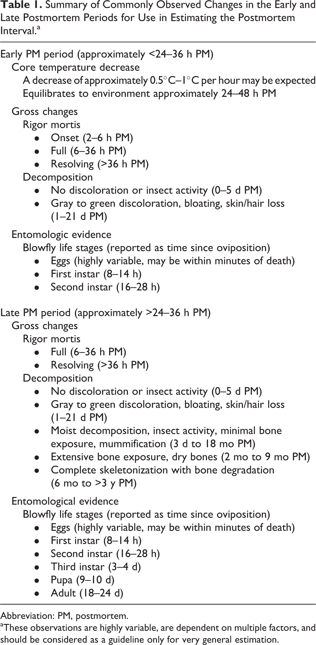
Abbreviation: PM, postmortem.
aThese observations are highly variable, are dependent on multiple factors, and should be considered as a guideline only for very general estimation.
The estimation of PMI remains a central topic in forensic medical research, and it is evident that much additional study is needed. Techniques demonstrating improved accuracy in the estimation of the PMI will undoubtedly assist in veterinary forensic investigations and would likely be investigated for their applicability to human forensic investigations.
Footnotes
Declaration of Conflicting Interests
The author(s) declared no potential conflicts of interest with respect to the research, authorship, and/or publication of this article.
Funding
The author(s) received no financial support for the research, authorship, and/or publication of this article.