
Abstract
Murine noroviruses (MNVs) are highly prevalent in laboratory mice, can cause persistent infections, and have been shown to infect macrophages, dendritic cells, and B cells. To address the potential impact of MNV infection on research outcomes, numerous studies have been conducted with various mouse models of human disease and have generated mixed results, ranging from no impact to significant disease. Many of these studies included histologic evaluations after MNV infection, and these results have similarly been variable in terms of whether MNV induces lesions, despite the fact that localization of MNV by viral culture and molecular techniques have demonstrated systemic distribution regardless of mouse immune status. The aim of this review is to summarize the histologic findings that have been reported with MNV infection in several mouse models. The studies demonstrate that experimental infection of MNV in wild-type mice results in minimal to no histologic changes. In contrast, immunodeficient mice consistently have detectable MNV-induced lesions that are typically inflammatory and, in the most severe cases, accompanied by necrosis. In these, the liver is commonly affected, with more variable lesions reported in the lung, gastrointestinal tract, mesenteric lymph nodes, brain, and spleen. In specific disease models including atherosclerosis, MNV infection had a variable impact that was dependent on the mouse model, viral strain, timing of infection, or other experimental variables. It is important to recognize the reported MNV lesions to help discern the possible influence of MNV infection on data generated in mouse models.
Keywords
Noroviruses are nonenveloped, single-stranded, positive-sense RNA viruses in the Caliciviridae family and are best known for their ability to cause acute gastroenteritis in humans. 8 These viruses are considered species specific and have been reported in animals, including pigs, cows, sheep, cats, dogs, and rats. 8,30,39,44,48,52 A norovirus infecting laboratory mice was first described in 2003 and designated murine norovirus 1 (MNV-1). 22 This virus caused lethal infection in mice deficient in signal transducer and activator of transcription 1 (Stat1-/- ) or the interferon αβ and γ receptors (IfnαβγR-/- ), both of which are important in the immune response against viral pathogens. 41 Evaluation by histopathology after infection of these mice with MNV-1 revealed pneumonia, hepatitis, encephalitis, meningitis, and vasculitis of the cerebral vessels. Interestingly, however, many strains of mice—including wild-type and other immunodeficient strains, such as recombination activating gene-deficient (Rag-/- ) mice, which lack T and B cells—did not succumb to lethal disease after infection with MNV-1. While this initial isolate of MNV caused only transient infection in mice, discovery and characterization of additional MNV strains revealed that the virus can cause persistent infections in laboratory mice and is shed in the feces for prolonged periods. 15
Prevalence
MNV was the first norovirus to be grown in cell culture. 50 The ability to grow the virus in vitro allowed for the development of diagnostic assays (including immunohistochemistry) to detect viral antigens, serologic assays for detection of anti-MNV antibodies, and molecular assays to detect viral RNA. 9,15,16,22,49 Initial large-scale prevalence data on MNV infection revealed that 22.1% of 12 639 serum samples from laboratory mice in the United States and Canada were positive for antibodies to MNV. 16 Another large-scale study likewise confirmed the high prevalence of MNV infection, with a rate of 32.4% for laboratory mice in North America and Europe, and other studies have reported rates as high as 64%. 26,27,32,40 These data indicate that MNV infection in laboratory mice is widespread and worldwide and therefore has the potential to interfere with research results.
Effects on Research
As a result of this novel discovery of a norovirus in laboratory mice, the variable clinical disease seen depending on the type of mouse infected, and the high prevalence of infection in research mice, further study is warranted to reveal and characterize the potential impact of this virus on research using MNV-infected mice. Importantly, MNV has a tropism to infect macrophages and dendritic cells and, more recently, has been reported to infect B cells. 18,50 Infection of immune cells therefore raises the possibility that MNV may be a confounding factor in mouse models of inflammatory disease. Surprisingly, results have been variable in that some mouse models are altered by MNV infection, while others do not show any changes. For example, MNV has been shown to cause Paneth cell abnormalities in a mouse model of Crohn’s disease and to exacerbate inflammatory bowel disease (IBD) in a Helicobacter bilis–driven mouse model of IBD. 4,29 Additionally, MNV has been reported to disrupt the epithelial barrier and increase inflammation in the stomach and colon of Il10-deficient mice. 3 Effects outside the gastrointestinal system have also been reported with MNV infection, resulting in increased atherosclerosis in low-density lipoprotein receptor–deficient mice. 36 MNV infection also altered another commonly used mouse model of atherosclerosis, the apolipoprotein E (Apoe)–deficient mice, but that effect was variable. 12 Interestingly, however, a number of studies have also reported a lack of effect of MNV infection on research mouse models. MNV infection did not alter IBD in Il10-deficient mice or the development of colon cancer in Smad3-deficient mice when inflammation was driven by H. bilis. 13,28 MNV infection did not disrupt the intestinal microbiota in Swiss Webster (SW) or C57BL/6 mice. 34 Infection of mice with MNV also had little or no impact on studies of mice infected with murine cytomegalovirus, Friend retrovirus, vaccinia virus, or influenza A virus and did not alter a mouse model of diet-induced obesity and insulin resistance. 1,5,10,35 Collectively, these studies highlight the variable effect that MNV has on mouse models of disease, suggesting that research using MNV-infected mice should be carefully scrutinized for potential confounding effects.
Histologic Lesions Caused by MNV Infection in Mice
As described above, MNV infection may alter some mouse models of disease but may have little or no impact on other models. This variable response may be due to a number of factors, including the particular disease model being studied or differences in pathogenicity of the infecting MNV strain. A large number of MNV strains have been described and are likely the result of genetic recombination and/or the lack of a proofreading activity in the RNA-dependent RNA polymerase of RNA viruses. 2,6,14,32 Therefore, determining whether MNV infection is a confounding factor in a particular research study using infected mice should be evaluated on a case-by-case basis.
A comprehensive summary has been compiled of whether histologic changes have been reported in particular tissues as a result of MNV infection in laboratory mice (Tables 1 –5). Additional information, including histologic changes, and detailed methods, including infectious challenge, the mouse genetic background, sex, health status, housing type (if reported), and any other infections or treatments, are provided in the Supplemental Tables 1–5. The intent of these summary tables is to assist the pathologist, veterinarian, and scientist with identifying the currently available and published information on the histologic findings in MNV-infected mice. The intent is not a comprehensive review of MNV itself, and so the reader is referred to a number of excellent reviews for more information on MNV and noroviruses in general. 20,21,23,51
Histologic Changes in Mice Naturally Infected With MNV.
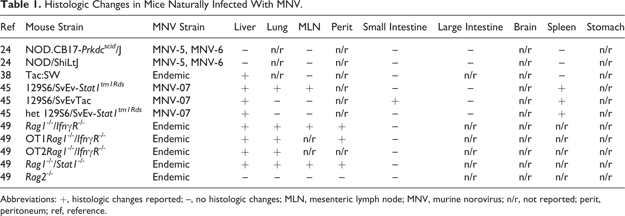
Abbreviations: +, histologic changes reported; –, no histologic changes; MLN, mesenteric lymph node; MNV, murine norovirus; n/r, not reported; perit, peritoneum; ref, reference.
Histologic Changes in Wild-Type Mice Experimentally Infected With MNV.
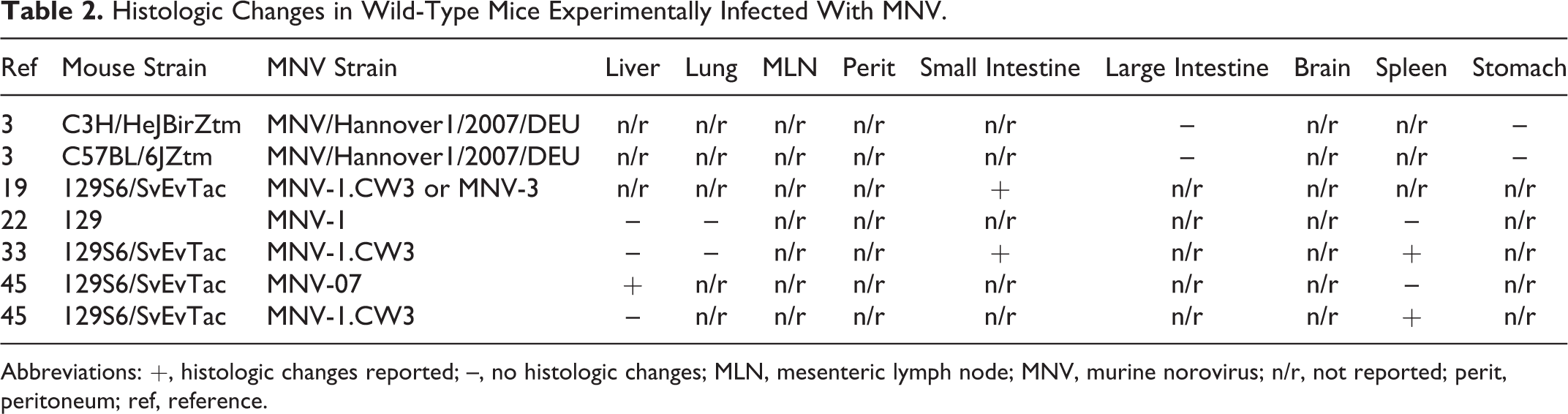
Abbreviations: +, histologic changes reported; –, no histologic changes; MLN, mesenteric lymph node; MNV, murine norovirus; n/r, not reported; perit, peritoneum; ref, reference.
Histologic Changes in Immunodeficient Mice Experimentally Infected With MNV.
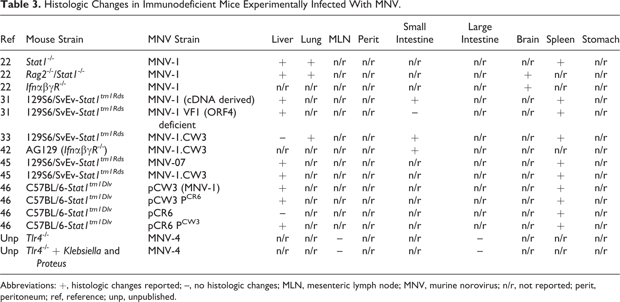
Abbreviations: +, histologic changes reported; –, no histologic changes; MLN, mesenteric lymph node; MNV, murine norovirus; n/r, not reported; perit, peritoneum; ref, reference; unp, unpublished.
Histologic Changes in Mice of Gastrointestinal Disease Models Experimentally Infected With MNV.
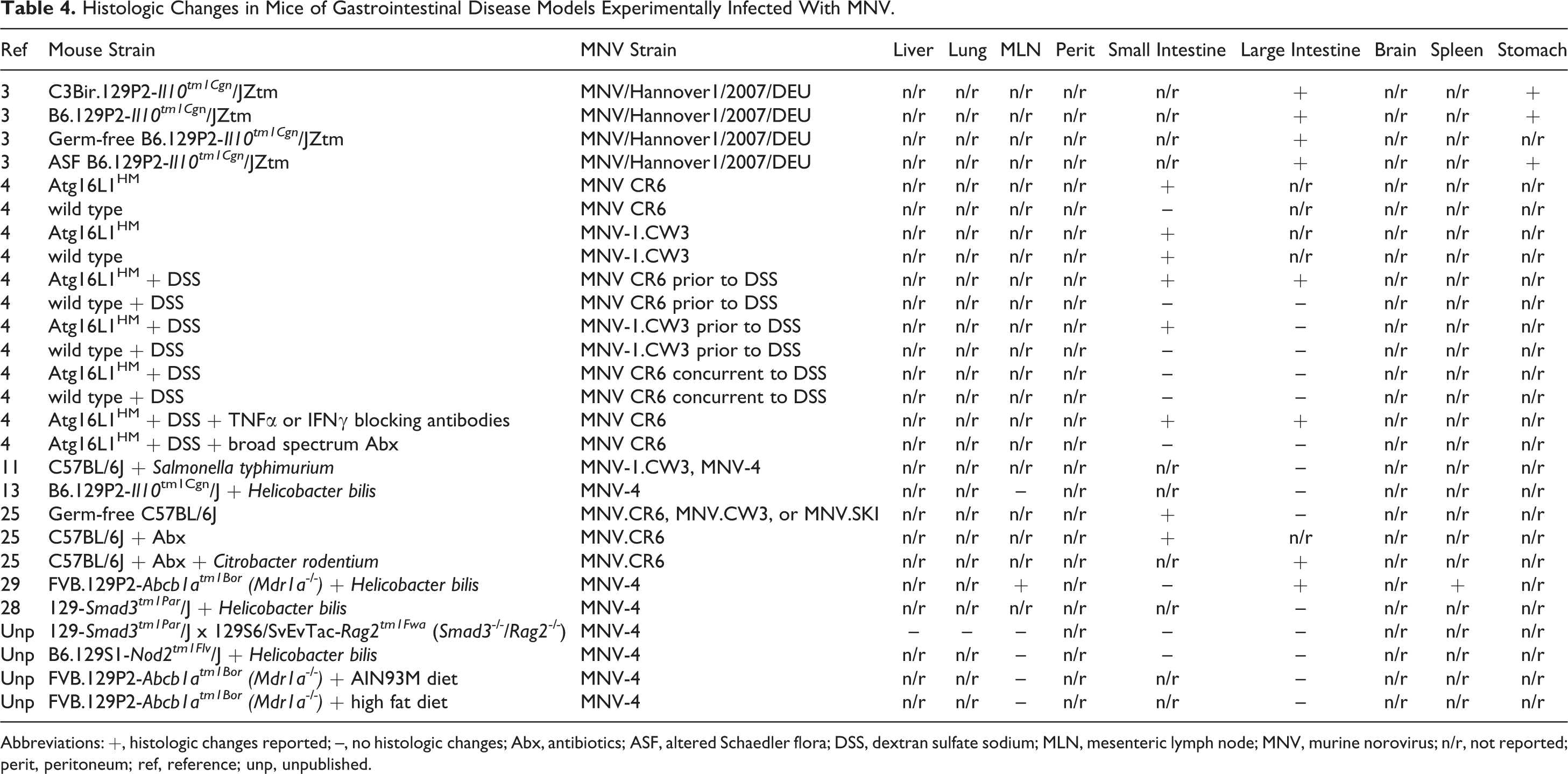
Abbreviations: +, histologic changes reported; –, no histologic changes; Abx, antibiotics; ASF, altered Schaedler flora; DSS, dextran sulfate sodium; MLN, mesenteric lymph node; MNV, murine norovirus; n/r, not reported; perit, peritoneum; ref, reference; unp, unpublished.
Histologic Changes in Mice of Other Disease Models Experimentally Infected with MNV.
Abbreviations: +, histologic changes reported; –, no histologic changes; MLN, mesenteric lymph node; MNV, murine norovirus; n/r, not reported; ref, reference; unp, unpublished.
The histologic changes seen in MNV-infected mice from endemic colonies with natural exposure (ie, via other infected animals) are summarized in Table 1. For these studies, the dose and route of inoculation are not controlled, and the MNV strain or isolate is often unknown or uncharacterized. Generally, MNV-associated liver lesions are significant only in immunodeficient Stat1-/- and IfnγR-/- mice (often in combination with Rag deficiency). These lesions are characterized as mild to severe, multifocal to diffuse inflammatory lesions with variable necrosis or fibrosis. 45,49 In contrast, only minimal to mild hepatic lesions have been reported in immunocompetent outbred SW mice and wild-type 129 mice. In SW mice, there were small foci of inflammatory cells, including neutrophils, lymphocytes, or macrophages in 80% of livers, and they were not consistently associated with MNV-positive IHC staining. 38 In 129 wild-type mice, 15% of livers had hepatic veins with adhesions of mononuclear leukocytes (diagnosed as mild vasculitis by the authors) with occasional mild inflammatory foci. 45 Pneumonia has been reported in naturally infected Stat1-/- and IfnγR-/- mice but not wild-type mice, indicating the importance of the interferon response in controlling inflammation against viral infections such as MNV. 45,49 Interestingly, although noroviruses are gastrointestinal pathogens, only 1 report described histologic changes in the intestines of 129 mice naturally infected with MNV, and these changes were limited to enlargement of Peyer patches with increased germinal centers. 45 This suggests that MNV is well adapted to its immunocompetent host and causes no or only subtle disease in the intestines after natural infection.
Controlled experimental MNV infections in wild-type mice allow for the evaluation of histologic lesions attributable to MNV without other confounding variables, such as immunodeficiency, unknown inoculating dose, viral strain, or timing of infection (Table 2). The majority of studies conducted in wild-type mice infected with MNV show no overt clinical signs of disease or histologic lesions. However, histologic changes were recently reported in wild-type 129 mice, including mild to moderate lesions in the liver (defined as 1–2 or 5–10 foci of inflammatory cells per 10 fields at 10× objective, respectively) and reactive changes in the lymphoid tissues, including enlargement of small intestinal Peyer patches with increased germinal centers, splenic red pulp hyperplasia, and activation of the white pulp. 45 Therefore, the strain or stock of the wild-type mouse and the particular infecting MNV strain or isolate are likely important determinants of whether histologic changes will be seen after infection.
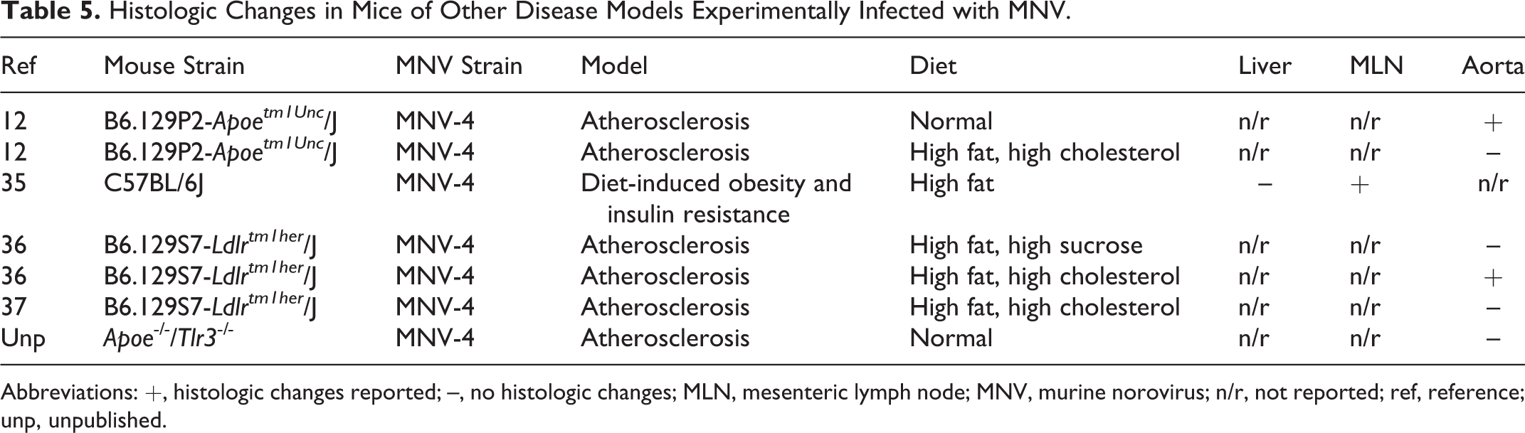
In contrast to immunocompetent mice, experimental infection of mice with known immunodeficiencies results in significant disease (Table 3). Most of these reports involve mice that lack genes important in the type I and type II interferon response to viruses, such as Stat1 and Ifn receptors. 41 In general, histologic changes, such as inflammation, necrosis, and apoptosis, were consistently reported in the liver (Figs. 1–4) and spleen of these immunodeficient mice following infection with MNV. Inflammation in the lungs and small intestines was also reported. Collectively, these reports indicate that experimental MNV infection in immunodeficient Stat1-/- and IfnαβγR-/- mice results in systemic lesions with significant hepatitis. This is in contrast to immunocompetent mice where experimental and natural infections lead to minimal lesions, such as small hepatic foci of inflammatory cells, including neutrophils, lymphocytes, or macrophages 38 or mild centrilobular accumulations of mononuclear leukocytes 45 ; neither group reported severe hepatitis in immunocompetent mice. Additionally, we report previously unpublished findings of MNV infection in mice with Toll-like receptor 4 (Tlr4) deficiencies with and without concurrent infection with the bacteria Klebsiella and Proteus. Tlr4 is a component of the innate immune system recognizing lipopolysaccharide from Gram-negative bacteria, but it has also been reported to be involved in the recognition of viruses. 17 In these Tlr4-deficient mice, MNV infection alone or concurrently with Klebsiella and Proteus infection did not appreciably increase lesions in the mesenteric lymph nodes or colitis (C. Hsu, unpublished data).
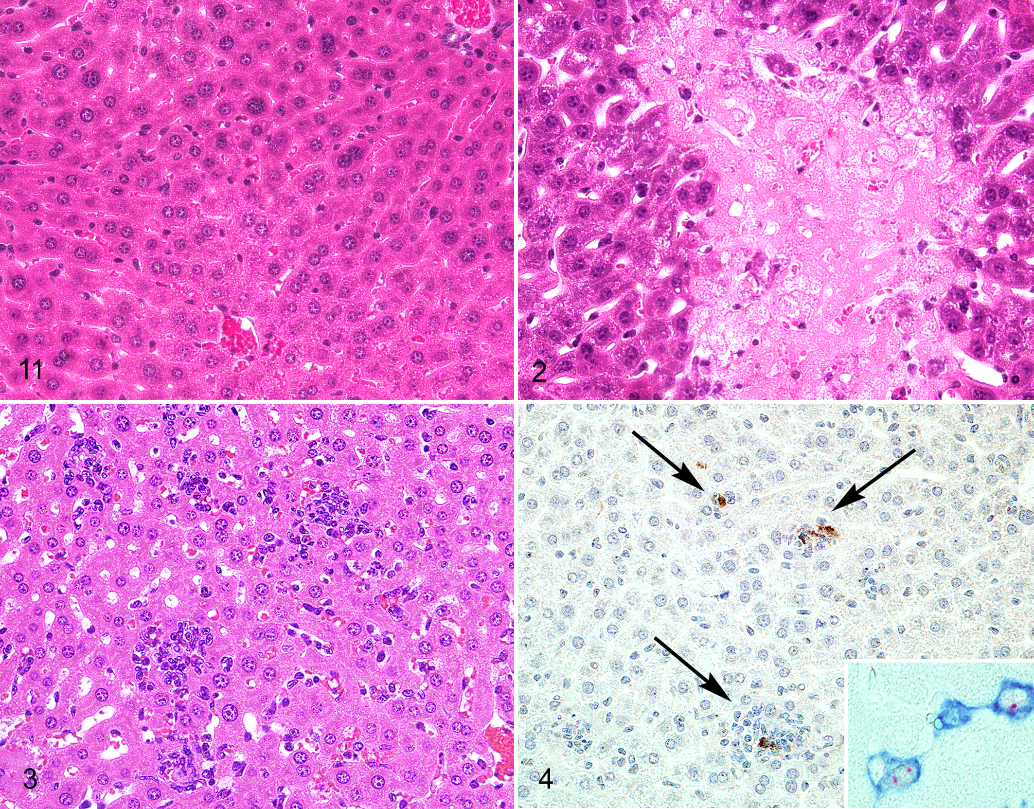
Since noroviruses are gastrointestinal pathogens, much of the research on the impact of MNV has been conducted on mouse models of gastrointestinal disease (Table 4), including dextran sodium sulfate (DSS)–induced colitis, mice with disrupted expression of the autophagy related 16–like 1 gene (Atg16L1), and mice deficient in multidrug resistance gene 1a (FVB.129P2-Abcb1atm1Bor; Mdr1a-/- ; Figs. 5–8) or in interleukin-10 (B6.129P2-Il10tm1Cgn /J;Il10-/- ). DSS is administered orally to mice to cause direct damage to the intestinal epithelium as a model of IBD, while mutations in the autophagy gene ATG16L1 have been associated with Crohn’s disease. 4,7,43 Mdr1a-/- mice have an altered intestinal epithelial barrier, while Il10-/- mice have impaired regulatory T cells resulting in IBD. 7,43 MNV infections in different mouse models of gastrointestinal disease varied in their ability to cause intestinal histologic lesions, perhaps due to the particular MNV strain used for infection, timing of MNV infection, or whether another experimental infectious agent was present. For example, we recently reported that MNV-4 did not alter IBD in mice lacking the immunoregulatory cytokine IL-10 when intestinal disease was driven by infection with H. bilis; however, others have reported histologic changes as a result of MNV infection in Il10-/- mice with a different strain of MNV and without H. bilis. 3,13 Additionally, mice with hypomorphic expression of the ATG16L1 protein (ATG16L1HM) displayed inflammatory hallmarks of Crohn’s disease after DSS treatment when infected with MNV CR6, which persistently infects mice, but not with MNV-1.CW3, which is not persistent. 4 This histologic change was also seen when ATG16L1HM mice were infected with MNV CR6 prior to DSS treatment but not when MNV CR6 was given concurrently with DSS administration, suggesting that timing of MNV infection is important. Finally, with MNV infection in 129-Smad3tm1Par /J x 129S6/SvEvTac-Rag2tm1Fwa (Smad3-/-/Rag2-/- ) double-knockout mice, B6.129S1-Nod2tm1Flv /J (Nod2-/- ) mice with and without H. bilis infection, and Mdr1a-/- mice fed a purified or high-fat diet, we found that MNV-4 infection did not cause histologic changes in these mice (unpublished data). Overall, the impact of MNV infection in mouse models of gastrointestinal disease varies widely, from having little to no impact to causing significantly increased inflammation in the gastrointestinal tract. Conversely, it has even been reported that MNV infection provides protection against inflammation induced by Citrobacter rodentium infection and antibiotic treatment. 25
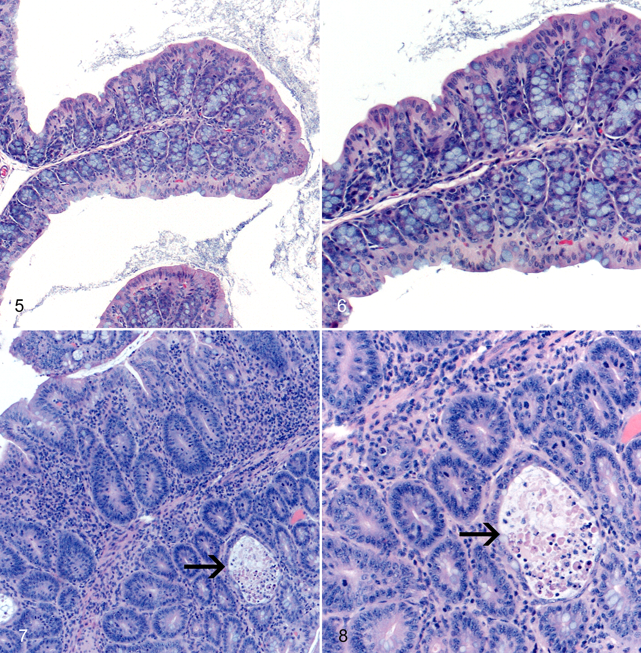
Despite the fact that noroviruses are gastrointestinal pathogens, MNV may infect research mice used to study a wide array of diseases beyond the gastrointestinal system. Since MNV is highly prevalent in many research institutions and has been reported to infect macrophages, dendritic cells, and B cells, MNV has the potential to alter research outcomes in diseases for which these cells are important, including atherosclerosis, diabetes, and obesity (Table 5). Our laboratory has evaluated the impact of MNV infection on atherosclerosis in B6.129P2-Apoetm1Unc /J (Apoe-/- ; Figs. 9, 10) and in low-density lipoprotein receptor (Ldlr)–deficient mice (B6.129S7-Ldlrtm1her /J), and we include previously unpublished data from our laboratory on the lack of impact that MNV infection had on the development of atherosclerosis in Apoe/Tlr3 double-knockout mice. 36,37 Interestingly, although a major component of atherosclerotic plaques is lipid-laden macrophages and even though MNV has as tropism for infecting macrophages, we found a variable influence of MNV infection on the development of atherosclerosis. These data underscore the importance of continuing to investigate the impact that MNV infection may have on various mouse models of disease.
Many studies have employed immunohistochemistry, immunofluorescence, in situ hybridization, and in situ reverse transcription polymerase-chain reaction to localize virus in tissue sections derived from MNV-infected mice (Figs. 4, 9c). These studies confirm that MNV can infect macrophages and dendritic cells in a variety of tissues, including liver, lung, mesenteric lymph node, peritoneum, small intestine, spleen, and aorta, indicating that systemic spread of the virus may occur in both immunocompetent and immunodeficient mice (Table 6). In the liver, MNV antigen was detected in Kupffer cells 38,47,50 and inflammatory cells, including F4/80+ macrophages. 49 MNV antigen was detected in mesenteric lymph node DC-like cells, F4/80+ cells in the medullary cords, and CD40+ paracortical cells in alveolar mononuclear cells and pleural inflammatory cells, peritoneal macrophages, and mesothelium. 49 Studies examining the spleen demonstrated MNV antigen in macrophages and macrophage-like cells, 47 red pulp and marginal zone, 50 and nonlymphoid cells in the white pulp. 50 Although the majority of MNV+ cells in the small intestine are inflammatory or antigen-presenting cells within the lamina propria or gastrointestinal-associated lymphoid tissues, enteric epithelium was also positive in some studies. 3,33,49
Localization of MNV by Immunohistochemistry, Immunofluorescence, In Situ Hybridization, or In Situ RT-PCR.
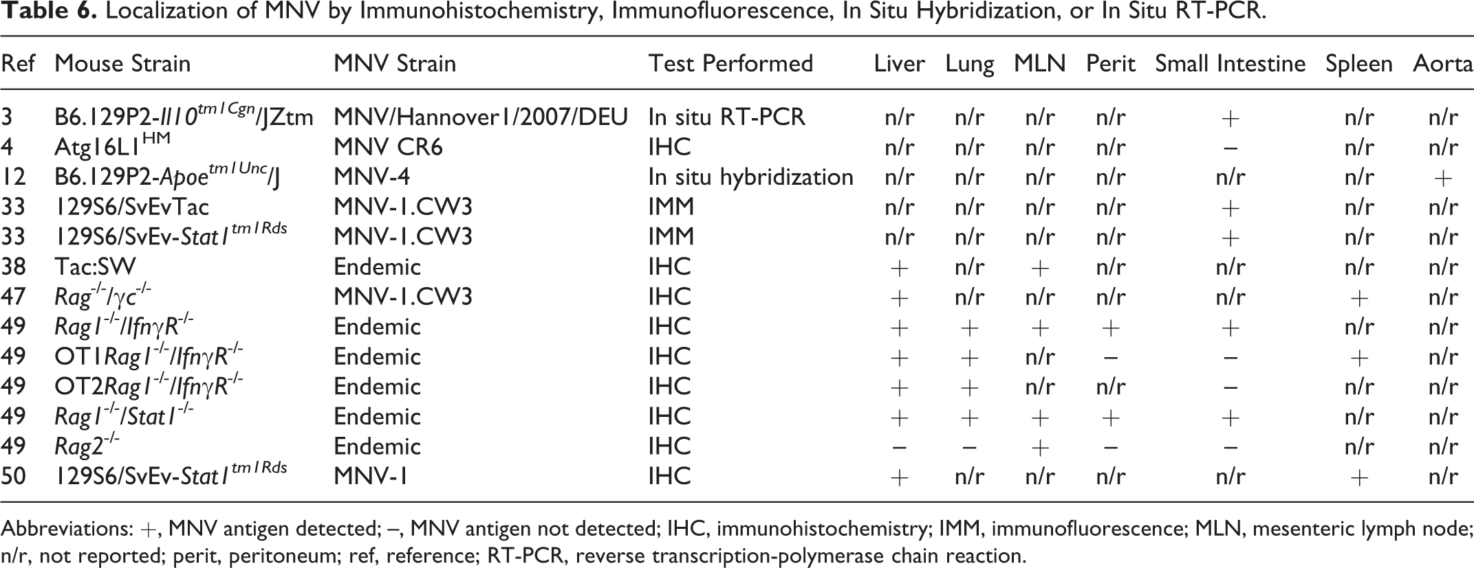
Abbreviations: +, MNV antigen detected; –, MNV antigen not detected; IHC, immunohistochemistry; IMM, immunofluorescence; MLN, mesenteric lymph node; n/r, not reported; perit, peritoneum; ref, reference; RT-PCR, reverse transcription-polymerase chain reaction.
Conclusion
MNV infection in laboratory mice continues to be endemic in many research colonies. Therefore, determining the impact that MNV infection may have on various mouse models of disease is important to ensure that research results are valid. While it is impractical to test whether MNV infection alters all mouse models of disease, more and more studies are being performed to answer the following question: “Will MNV infection alter and confound my research results?” MNV is detected and may induce lesions in a wide variety of tissues, principally liver, spleen, and gastrointestinal tract, from immunocompetent and immunodeficient mice where it infects mononuclear cells, including macrophages and dendritic cells. Importantly, MNV has a variable impact on mouse models, in some cases increasing lesions while in other cases having no impact. Consequently, it is important to recognize reported MNV lesions to help discern the possible influence of MNV infection on data generated in mouse models.
Footnotes
Acknowledgements
We thank Claudia Monaco from the University of Oxford for sharing Apoe/Tlr3 double-knockout mice. We thank Herbert “Skip” Virgin from Washington University and Jerrold Ward from Global VetPathology for sharing previously published figures for reprint. We also thank Jisun Paik, Thea Brabb, and Audrey Seamons from the University of Washington for their assistance with the unpublished studies.
Declaration of Conflicting Interests
The author(s) declared no potential conflicts of interest with respect to the research, authorship, and/or publication of this article.
Funding
The author(s) disclosed receipt of the following financial support for the research, authorship, and/or publication of this article: This work was supported by the National Institutes of Health (grants R21-OD011135, R01-OD011149) and the University of Washington’s Nutrition Obesity Research Center (grant P30-DK035816).
References
Supplementary Material
Please find the following supplemental material available below.
For Open Access articles published under a Creative Commons License, all supplemental material carries the same license as the article it is associated with.
For non-Open Access articles published, all supplemental material carries a non-exclusive license, and permission requests for re-use of supplemental material or any part of supplemental material shall be sent directly to the copyright owner as specified in the copyright notice associated with the article.