
Abstract
Miniature dachshund dogs are a common breed in Japan and are known to be predisposed to granulomatous diseases. Here we report the pathologic features of multiple lingual nodules in 7 miniature dachshunds. Seven dogs had multiple nodules of variable sizes mainly on the ventral and lateral surface of the tongue. In addition, 1 dog also had masses on the left oral mucosa. Three cases had recurrence after surgical resection. Histologically, the lingual nodules were composed of aggregates of foam cells with clear vacuolated cytoplasm that were negative for oil red O, PAS, and alcian blue. They stained positively for CD204 (macrophage scavenger receptor) and MHC class II and negatively for Iba-1, E-cadherin, adipophilin, cytokeratins, S-100, and nestin. These findings indicate that the multiple lingual nodules in miniature dachshunds are an unusual, unique lesion consisting of macrophage-derived foam cells, which does not correspond to canine lingual diseases reported to date.
Miniature dachshund dogs are one of the most popular breeds in Japan (the third from 2008 to 2013; data by Japan Kennel Club http://www.jkc.or.jp/modules/publicdata/index.php) and are known to be predisposed to granulomatous diseases such as suture granuloma, panniculitis, and granulomatous gastroenteritis. 5 However, lingual disease specific to this breed has not been reported to date. Here we describe the histopathological features of multiple lingual nodules characterized by accumulation of foam cell macrophages in 7 miniature dachshunds. All case material was submitted as biopsies to the Laboratory of Veterinary Pathology, Osaka Prefecture University.
Case histories of the 7 miniature dachshunds are listed in Table 1. Age of the dogs at initial biopsy ranged from 6 to 13 years (median, 9.7 years). Seven dogs had multiple lingual nodules of variable size, distributed mainly on the ventral and lateral surface of the tongue (Fig. 1). One dog (dog No. 1) had a bleeding nodule. There were no other clinical signs in the other 6 dogs. In 2 dogs (dog Nos. 1, 5), the nodules were noticed by the owners, while they were noticed by the veterinarians in 5 dogs (dog Nos. 2, 3, 4, 6, 7). Involvement of the oral mucosa (the left oral vestibule) with suspected oral pain was present in 1 dog (dog No. 4, Supplemental Fig. 1). Four dogs were followed up for more than 2 years; 3 of the 4 had recurrence of the lingual nodules (dog Nos. 1, 4, 5). All cases were treated by surgical excision. Three dogs had a past history of surgery (dog Nos. 1, 4, 6). Dog No. 1 had an earlier duodenal biopsy due to vomiting and hematochezia 4 years before the first surgery for the lingual nodule; mild infiltration of neutrophils and lymphocytes was observed. The dog had subcutaneous panniculitis 2 years after the second surgery for the lingual nodules. There were no specific lesions that occurred concurrently with the lingual nodules.
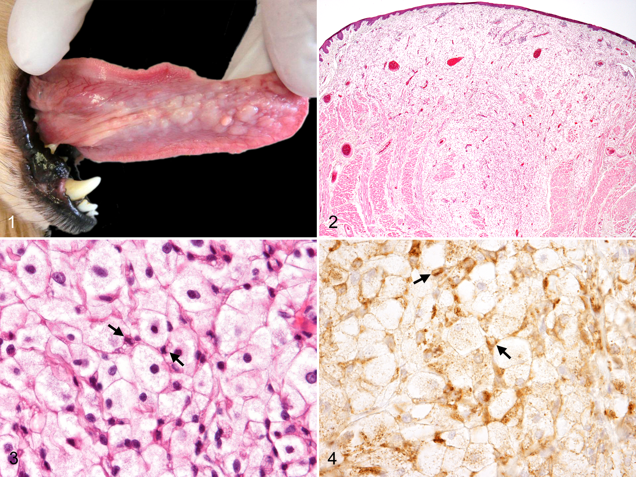
Histiocytic foam cell nodules, dog No. 1.
Clinical and Additional Pathologic Findings in Miniature Dachshunds With Histiocytic Foam Cell Nodules of the Tongue.
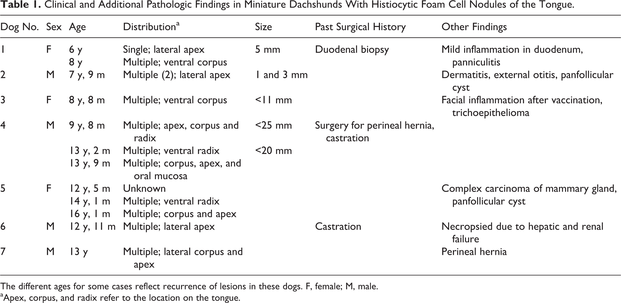
The different ages for some cases reflect recurrence of lesions in these dogs. F, female; M, male.
aApex, corpus, and radix refer to the location on the tongue.
Specimens were fixed in 10% neutral buffered formalin, embedded in paraffin, cut at 4 μm, and stained with hematoxylin and eosin. Selected slides were subjected to immunohistochemistry. Primary antibodies used are listed in Suppl. Table 1. The sections were reacted with each primary antibody for 1 hour at room temperature and were visualized with horseradish peroxidase-conjugated secondary antibody (Histofine Simplestain MAX-PO; Nichirei, Tokyo, Japan) and 3, 3’-diaminobenzidine tetrahydrochloride (DAB; Nichirei). Specimens of 3 cases (dog Nos. 1, 4, 5) were immediately frozen at –80°C and were cut at 10 μm in a cryostat and stained with oil red O, Sudan III, alcian blue (pH 1.0 and pH 2.5), and periodic acid Schiff (PAS). The formalin-fixed sample of dog No. 1 was postfixed in osmium tetrachloride, dehydrated through graded alcohols, and embedded in epoxy resin. Ultrathin sections were stained with uranyl acetate and lead citrate and examined with a Hitachi H-7500 electron microscope (Hitachi, Tokyo, Japan).
Microscopically, the lingual nodules were located in the lamina propria, occasionally involving the muscle tissue (Fig. 2). They were composed of an accumulation of large foam cells with distinct cell borders, clear and vacuolated cytoplasm, and centrally located oval nuclei (Fig. 3). Between the foam cells, there were small round to polygonal cells with clear cytoplasmic vacuoles, interpreted as macrophages (Fig. 3 arrows). Mitotic figures were not observed in the foam cells. Mucosal hyperplasia and ulceration were common overlying the lesions (Suppl. Fig. 2). Aggregation of the foam cells just beneath the mucosa was occasionally seen (Suppl. Fig. 3).
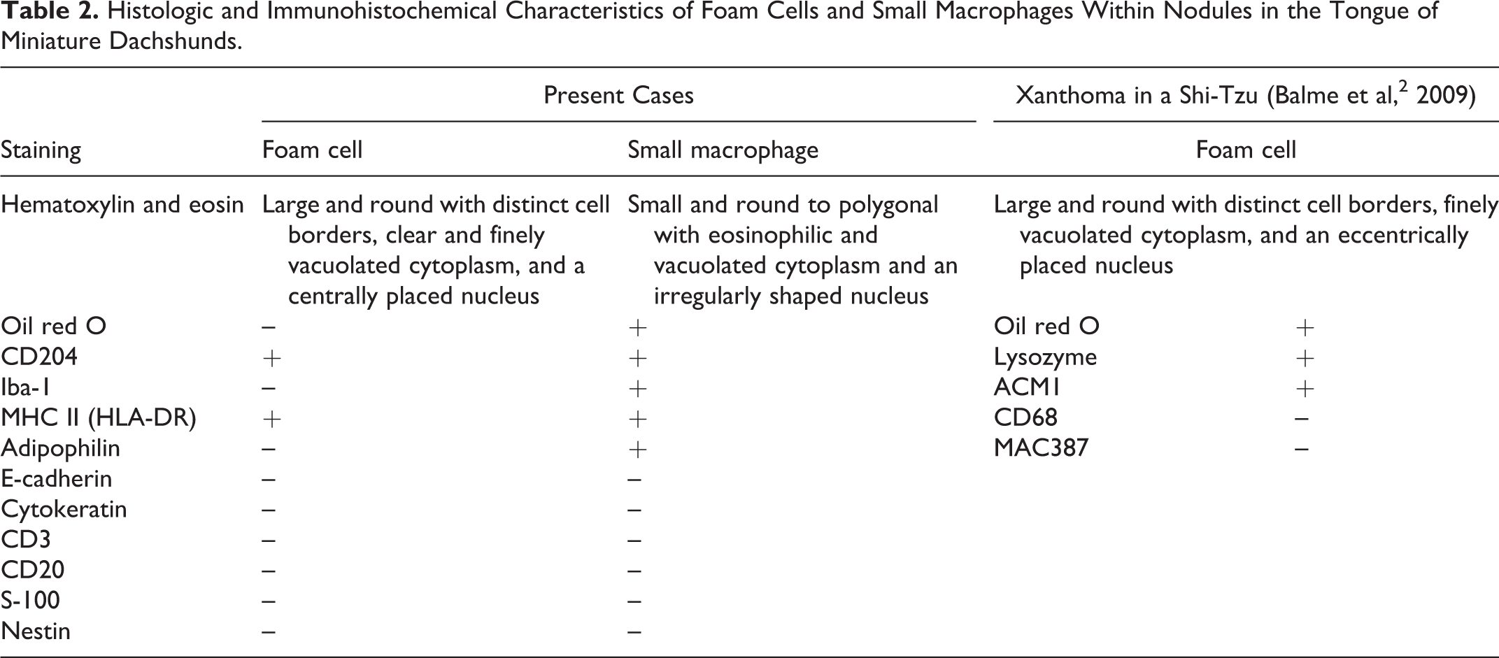
The foam cells were negative for oil red O, Sudan III, PAS, and alcian blue, whereas the small macrophages with cytoplasmic vacuoles were positive for oil red O and Sudan III. Immunohistochemically, the foam cells were moderately positive for macrophage scavenger receptor CD204 (Fig. 4) and MHC class II (HLA-DR) and negative for Iba-1, E-cadherin, adipophilin, CD3, CD20, cytokeratins (AE1/AE3), S-100, and nestin (Table 2). The small macrophages were strongly positive for CD204 (Fig. 4 arrows) and Iba-1. The cytoplasmic vacuoles of these cells stained with adipophilin (Suppl. Figs. 4, 5), which, along with the oil red O- and Sudan III-positive staining, is consistent with lipid droplets. Only a few foam cells were positive for Ki-67 (0.5%), less frequent than the basal mucosal epithelia (20.3%). Ultrastructurally, the cytoplasmic vacuoles in the foam cells were composed of homogenous, electron-lucent material surrounded by a single limiting membrane (Suppl. Fig. 6). Other characteristic structures were not observed in the foam cells.
Histologic and Immunohistochemical Characteristics of Foam Cells and Small Macrophages Within Nodules in the Tongue of Miniature Dachshunds.
Miniature dachshunds are known to be predisposed to granulomatous diseases. Suture granuloma, granulomatous gastroenteritis, panniculitis, and sterile granuloma have been reported after ovariohysterectomy in female miniature dachshunds in Japan. 5 Male and intact female dogs are also affected with granulomatous diseases in the gastrointestinal tract and epidural region. 1,7,9 It is suspected that these granulomatous diseases in miniature dachshunds involve an immune dysregulation since some cases are responsive to immunosuppressive therapy.
Lingual nodular lesions in dogs represent infection, foreign material, immune dysregulation, xanthoma, and neoplasia. 4 In dogs, there are some granulomatous and histiocytic diseases of unknown cause, including eosinophilic granuloma, sterile granuloma/pyogranuloma, and reactive histiocytosis. 3,6,8 Immune dysregulation is suspected in their pathogenesis.
Granular cell tumor (GCT) should be considered in the differential diagnosis, as the foam cells in the present cases are cytologically similar to neoplastic cells in canine lingual GCTs. GCT is a neoplasm composed of round to polygonal cells with PAS-positive, eosinophilic granular cytoplasm. 10 A recent study showed that cultured and transplanted cells from lingual GCT are positive for S-100, nestin, and CD133, suggesting neural crest origin. 11 The foam cells in the present cases did not have PAS-positive cytoplasmic granules and were negative for S-100 and nestin. Ectomesenchymal chondromyxoid tumor is a benign neoplasm involving the anterior dorsal tongue in humans. 12 It is composed of round to polygonal cells with uniform small nuclei and moderate amount of faintly basophilic cytoplasm, sharing some histologic features with the foam cells in the present cases. The neoplastic cells stain with alcian blue, GFAP, and cytokeratins in most cases; these phenotypes are different from those of the foam cells.
The multiple lingual lesions were composed of an accumulation of foam cells expressing CD204 and MHC class II admixed with infiltrating macrophages that stained positively for CD204, Iba-1, and adipophilin. These findings indicate a granulomatous disorder. Multiple xanthomas similar to human verruciform xanthoma are reported in the oral cavity (tongue and inner lip) and upper alimentary tract of a Shi-Tzu dog (Table 2). 2 The gross and histopathologic features of the lingual xanthomas are similar to those in the present cases. However, the foam cells in the present cases do not have the oil red O-positive lipid droplets that are a hallmark of xanthomas.
Other granulomatous disorders occurring in the tongue are eosinophilic granuloma and foreign body stomatitis (glossitis). 3 Eosinophilic granuloma (linear granuloma) in dogs occurs as a familial disease in young Siberian huskies and sporadically in other breeds. Affected dogs have single or multiple plaques on the surface of the tongue. Eosinophil infiltration and collagenolysis are the histopathologic features; however, they were not found in the present cases. Foreign body stomatitis is characterized by accumulation of multinucleated giant cells and macrophages with foreign material (eg, plant material) embedded in the lesions. There are neither multinucleated giant cells nor foreign material in the lingual lesions of the present cases.
The multiple lingual lesions in miniature dachshunds are a unique entity that do not correspond to other lingual lesions in dogs reported to date. Although the accumulated materials are still unspecified, the foam cells are considered to originate from macrophages/histiocytes. Their distribution pattern and low proliferative activity indicate a reactive rather than neoplastic disease. Miniature dachshunds are predisposed to granulomatous diseases that are thought to be caused by immune dysregulation. Accumulation of further cases and therapeutic study (ie, immunosuppression) would be useful to further understand the pathogenesis of this disease.
Footnotes
Declaration of Conflicting Interests
The author(s) declared no potential conflicts of interest with respect to the research, authorship, and/or publication of this article.
Funding
The author(s) received no financial support for the research, authorship, and/or publication of this article.
References
Supplementary Material
Please find the following supplemental material available below.
For Open Access articles published under a Creative Commons License, all supplemental material carries the same license as the article it is associated with.
For non-Open Access articles published, all supplemental material carries a non-exclusive license, and permission requests for re-use of supplemental material or any part of supplemental material shall be sent directly to the copyright owner as specified in the copyright notice associated with the article.