
Abstract
Domestic ducks can be a key factor in the regional spread of H5N1 highly pathogenic avian influenza (HPAI) virus in Asia. The authors performed experimental infections to examine the relationship between corneal opacity and H5N1 HPAI virus infection in domestic ducks (Anas platyrhyncha var domestica). A total of 99 domestic ducks, including 3 control birds, were used in the study. In experiment 1, when domestic ducks were inoculated intranasally with 2 H5N1 HPAI viruses, corneal opacity appeared more frequently than neurologic signs and mortality. Corneal ulceration and exophthalmos were rare findings. Histopathologic examinations of the eyes of domestic ducks in experiment 2 revealed that corneal opacity was due to the loss of corneal endothelial cells and subsequent keratitis with edema. Influenza viral antigen was detected in corneal endothelial cells and some other ocular cells by immunohistochemistry. Results suggest that corneal opacity is a characteristic and frequent finding in domestic ducks infected with the H5N1 HPAI virus. Confirming this ocular change may improve the detection rate of infected domestic ducks in the field.
Keywords
Domestic ducks have played an important epidemiologic role in the regional spread of the H5N1 highly pathogenic avian influenza (HPAI) virus in Southeast Asia. 11,12,23,38 In contrast to chickens, in which infection is usually fatal, clinical signs in domestic ducks infected with the H5N1 HPAI virus greatly vary, ranging from asymptomatic to a fatal infection. 29,30,40,54 The main epidemiologic problem is that asymptomatically infected ducks can shed the virus for a particular period. 45,53 Therefore, the expansion of our ability to detect infected domestic ducks with few clinical signs would be valuable for containing an epidemic of this disease.
In general, the major clinical signs of domestic ducks infected with the H5N1 HPAI virus are depression, neurologic signs, and mortality. 30,54 In addition, we have found that domestic ducks frequently exhibit corneal opacity in experimental infections. 51,53,54 Similar ocular changes in ducks—termed cloudy eye, ocular opacity, or blindness—have been reported in related studies on the H5N1 HPAI virus. 2,20,27,29 If corneal opacity is a frequent finding in domestic ducks infected with the H5N1 HPAI virus, checking for this ocular change could be a useful clinical approach to detect infected domestic ducks. In the present study, we performed experimental infections to elucidate the pathogenesis of and association between corneal opacity and H5N1 HPAI virus infection in domestic ducks.
Materials and Methods
Animals
Japanese domestic ducks (Anas platyrhyncha var domestica), a half-breed of wild mallard and domesticated ducks, were obtained from a breeder at 1 day of age and raised on commercial feed in an isolated facility. The birds were moved into negative-pressure isolators in a biosafety level 3–approved laboratory for acclimatization 1 week before virus inoculation. They were provided feed and water ad libitum. Preinoculation sera from the birds were assessed by hemagglutination inhibition (HI) tests and were negative for antibodies against the viruses used in the present study. All experimental procedures involving birds were approved by the Ethics Committee of the National Institute of Animal Health, Japan (authorization Nos. 07-118, 08-139).
Virus
We used 2 H5N1 HPAI virus strains: clade 2.2 virus A/chicken/Miyazaki/K11/2007 (Ck/Miya/K11/07) and clade 2.3.2 virus A/whooper swan/Akita/1/2008 (Ws/Akita/1/08). These viruses were isolated from past Japanese outbreaks. 44 The stock virus was propagated for 2 days in the allantoic cavity of 10-day-old embryonated chicken eggs at 37°C. Fresh infectious allantoic fluid was harvested and stored at −80°C until use. The inoculum was prepared by diluting the infectious allantoic fluid in phosphate-buffered saline on the day of inoculation.
Experimental Infection 1
In experiment 1, we examined the prevalence of corneal opacity in 4-week-old domestic ducks experimentally infected with the H5N1 HPAI virus. Six inoculation groups of domestic ducks (4 birds per group; Nos. 1–24) were established for 6 inoculation doses of Ck/Miya/K11/07. Similarly, 6 groups of domestic ducks (Nos. 25–48) were established for inoculation with Ws/Akita/1/08. The domestic ducks in each group were intranasally inoculated with 101 to 106 50% egg infectious doses (EID50) of the corresponding virus. Clinical findings, including mortality, depression, neurologic signs, and ocular changes, were recorded daily for 14 days. On day 14 postinoculation (PI), the inoculated ducks were euthanized with an overdose intravenous injection of pentobarbital sodium (100 mg/kg body weight). H5N1 HPAI virus infection in inoculated ducks was confirmed by the presence of HI antibody on day 14 PI in the present study. Birds that died before day 14 PI were not examined by HI test.
Experimental Infection 2
Experiment 2 was conducted to examine the pathogenesis of ocular lesions in 2-week-old domestic ducks through a histopathologic evaluation. Twenty-four ducks (Nos. 49–72), as the first group, were intranasally inoculated with 105 EID50 of Ck/Miya/K11/07. As the second group, 24 ducks (Nos. 73–96) were intranasally inoculated with 105 EID50 of Ws/Akita/1/08. Three birds in each group were euthanized on days 1, 2, 3, 4, 5, 7, 10, and 14 PI. Because of unexpected mortality of some birds inoculated with Ws/Akita/1/08, 2 birds (Nos. 88, 89) were examined on day 6 PI, and only 1 bird (No.90) was examined on day 7 PI. The manifestation of corneal opacity in each duck was determined by daily observation. The eyes and brains of all ducks were collected at necropsy and histopathologically examined. The eyes of 2 domestic ducks (Nos. 12, 40), which developed exophthalmos in experiment 1, were also included in the analysis.
HI Antibody Test
Preinoculation sera and the sera of birds euthanized on day 14 PI of experiment 1 were collected for HI tests using 4 hemagglutination units of the corresponding virus as antigen and 0.5% chicken red blood cell suspension. 49 All sera were pretreated at 56°C for 30 minutes. An HI antibody titer <1:16 was considered negative for antibody production. 49 The sera of three 4-week-old uninoculated control ducks (Nos. 97–99) were prepared as negative controls.
Histopathologic Analysis
The eyes and brains from ducks in experiment 2 were fixed in 10% neutral-buffered formalin. After removal of the nictitating membrane and Harderian gland from the eyes, decalcification was performed with 20% formic acid solution, as necessary. Samples were embedded in paraffin, sectioned at 2 μm, and stained with hematoxylin and eosin, the periodic acid–Schiff reaction, and the von Kossa method. Uninoculated ducks (Nos. 97–99) were used as histopathologic controls.
Immunohistochemistry was performed to detect influenza viral antigen using a Histofine Simple Stain MAX PO (M) Kit (Nichirei Inc, Tokyo, Japan) and mouse monoclonal antibody specific for the type A influenza virus matrix protein (1:500, clone GA2B; AbD Serotec, Kidlington, UK). The sections were pretreated with 10 mM citrate buffer (pH 6.0) in a microwave oven at 500 W for 15 minutes for antigen retrieval. Primary antibodies diluted in phosphate-buffered saline containing 1% bovine serum albumin were added overnight at 4°C. 3′-3-Diaminobenzidine tetrahydrochloride was used as the chromogen. All slides were counterstained with Mayer’s hematoxylin. Positive control sections were obtained from samples in previous experiments. 54 Ocular and brain tissues of uninoculated ducks (Nos. 97–99) were prepared as negative controls for immunohistochemistry. We did not observe any false-positive reaction in the negative control sections.
Results
Experiment 1
Corneal opacity was the most frequent clinical sign in both inoculation groups (Table 1; Figs. 1, 2).
Clinical Signs of Domestic Ducks Inoculated With H5N1 Highly Pathogenic Avian Influenza Viruses in Experiment 1.a
Abbreviations: CO, corneal opacity; D, depression; E, exophthalmos; HI, hemagglutination inhibition antibody production in ducks euthanized on day 14 postinoculation; M, mortality; NS, neurologic symptoms; U, corneal ulceration; V, virus strain and inoculation dose (50% egg infectious doses).
aData are presented as number of positive birds. The total number of birds per cell is 4, unless noted otherwise.
bTotal number of birds, n = 3.
cTotal number of birds, n = 1.
dTotal number of birds, n = 2.
Highly pathogenic avian influenza; domestic ducks.
In the Ck/Miya/K11/07 inoculation groups, 1 bird (No. 17) inoculated with 105 EID50 died after exhibiting depression on day 5 PI (Table 1). Sixteen domestic ducks developed corneal opacity after inoculation with 103 EID50 or more of Ck/Miya/K11/07. Bilateral corneal opacity was observed, although in some cases opacity started first in 1 eye and became bilateral 1 to 2 days later. Different severity was occasionally observed between eyes of the same animal. The mean time for manifesting corneal opacity in 16 ducks was 4.8 days PI (range, 4–6 days), and the results in each inoculation group are shown in Table 2. The manifestation of corneal opacity occurred sooner with higher inoculation doses. Except for 1 duck (No. 17), which died on day 5 PI, 9 of 15 ducks recovered from opacity on days 9–13 PI (Table 2). One duck (No. 12) in the 103 EID50 inoculation group exhibited bilateral exophthalmos on days 12–14 PI (Table 1, Fig. 3). Corneal ulceration was found in 1 duck (No. 19) in the 105 EID50 inoculation group on day 14 PI. Ulceration was difficult to determine by clinical observation and was identified more clearly after formalin fixation. Only 1 duck (No. 14) in the 104 EID50 inoculation group exhibited mild torticollis as a neurologic sign, on days 10–14 PI.
Mean Time to Manifestation and Recovery of Corneal Opacity of Domestic Ducks Inoculated With H5N1 Highly Pathogenic Avian Influenza Viruses in Experiment 1.
Abbreviations: CO, the number of ducks with corneal opacity; COR, the number of ducks recovered from corneal opacity; MTTM, mean time to manifestation of corneal opacity (data range in parentheses expressed as days postinoculation); MTTR, mean time to recovery from corneal opacity (data range in parentheses expressed as days postinoculation); n/a, not applicable; V, virus strain and inoculation dose (50% egg infectious doses).
The domestic ducks in the Ws/Akita/1/08 inoculation groups exhibited higher rates of mortality and morbidity than Ck/Miya/K11/07-inoculated ducks (Table 1). Mortality occurred from day 6 to 9 PI. Bilateral corneal opacity was observed in 12 ducks inoculated with 104 EID50 or a higher dose of Ws/Akita/1/08. The mean time for manifesting corneal opacity in these 12 ducks was similar to that in Ck/Miya/K11/07-inoculated birds at 4.4 days PI (range, 3–7 days; Table 2). The manifestation of corneal opacity became slightly faster in parallel with higher inoculation dose. Recovery from opacity was not observed in these 12 ducks until their death on days 6–9 PI or until day 14 PI, the final day of the experiment. Mild lateral exophthalmos of the left eye was noticed in 1 duck (No. 40) in the 104 EID50 inoculation group on day 14 PI (Table 1). Corneal ulcerations were confirmed in 1 duck (No. 41) that died on day 8 PI in the 105 EID50 inoculation group and in 2 ducks (Nos. 37 and 40) on day 14 PI in the 104 EID50 inoculation group (Fig. 4). Severe neurologic signs composed of torticollis, circling movement, and intermittent tremor started from day 5 PI. Four ducks (Nos. 39, 41–43) with severe neurologic signs in the 104 and 105 EID50 inoculation groups died on days 6–9 PI, while 1 duck (No. 47) with torticollis in 106 EID50 inoculation group survived for 14 days.
HI antibody test was performed for domestic ducks euthanized on day 14 PI. HI antibody production was confirmed in all domestic ducks that exhibited corneal opacity (Table 1), while all domestic ducks without corneal opacity were negative for HI antibody. The geometric means of HI antibody titers of the 103 to 106 EID50 inoculation groups of Ck/Miya/K11/07 were 32 (n = 4), 45.3 (n = 4), 25.4 (n = 3), and 76.1 (n = 4), respectively. The geometric mean titers of the 104 to 106 EID50 inoculation groups of Ws/Akita/1/08 were 50.8 (n = 3), 64 (n = 1), and 128 (n = 2), respectively.
Experiment 2
The clinical signs of the inoculated ducks were similar to those in experiment 1. Corneal opacity emerged on day 3 or 4 PI in both inoculation groups (Table 3). Recovery from corneal opacity was not observed in experiment 2. Mortality was found only in the Ws/Akita/1/08 inoculation group.
Corneal Opacity and Mortality of Domestic Ducks Inoculated With H5N1 Highly Pathogenic Avian Influenza Viruses in Experiment 2.a
Abbreviations: CO, corneal opacity; DPI, day postinoculation; M, mortality; n/a, not applicable.
aData are presented as number of positive birds. The total number of birds per cell is 3, unless noted otherwise.
bTotal number of birds, n = 2.
cTotal number of birds, n = 1.
The histopathologic changes in ocular tissue were almost identical between the Ck/Miya/K11/07 and Ws/Akita/1/08 inoculation groups (Table 4), except for corneal epithelial necrosis and thinning found only in the Ws/Akita/1/08 inoculation group. Marked histologic lesions were observed in the cornea, iris, and iridocorneal angle. Some histologic findings in the Ws/Akita/1/08 inoculation group emerged slightly earlier than those of the Ck/Miya/K11/07 inoculation group.
Ocular Histologic Findings of Domestic Ducks Inoculated With H5N1 Highly Pathogenic Avian Influenza Viruses in Experiment 2.a
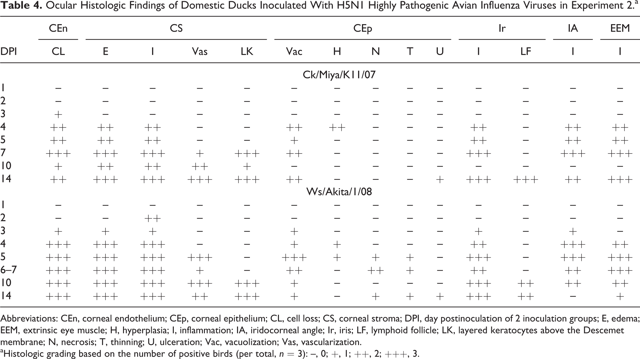
Abbreviations: CEn, corneal endothelium; CEp, corneal epithelium; CL, cell loss; CS, corneal stroma; DPI, day postinoculation of 2 inoculation groups; E, edema; EEM, extrinsic eye muscle; H, hyperplasia; I, inflammation; IA, iridocorneal angle; Ir, iris; LF, lymphoid follicle; LK, layered keratocytes above the Descemet membrane; N, necrosis; T, thinning; U, ulceration; Vac, vacuolization; Vas, vascularization.
aHistologic grading based on the number of positive birds (per total, n = 3): –, 0; +, 1; ++, 2; +++, 3.
Influenza viral antigen was detected in the ocular tissue on days 3–5 PI in the Ck/Miya/K11/07 group and on days 2–6 PI in the Ws/Akita/1/08 inoculation group (Table 5). The distribution of the viral antigen was similar between groups, although the corneal epithelium was positive for viral antigen in only the Ws/Akita/1/08 inoculation group. The histologic and immunohistochemical findings in the various parts of the eye are described in Tables 4 and 5.
Influenza Viral Antigen Distribution by Immunohistochemistry in the Ocular Tissue of Domestic Ducks Inoculated With H5N1 Highly Pathogenic Avian Influenza Viruses in Experiment 2.a
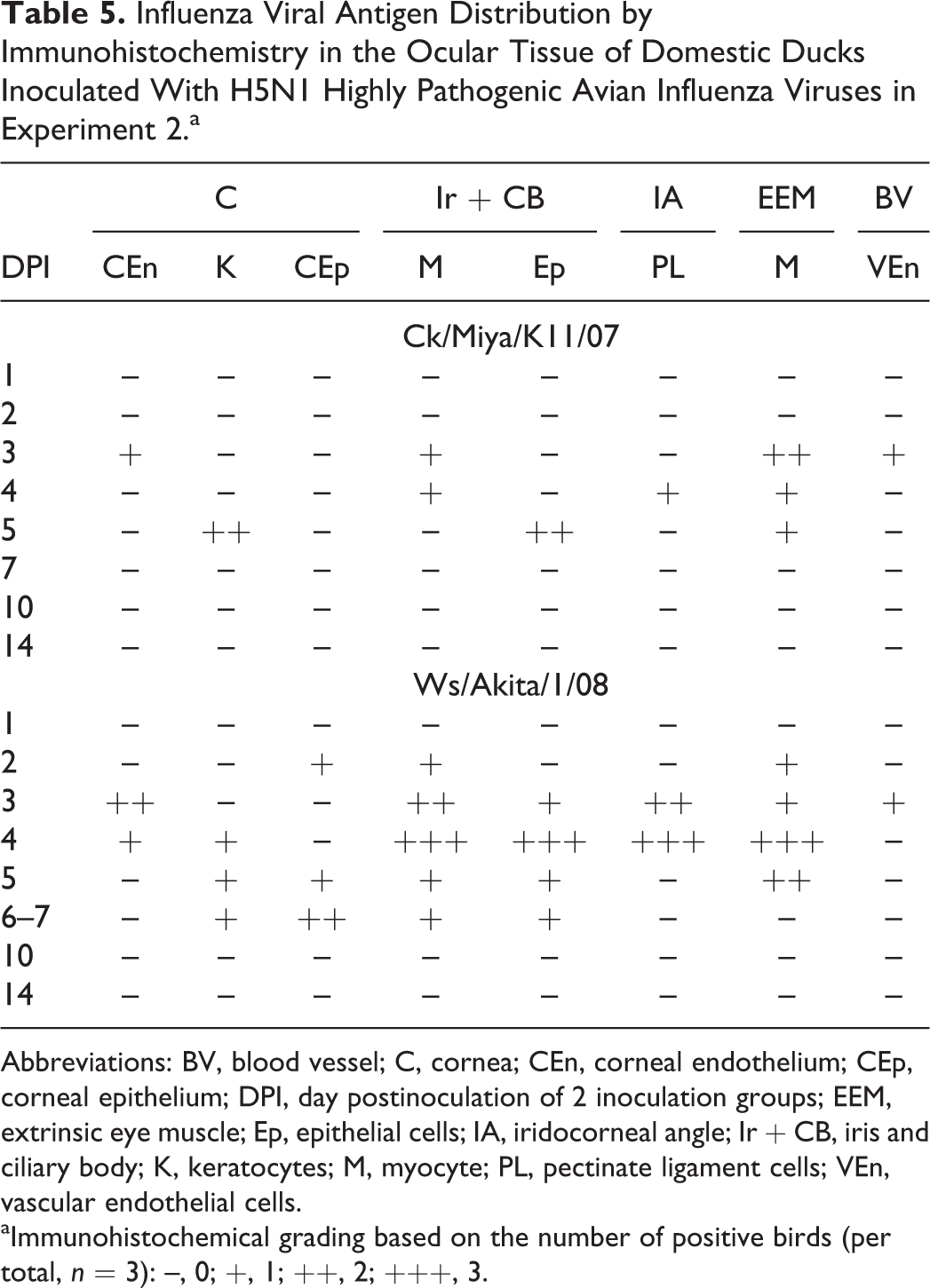
Abbreviations: BV, blood vessel; C, cornea; CEn, corneal endothelium; CEp, corneal epithelium; DPI, day postinoculation of 2 inoculation groups; EEM, extrinsic eye muscle; Ep, epithelial cells; IA, iridocorneal angle; Ir + CB, iris and ciliary body; K, keratocytes; M, myocyte; PL, pectinate ligament cells; VEn, vascular endothelial cells.
aImmunohistochemical grading based on the number of positive birds (per total, n = 3): –, 0; +, 1; ++, 2; +++, 3.
Cornea
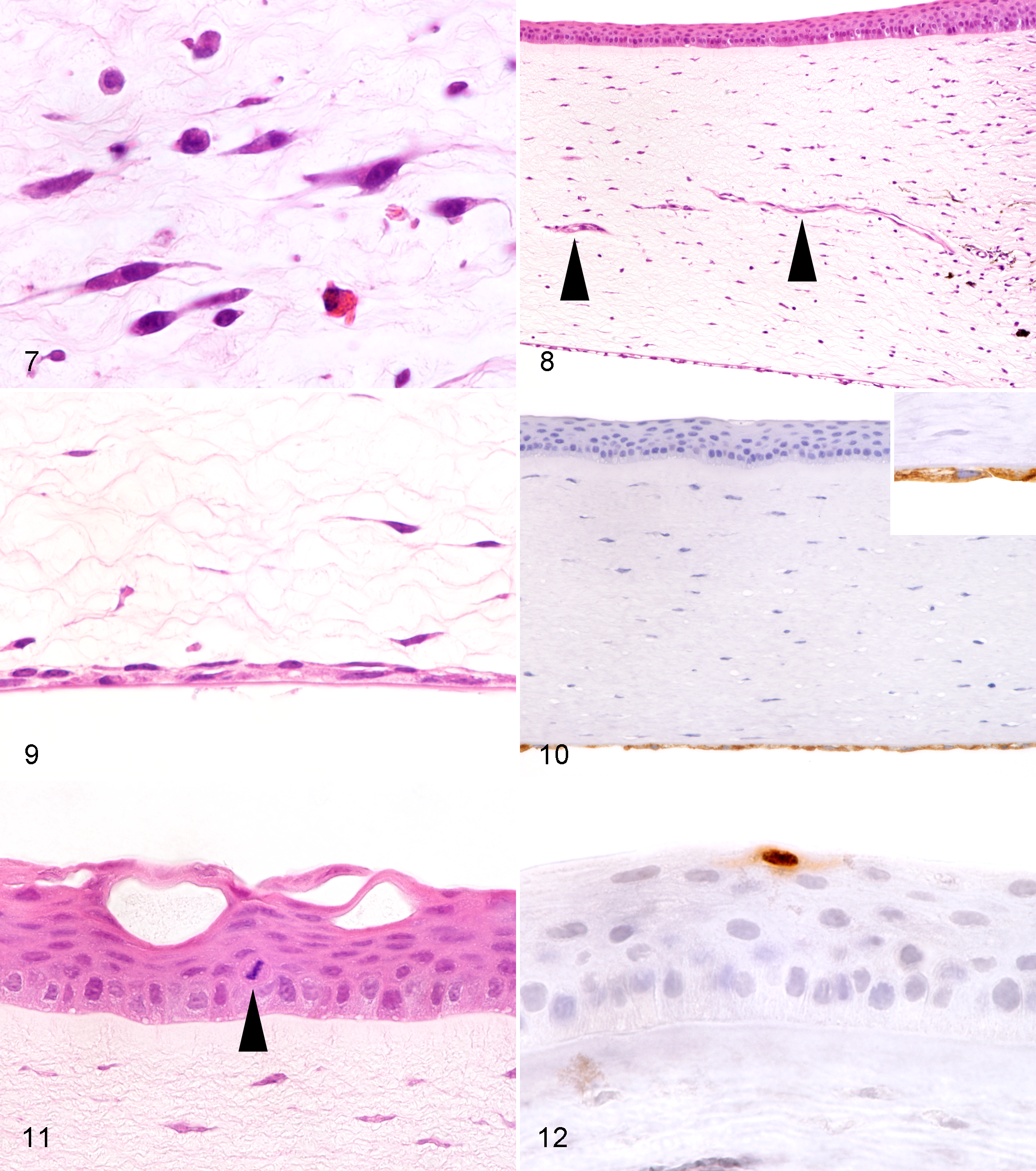
The cornea was a consistently affected tissue in both inoculation groups from day 2 or 3 PI and later. The early change in the cornea was focal or diffuse loss of corneal endothelial cells (Fig. 5). Coincident with or subsequent to the loss of corneal endothelium, an inflammatory reaction and edema occurred in the corneal stroma. Inflammatory cells initially emerged at the corneal limbus and then were predominantly located in the middle to deep layers of the stroma (Fig. 6), suggesting that this inflammation was related to loss of the corneal endothelium. The major inflammatory cell type was heterophils on days 2–4 PI. Plasma cells, lymphocytes, and macrophages became gradually more predominant in the lesions, accompanied by scattered cell debris and the swelling of corneal stromal cells (keratocytes; Fig. 7). Heavy vascularization was observed on day 5 PI and later (Fig. 8). Many fibroblast-like cells, which were considered activated keratocytes, were present above the Descemet membrane and formed single to multiple cell layers in the later stage of the experiment (Fig. 9). This accumulation of keratocytes was considered a compensatory response against the loss of the corneal endothelium. The corneal endothelial loss became milder on day 14 PI and was observed only in small areas of the cornea. The Descemet membrane appeared normal in tissues stained with periodic acid–Schiff. Influenza viral antigen was detected in corneal endothelial cells on day 3 PI in the Ck/Miya/K11/07 group and on days 3 and 4 PI in the Ws/Akita/1/08 group (Fig. 10), which was the time that corneal endothelial loss began. A few keratocytes were positive for viral antigen in the Ck/Miya/K11/07 group on day 5 PI and in the Ws/Akita/1/08 group on days 4–6 PI.
Highly pathogenic avian influenza; domestic ducks.
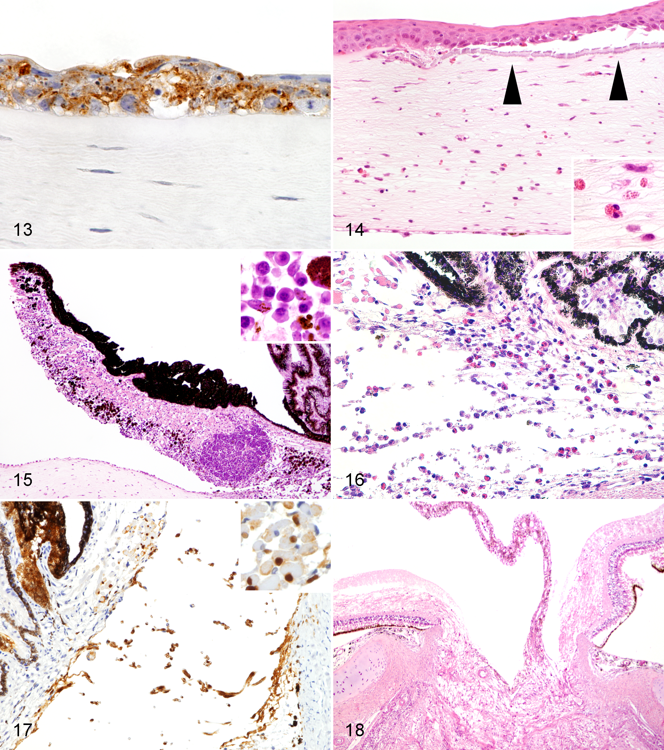
Lesions of the corneal epithelium became evident on days 4–5 PI, 1–2 days after the loss of the corneal endothelium. Vacuolization and epithelial hyperplasia were common in both inoculation groups (Fig. 11). Single to multiple extracellular vacuoles, occasionally containing weak eosinophilic fluid, formed in the epithelium. Small intracytoplasmic vacuoles were often found in the basal cells. The hyperplastic epithelium occasionally exhibited a disorganized cell layer. Focal to diffuse epithelial necrosis and thinning were observed in some ducks but only in the Ws/Akita/1/08 inoculation group on day 5 PI or later. Influenza viral antigen was detected in the corneal epithelium only in the Ws/Akita/1/08 inoculation group. Two characteristic types of viral antigen distribution were noted in the corneal epithelium. One type included viral antigen that was detected in a few superficial epithelial cells without morphologic changes observed on day 2 PI (Fig. 12). The other type included viral antigen that was abundantly detected mostly in association with epithelial necrosis on days 5–6 PI (Fig. 13). The ulcerations on day 14 PI caused the calcification of Bowman membrane and heterophilic infiltration into the stroma (Fig. 14). The calcified Bowman membrane was positive with the von Kossa method.
Highly pathogenic avian influenza; domestic ducks.
Iris and Ciliary Body
Iritis started on day 4 PI in the Ck/Miya/K11/07 group and on day 3 PI in the Ws/Akita/1/08 group. Lymphoplasmacytic inflammation and focal loss of iris muscle were characteristics of the iris. The lesions were accompanied by melanin-laden macrophages and scattered pigmented epithelial cells, which probably dropped from the upper epithelial layer. Lymphoid follicles formed on days 10 and 14 PI (Fig. 15). Viral antigen was detected in the iris on days 3–5 PI in the Ck/Miya/K11/07 group and on days 2–6 PI in the Ws/Akita/1/08 inoculation group. The iridal muscle cells and pigmented epithelial cells were positive for viral antigen. Although the inflammation was not apparent in the ciliary body, viral antigen was infrequently detected in the ciliary muscle cells and in the pigmented and nonpigmented epithelial cells of the ciliary process.
Iridocorneal Angle
Inflammatory infiltrations emerged in the iridocorneal angle from day 3 or 4 PI in both inoculation groups. Inflammatory cells including heterophils, lymphocytes, plasma cells, and melanin-laden macrophages were trapped in the pectinate ligament of the iridocorneal angle with a collapse of the histologic structure (Fig. 16). Pigmented cells similar to iris epithelial cells were also rarely trapped. Lymphoplasmacytic inflammation occasionally extended into the trabecular meshwork and around Schlemm canal. Viral antigen was detected in both inoculation groups on days 3 and 4 PI. The cells of the pectinate ligament and stromal cells of the trabecular meshwork were positive for viral antigen (Fig. 17).
Other Eye Sites
Focal lymphocytic infiltration was frequently observed in the extrinsic eye muscle on day 4 PI and later in both inoculation groups. Viral antigen was detected in skeletal myocytes on days 3–5 PI in the Ck/Miya/K11/07 group and on days 2–5 PI in the Ws/Akita/1/08 inoculation group. Vascular endothelial cells of the capillaries in the pecten and conjunctiva were positive for viral antigen on day 3 PI in both inoculation groups. The pecten of ducks (Nos. 60, 83, 87, 88, 93) from days 4 to 10 PI occasionally exhibited congestion with heterophils, lymphocytes, and plasma cells in the lumen. Viral antigen was detected in a few melanocytes in the pecten on days 3 and 4 PI in 3 ducks (Nos. 55, 60, 79) in both inoculation groups. A very few small aggregates of undetermined cells were found in the optic nerve only in the Ck/Miya/K11/07 inoculated ducks (Nos. 57, 60, 64) on days 3, 4, and 7 PI. No noticeable histopathologic changes were observed in the retina, choroid, sclera, lens, or conjunctiva, although viral antigen was detected in a few conjunctival epithelial cells on days 2–3 PI in 2 ducks (Nos. 78 and 79) in the Ws/Akita/1/08 inoculation group. A few osteoblasts around the scleral ossicle were positive for viral antigen on days 3 and 5 PI in 2 ducks (Nos. 82 and 85) in the Ws/Akita/1/08 inoculation group.
Viral Antigen in the Brain
Small aggregates of glial cells with occasional neuronal necrosis and a few heterophils were randomly distributed in the brain on days 2–3 PI in both inoculation groups. These lesions were subsequently replaced by severe lymphoplasmacytic encephalitis until day 14 PI. Immunohistochemistry was performed on the brain to determine virus spread in the body. Viral antigen was detected in the neurons, glial cells, and ependymal cells on days 3–5 PI in the Ck/Miya/K11/07 inoculation group and from days 2 to 7 PI in the Ws/Akita/1/08 inoculation group. Vascular endothelial cells in the brain were positive for viral antigen on day 3 PI in 1 duck (No. 81) in the Ws/Akita/1/08 inoculation group.
Histopathology of Ducks With Exophthalmos
Two ducks (Nos. 12, 40) exhibited exophthalmos in experiment 1. The eyes of 2 ducks on day 14 PI were examined histologically. A cupping of the optic disk was evident in 1 duck (No. 12; Fig. 18). The corneas of 2 ducks had focally blurred Descemet membrane, loss of the corneal endothelium, and an attachment of thin fibrous tissue on the posterior surface. Periodic acid–Schiff reaction revealed the thickening and focal defect of the Descemet membrane. Viral antigen was not detected in the ocular samples of these 2 ducks.
Discussion
Our results suggest that corneal opacity is one of the characteristic and frequent findings in domestic ducks infected with the H5N1 HPAI virus. In experiment 1, corneal opacity appeared more frequently than neurologic signs or mortality. All domestic ducks that exhibited corneal opacity produced HI antibody against the virus on day 14 PI, while all domestic ducks that did not show corneal opacity were negative for HI antibody 14 days after virus inoculation. These results indicate that corneal opacity can be the most common clinical finding in domestic ducks infected with H5N1 HPAI virus. In fact, corneal opacity was useful for a presumptive diagnosis of viral infection in our experimental infection. Although corneal opacity was recoverable in some domestic ducks in experiment 1, this corneal lesion persisted in the other ducks until day 14 PI, the final day of the experiment. Regarding the characteristic appearance and persistence of corneal opacity, checking for this corneal lesion may be useful for detecting domestic ducks infected with the H5N1 HPAI virus in the field. Nonexperts such as poultry farmers may be able to identify corneal opacity in areas where the H5N1 HPAI virus is endemic in domestic duck populations. In experimental studies using the H5N1 HPAI virus, corneal opacity have been recorded in some waterfowl species, including bar-headed geese, 3,27 cracking geese, 3 herring gulls, 4 laughing gulls, 2 mallard ducks (including domesticated cherry valley and Pekin breeds), 17,20,28,29,40 swan geese, 52 and wood ducks. 2 Unknown domestic duck species have had cloudy corneas in natural outbreaks in Thailand. 38 These reports raise the possibility that corneal opacity may be a common clinical finding in some waterfowl species infected with the H5N1 HPAI virus.
The results of the histopathologic examination in experiment 2 suggest that corneal opacity could be attributed to keratitis induced by the H5N1 HPAI virus. The loss of corneal endothelial cells was characteristically and consistently observed in both inoculation groups in experiment 2. The corneal endothelium, sometimes referred to as the posterior epithelium, 1,16,37 is a functional cell monolayer that maintains corneal transparency. 10,35,42 The corneal endothelium transports water from the stroma to the anterior chamber and functions as a physical barrier to water movement. 10,35,42 Although controversial, corneal endothelial cells are thought to have a limited capacity to regenerate. 10,15,35,36,42 Therefore, an extensive loss of these cells can lead to stromal edema resulting in corneal opacity. 35 Our histopathologic findings of stromal edema, inflammatory infiltration, and the migration of keratocytes covering the Descemet membrane suggest that a strong link exists between the loss of corneal endothelium and keratitis for the manifestation of corneal opacity. The proliferation of keratocytes after corneal injury has been reported in chickens. 24,34 In experiment 1, corneal opacity in some ducks inoculated with Ck/Miya/K11/07 resolved before day 14 PI. This recovery may have resulted from a milder form of corneal endothelial loss or compensatory adaptation by the corneal endothelial cells. 10,35 Recovery from ocular opacity has been reported in bar-headed geese experimentally infected with the H5N1 HPAI virus. 27
Viral antigen was detected in corneal endothelial cells on days 3 and 4 PI in experiment 2. This period coincided with the time of corneal opacity in experiments 1 and 2. In addition, detection of viral antigen in the cells of the pectinate ligament, iris, ciliary process, pecten, and corneal epithelium suggest that the H5N1 HPAI virus can replicate in various types of ocular cells in domestic ducks. Detection of viral antigen in the brain and vascular endothelial cells as early as days 2 or 3 PI in experiment 2 suggest that viremia may have occurred in the domestic ducks at this time. The H5N1 HPAI virus may have reached the corneal endothelium through the blood circulation.
Histopathologic changes in the corneal epithelium were observed in both inoculation groups in experiment 2. However, the frequency and variety of lesions on the corneal epithelium were higher in the Ws/Akita/1/08 inoculation group, and viral antigen was detected in the corneal epithelium only in this group. This difference may have been attributed to the different virulence of the H5N1 HPAI viruses, as observed in the clinical disease severity of the ducks in experiment 1. According to histopathologic analysis, vacuolization and hyperplasia of the corneal epithelium emerged mostly 1 to 2 days after corneal endothelial loss, suggesting that these lesions were subsequent to lesions formed in the deeper cornea. Intra- and intercytoplasmic vacuoles in the corneal epithelium can be caused by edema in the corneal stroma. 10 In the present study, viral antigen was not detected in the corneal epithelium in association with vacuolar changes or hyperplasia. In contrast, the corneal epithelial necrosis observed only in the Ws/Akita/1/08 inoculation group was accompanied by viral antigen, suggesting that necrotic changes in the corneal epithelium were caused by a direct effect of virus replication. Severe necrosis may have resulted in ulceration or epithelial thinning.
Two routes of corneal epithelium infection are possible, although it is difficult to determine in individual cases. One possibility is that the virus reached the corneal epithelium through the stroma after infecting the corneal endothelium. The other is that the virus directly infected the most superficial corneal epithelium in the isolator. A few superficial epithelial cells positive for viral antigen on day 2 PI may support the latter possibility in some ducks in our study. Similar staining pattern of the viral antigen in the superficial corneal epithelium was recorded in Muscovy ducks. 31 When 3 virus inoculation routes were applied in the experimental infection using Muscovy ducks and H5N1 HPAI virus, only after intraocular inoculation was the superficial corneal epithelium positive for viral antigen on day 2 PI. 31 This immunohistochemical finding suggests that the superficial corneal epithelium of ducks can be susceptible to H5N1 HPAI virus and may serve as a site for viral entry into the host. 31
Iritis was another histopathologic feature observed in the present study. In experiment 2, lymphoplasmacytic infiltration, often accompanied by lymphoid follicles at the later stage of the infection, was primarily associated with virus replication in the iris muscle. Unlike that in mammals, skeletal muscle predominates in the iris of birds. 55 As observed in the inflamed extrinsic eye muscle in the present study, skeletal muscles can be target cells of H5N1 HPAI virus in domestic ducks. 43,54
Two domestic ducks exhibited exophthalmos in the latter stage of experiment 1. The histopathologic results of experiment 2 suggest that these ducks may have developed acute secondary glaucoma, although intraocular pressure was not measured in our study. Inflammation at the iridocorneal angle can lead to the obstruction of aqueous humor outflow. 13,46 Cupping of the optic disc, which was observed in a domestic duck with exophthalmos on day 14 PI, can be induced by the elevated intraocular pressure of glaucoma. 13,46
Some related studies have reported blindness as a clinical sign in ducks infected with the H5N1 HPAI virus. 20,38 We did not confirm complete loss of vision in our domestic ducks, because domestic ducks exhibiting corneal opacity could eat feed and drink water during the experiment.
Few reports are available on the ocular histopathology of birds infected with H5N1 HPAI virus, although corneal opacity has been clinically recorded in some species of waterfowl. 2,3,4,17,20,27 –29,40,52 Except for our previous reports using the same duck species as in the present study, 51,54 to our knowledge only 1 report of immunohistochemical findings in waterfowl is available, where viral antigen was detected in the corneal and ciliary epithelial cells of Muscovy ducks experimentally infected with H5N1 HPAI virus. 31 However, Muscovy ducks were histologically examined only on day 2 PI in this study and lacked the description of morphologic change of the eye. 31 Therefore, it is difficult to comparatively discuss the histopathologic lesions and viral antigen distribution of the eye among the different waterfowl species infected with H5N1 HPAI virus.
Conjunctivitis or conjunctival hemorrhage, which were not apparent in domestic ducks in the present study, have been recorded in chickens infected with H5N1 HPAI virus. 26,54 Hemorrhagic conjunctivitis in chickens was attributed to the destruction of capillaries by virus replication in the vascular endothelial cells. 26,54 The conjunctivitis was also macroscopically recorded in swans and domestic ducks infected with H5N1 HPAI virus 22,41 and in chickens infected with H9N2 low pathogenic avian influenza virus, 19 although histologic examination of the eye was not performed for these birds. Conjunctivitis can be differentiated clinically from corneal opacity.
The temporal relationship between virus excretion and corneal opacity of domestic ducks should be taken into account. We previously reported the time course of H5N1 HPAI virus excretion from experimentally infected domestic ducks, where virus was isolated from oropharyngeal and cloacal swabs from days 2 to 5 PI, with peak virus titers on days 3–4 PI. 53 Corneal opacity of domestic ducks in this experiment emerged on day 3 PI and persisted until euthanasia on day 10 PI. 53 Similarly, the mean time for manifesting corneal opacity was about 4 days after inoculation in experiment 1 of the present study. In related experimental studies performing daily virus isolation from swabs, virus excretion from some duck species and bar-headed geese started on day 1 or 2 PI and continued until 5–8 days after H5N1 HPAI virus inoculation. 5,27,45 These data indicate that virus excretion from infected ducks could start a few days before the manifestation of corneal opacity. Immunohistochemical analysis in experiment 2 revealed that corneal opacity was related to the loss of corneal endothelial cells following the virus spread in the body. Therefore, it is reasonable that the manifestation of corneal opacity takes a little longer than the beginning of virus excretion through the respiratory and gastrointestinal tract. When domestic ducks with corneal opacity are observed in the area where H5N1 HPAI virus exists, people should be careful about the risk of virus excretion from suspected ducks. At the same time, the virus detection rate from suspected ducks may be improved if virologic sampling is performed as soon as possible after the manifestation of corneal opacity.
Possible virus excretion from the ocular tissue of infected domestic ducks may be an interesting topic to discuss, although virus isolation was not performed in the present study. According to the immunohistochemical analysis, viral antigen was detected in the corneal and conjunctival epithelium from days 2 to 7 PI. This result may support the possible virus shedding through the ocular tissue. In this regard, Bui et al reported that H5N1 HPAI virus was consistently isolated on days 1–7 PI from the conjunctival swab of domestic ducks intranasally inoculated with the virus. 5 In addition, the H5N1 HPAI virus has been isolated from conjunctival swabs of diseased whooper swans naturally infected with the virus. 6 These findings suggest that H5N1 HPAI virus can be present on the ocular surface of domestic ducks and wild swans infected with the virus. 5,6
One of the unique features of Asian-lineage H5N1 HPAI virus is that it can cause severe clinical signs to waterfowl, which have been considered natural reservoirs of avian influenza virus in nature. 39 Aside from rare exceptions, 7 clinical disease severity by low pathogenic or HPAI virus infection has been considered very mild in ducks until the emergence of H5N1 HPAI virus virulent to waterfowl. 25,33,39,48 When these factors are considered together, domestic ducks may manifest corneal opacity only in H5N1 HPAI virus infection. H5N8 HPAI virus, which has hemagglutinin protein derived from H5N1 HPAI virus, is also currently epidemic and produces severe clinical signs to ducks. 21,50 Because hemagglutinin protein of avian influenza virus is a critical factor for its virulence, 32 clinical signs—such as neurologic signs and corneal opacity, which are similar to those caused by H5N1 HPAI virus—may be found in domestic ducks infected with H5N8 HPAI virus.
Our results indicate that the H5N1 HPAI virus should be included in the differential diagnosis when corneal opacity is clinically observed in domestic ducks. Corneal opacity may be difficult to confirm in carcasses because of autolysis and dehydration of tissue. Except for H5N1 HPAI virus, to our knowledge there is no report on association of corneal opacity to any other viral disease in ducks. 18,37 Pox virus infection of the corneal epithelium has been recorded in some bird species other than ducks. 18,37,47 In general avian medicine, infection by bacteria and fungi, traumatic wounds, congenital corneal anomalies, vitamin A deficiency, and intoxication such as ammonia toxicity and plant-induced photosensitization may cause corneal disease. 18,37,47
We have an interest in the disease similarities observed in the present study and corneal opacity in dogs infected with infectious canine hepatitis virus. Corneal opacity of this canine disease is triggered by virus replication in the corneal endothelium and becomes more apparent during the recovery phase because of type III hypersensitivity. 8,9,14 In the domestic ducks in our study, corneal opacity caused by the H5N1 HPAI virus immediately emerged after infection, around 3–4 days PI when it was considered too early for an antibody response, suggesting that the corneal opacity observed in the present study may have been largely attributed to the direct effect of virus replication rather than hypersensitivity. In this regard, further study is necessary to elucidate the pathogenesis of corneal opacity in domestic ducks infected with the H5N1 HPAI virus.
In conclusion, our results indicate that corneal opacity is an indicator of Asian-lineage H5N1 HPAI virus infection in domestic ducks. Confirming this ocular change may improve the detection rate of infected domestic ducks in the field.
Footnotes
Acknowledgements
We thank Megumi Shimada and Masaru Kobayashi for their technical assistance.
Declaration of Conflicting Interests
The author(s) declared no potential conflicts of interest with respect to the research, authorship, and/or publication of this article.
Funding
The author(s) received no financial support for the research, authorship, and/or publication of this article.