
Abstract
The authors herein describe the morphologic and immunohistochemical features of normal Merkel cells as well as the clinicopathologic findings of Merkel cell carcinoma in cats. Merkel cells were characterized as vacuolated clear cells and were individually located in the epidermal basal layer of all regions examined. Clusters of Merkel cells were often observed adjacent to the sinus hair of the face and carpus. Immunohistochemically, Merkel cells were positive for cytokeratin (CK) 20, CK18, p63, neuron-specific enolase, synaptophysin, and protein gene product 9.5. Merkel cell carcinoma was detected as a solitary cutaneous mass in 3 aged cats (13 to 16 years old). On cytology, large lymphocyte-like cells were observed in all cases. Histologic examinations of surgically resected tumors revealed nests of round cells separated by various amounts of a fibrous stroma. Tumor cells were commonly immunopositive for CK20, CK18, p63, neuron-specific enolase, and synaptophysin, representing the characteristics of normal Merkel cells.
Keywords
Merkel cells are located in the basal layer of the epidermis and mucosa of vertebrates. 29 Merkel cells are located in the hair discs (Haarscheiben), hair follicles, interfollicular epidermis, and glabrous skin in humans. 20,21 However, a previous study reported that Merkel cells were absent in hair follicles in the body skin of mice and present only in the epidermal hair discs and whisker hair follicles. 20 Most Merkel cells possess synaptic connections with the enlarged terminal endings of myelinated sensory nerve fibers. Neurotransmitters or neuromodulators are secreted from Merkel cells through exocytosis in response to mechanical stimuli to the skin, thereby suggesting a function of Merkel cells as a mechanoreceptor of tactile sensation. 10,12
Difficulties have been associated with identifying Merkel cells on routine hematoxylin and eosin (HE)–stained sections. 20 Merkel cells express various neuropeptides and neuroendocrine markers, such as neuron-specific enolase (NSE), chromogranin A, synaptophysin, and protein gene product 9.5 (PGP9.5) in humans, making them identifiable by immunohistochemistry. 7,9,17,24 Cytokeratin (CK) 20 is assumed to be the most specific marker for Merkel cells in the skin of humans and mice. 5 In humans, CK20-positive Merkel cells are distributed in the palms and hairy skin of embryos and fetuses and in the ventral midline of adults (the incision line of a routine autopsy). 14,20 To the best of our knowledge, the presence and distribution of CK20-positive Merkel cells have not yet been examined in normal cat skin.
Merkel cell carcinoma is a rare cutaneous tumor showing both epithelial and neuroendocrine differentiation. Although Merkel cell carcinoma is considered to arise from Merkel cells, recent findings indicated that primitive epidermal stem cells or early B cells were the origin of Merkel cell carcinoma. 13,27,30 Only 3 cases of feline Merkel cell carcinoma have been reported to date and were diagnosed by examining biopsy samples. Two cases were accompanied by pulmonary and lymph node metastases showing malignant behaviors, 22,23 while the other had a benign nature (no relapse or metastasis during a 2-year follow-up period). 4
We herein characterized the cytologic, histopathologic, and immunohistochemical findings of 3 cases of feline Merkel cell carcinoma with different clinical outcomes together with immunohistochemical phenotypes of normal Merkel cells in the cat.
Materials and Methods
Three feline Merkel cell carcinoma cases were acquired from biopsy samples submitted to the Department of Pathology, The University of Tokyo. To understand the general distribution of Merkel cells in the cat, normal skin samples of the head, neck, forelimb (carpal region and pad), and ventral and dorsal trunk were collected from 3 cats through routine necropsies and biopsies performed at the Veterinary Medical Center, The University of Tokyo.
Formalin-fixed tissues from normal skin and Merkel cell carcinomas were routinely processed, and 4-µm-thick serial sections were cut. HE staining was performed for an initial microscopic evaluation. The number of mitotic figures per 10 fields (×400) was counted in each case. Consecutive sections were stained using an immmunoenzyme technique. To inactivate endogenous peroxidase, deparaffinized sections were immersed in 1% hydrogen peroxide in methanol for 20 minutes. After washing with Tris-buffered saline, the slides were blocked with 5% skim milk in Tris-buffered saline. The primary antibodies that were used in the present study are listed in Table 2. After incubation with the primary antibody at 4°C overnight, immunolabeled antigens were visualized using the Dako Envision+ System (Dako, Carpinteria, CA). The sections were incubated with a HRP-labeled polymer at 37°C for 40 minutes, reacted with 0.05% 3′3-diaminobenzidine plus 0.03% hydrogen peroxide in Tris-hydrochloric acid buffer, and then counterstained with hematoxylin. Negative controls were obtained by omitting the primary antibodies. Two experienced, Japanese College of Veterinary Pathologists–certified veterinary pathologists (J.K.C. and K.U.) independently reviewed the specimen slides.
Feline Merkel Cell Carcinoma Cases in the Present Study and Previous Studies.
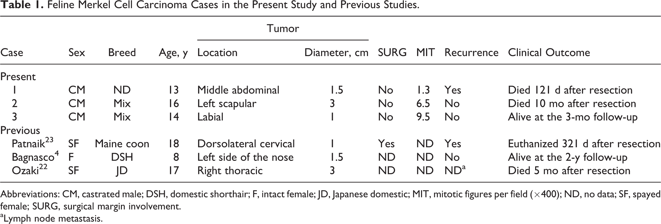
Abbreviations: CM, castrated male; DSH, domestic shorthair; F, intact female; JD, Japanese domestic; MIT, mitotic figures per field (×400); ND, no data; SF, spayed female; SURG, surgical margin involvement.
aLymph node metastasis.
Antibodies Used for Immunohistochemistry.
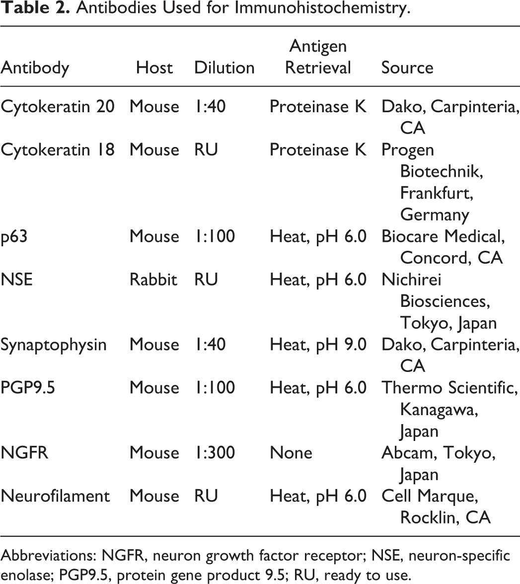
Abbreviations: NGFR, neuron growth factor receptor; NSE, neuron-specific enolase; PGP9.5, protein gene product 9.5; RU, ready to use.
Results
Characterization of Normal Merkel Cells in Cats
Morphologic observations of consecutive sections stained with HE or immunostained for CK20 and CK18 revealed normal Merkel cells in the epidermal basal layer. Merkel cells were observed at all sites examined (head, neck, forelimbs, pads, and ventral and dorsal trunk). Although most Merkel cells were individualized and randomly located (2 or 3 Merkel cells per histologic section), clusters of Merkel cells were often observed in the epidermis above the sinus (tactile hair follicle) of the facial and carpal regions (Figure 1). 18,20 Merkel cells were often vacuolated on HE-stained sections, giving a “clear cell” appearance, and protruded (rete peg) to the dermis (Fig. 1a). These morphologic features of Merkel cells were similar to those described previously in humans. 20 Normal Merkel cells were specifically positive for CK20 and CK18 with a distinct paranuclear pattern (Fig. 1b, c). All nuclei of the basal layer, including the Merkel cells, were positive for p63 (Table 3). Merkel cells were also positive for NSE (Fig. 1d). The granular staining pattern of synaptophysin and weak immunoreactivity to PGP9.5 were observed in the cytoplasm of Merkel cells (Fig. 1e, f). Neuritic processes positive for nerve growth factor receptor (NGFR) and neurofilament (NF) were observed on the dermal side of Merkel cells (Fig. 1g, f).
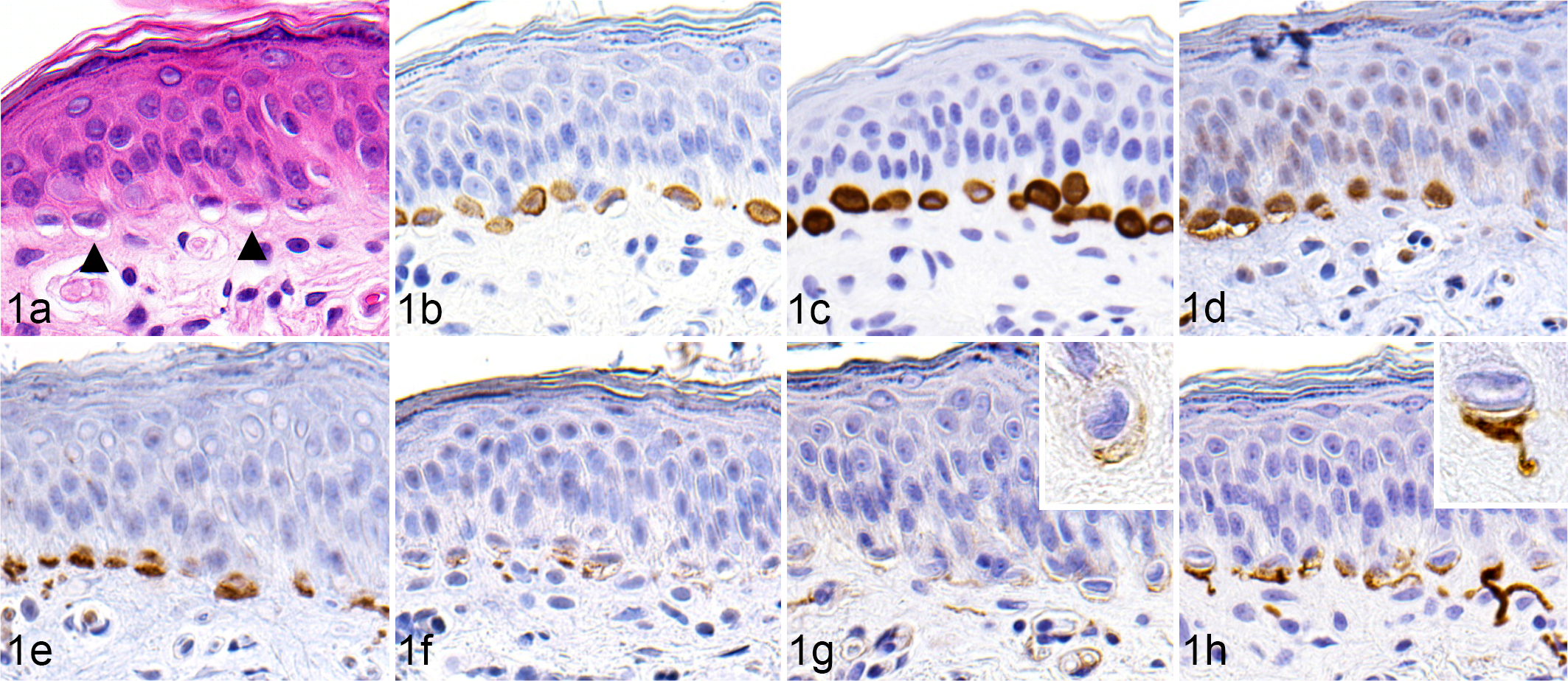
Skin (carpus); cat. Merkel cells in normal cat skin. (a) Merkel cells are often vacuolated (arrowheads) and protrud into the dermis. (b–f) Immunohistochemical labeling of Merkel cells for cytokeratin 20 (b), cytokeratin 18 (c), neuron-specific enolase (d), synaptophysin (e), protein gene product 9.5 (f). (g, h) Neuritic processes beneath Merkel cells are positive for nerve growth factor receptor (g; inset, higher magnification) and neurofilament (h; inset, higher magnification).
Results of Immunohistochemistry for Merkel Cell Carcinoma Cases and Normal Merkel Cells.a
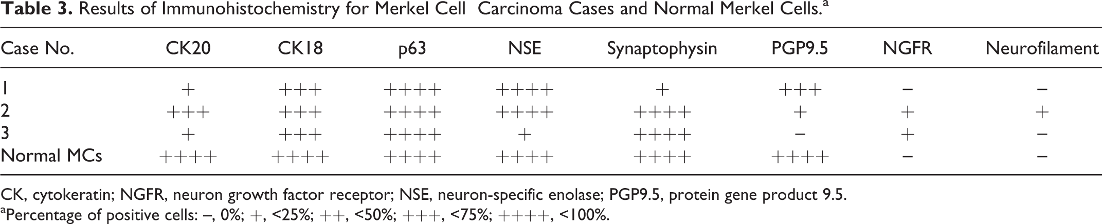
CK, cytokeratin; NGFR, neuron growth factor receptor; NSE, neuron-specific enolase; PGP9.5, protein gene product 9.5.
aPercentage of positive cells: –, 0%; +, <25%; ++, <50%; +++, <75%; ++++, <100%.
Clinical Case Histories: Gross and Cytologic Findings of Merkel Cell Carcinoma
General information (breed, sex, age, tumor location, tumor size) regarding the 3 Merkel cell carcinoma cases is listed in Table 1. Case No. 1, a 13-year-old castrated male cat, presented for a regular health checkup. The clinician detected a solitary, firm, dome-shaped red mass, measuring 1.5 cm in diameter, in the middle of the abdominal skin (Fig. 2). The surface was alopecic and partly ulcerated in the center. On cytology, many large discrete round-shaped cells (15 to 25 μm in diameter) with high nuclear:cytoplasmic ratio were observed. These cells had a basophilic cytoplasm, and the nucleus often had a large nucleolus in the center, resembling immunoblasts. The mass was surgically resected and fixed in 10% formalin for a histopathologic examination. The clinician confirmed local recurrence 3 weeks after surgery, leading to second and third surgeries. The cat died 121 days after the first surgery. The clinician confirmed multiple masses in the skin, including the primary site and the lung by radiograph, suggesting recurrence and metastasis. Permission was not given for necropsy.
Case No. 2, a 16-year-old castrated male mix-breed cat, presented with a solitary, irregularly raised, reddish mass measuring 3 cm in diameter in the left scapular region (Fig. 3). The surface was partly ulcerated. Fine needle aspiration cytology revealed many discrete round-shaped cells (10 to 15 μm in diameter) with a scant cytoplasm and 1 or 2 small nucleoli. The mass was resected and fixed in 10% formalin for a histopathologic examination. The cat died 10 months after surgery due to renal failure. Neither relapse nor metastasis was detected. Necropsy was not permitted.
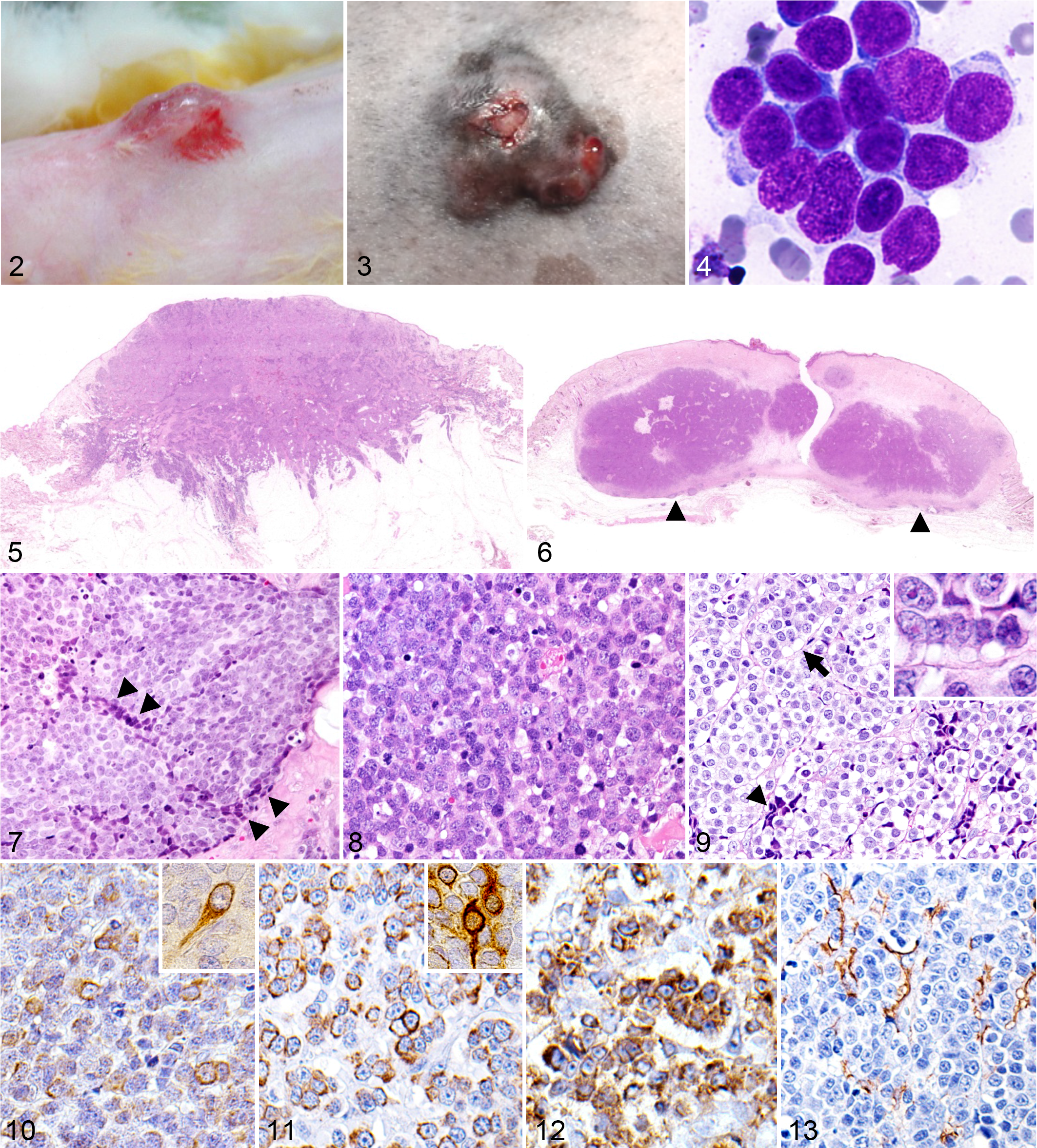
Case No. 3, a 14-year-old castrated mix-breed cat, presented with a solitary elastic mass measuring 1 cm in diameter on the labial surface. The tumor enlarged rapidly and was surgically resected. Touch smear cytology revealed many large round-shaped cells (15 to 25 μm in diameter) discretely or in loose clumps (Fig. 4). These cells had a scant cytoplasm and 1 or 2 small nucleoli. The resected tissue was fixed in 10% formalin for histopathologic examination. Neither relapse nor metastasis was detected at the 3-month follow-up. In all 3 cases, veterinary clinicians suspected lymphoma based on the cytologic findings.
Histopathology of Merkel Cell Carcinoma
In case No. 1, the neoplasm extended from the superficial dermis to the subcutaneous fatty tissue (Fig. 5). The aggressive infiltration of neoplastic cells into the subcutaneous tissue resulted in an indistinct margin of the neoplasm, in contrast to case Nos. 2 and 3 (Figs. 5, 6). Neoplastic tissue was not observed at the excision margin. Round neoplastic cells formed nests separated by an abundant fibrovascular stroma (Fig. 7). Neoplastic cells had distinct cell borders, a scant to moderate amount of cytoplasm, and finely dispersed chromatin with 1 prominent nucleolus, which was consistent with the cytologic observations. Anisokaryosis was moderate. The mean number of mitotic figures was 1.3 per field (×400). Neoplastic cells in the periphery of each nest were smaller and darker than those in the center (Fig. 7).
In case No. 2, an encapsulated neoplasm was located in the dermis (Fig. 6). Neoplastic tissue was composed of dense sheets of round-shaped cells with scant cytoplasm (Fig. 8), which was consistent with the cytologic observations. Nuclei were round and had finely stippled or dispersed chromatin. Nucleoli were rarely detected, and anisokaryosis was moderate. The mean number of mitotic figures was 6.5 per field (×400). Focal necrosis was randomly observed. In addition, apoptotic cells were widely scattered. Perivascular lymphocytic infiltration was detected around the neoplasm, which was fully encapsulated with a thick fibrotic wall.
In case No. 3, a noncapsulated neoplasm was located in the dermis, adjacent to a sinus hair. Neoplastic cells were densely packed in solid nests separated by a basement membrane–like thin fibrous stroma (Fig. 9). Intracellular vacuoles were often observed at the basal side, similar to normal Merkel cells. The nuclei had dispersed and stippled chromatin and one nucleolus. Anisokaryosis was moderate. Marked proliferation of neoplastic cells was observed at the dermoepidermal junction and in the epidermis. The mean number of mitotic figures was 9.5 per field (×400).
Immunohistochemistry of Merkel Cell Carcinoma
Table 3 summarizes the immunohistochemical results of the 3 Merkel cell carcinoma cases and normal Merkel cells in the cat. Neoplastic cells expressed CK20 with a perinuclear cytoplasmic staining pattern in all Merkel cell carcinoma cases (Fig. 10). Neoplastic cells also expressed CK18 with a similar staining pattern to CK20 (Fig. 11), although the positivity of CK18 staining was higher than that of CK20 (Table 3). CK20- or CK18-positive cell processes extended from neoplastic cells in case Nos. 1 and 3 (Figs. 10, 11). The nuclei of neoplastic cells were positive for p63 (Table 3). Furthermore, most neoplastic cells were positive for NSE with a distinct granular to diffuse pattern in all cases and also for synaptophysin in case Nos. 2 and 3 (Fig. 12). However, only a few neoplastic cells expressed synaptophysin in case No. 1. The majority of cells in case No. 1 and only a few cells in case No. 2 expressed PGP9.5, whereas case No. 3 was negative for PGP9.5. Although no cell was positive for NGFR and NF in case No. 1, a moderate number of cells expressed NGFR in case Nos. 2 and 3, and a small number of cells expressed NF in case No. 2. Thin processes between neoplastic cells were positive for NGFR similar to neuritic fibers (Fig. 13).
Discussion
Merkel cells are derived from the neural crest and show both epithelial and neuroendocrine differentiation. 26 As demonstrated in humans and mice, normal Merkel cells can be characterized by the specific expression of CK20 in the epidermal basal layer (Fig. 1b). CK20-positive Merkel cells were randomly located at all sites examined (ie, head, neck, forelimbs, pads, ventral and dorsal trunk). The wide distribution of Merkel cells may explain the various locations of Merkel cell carcinomas in cats (Table 1). However, additional studies that include more animals from different breeds and that sample more locations will be useful to provide a comprehensive view of the distribution of Merkel cells in feline skin. CK20-positive Merkel cells also expressed CK18, p63, synaptophysin, NSE, and PGP9.5 (Fig. 1c–f), indicating both epithelial and neuroendocrine differentiation. Immunohistochemistry for NGFR and NF revealed the existence of the neurites of sensory nerves beneath the Merkel cells (Fig. 1g, h).
All 3 Merkel cell carcinoma cases in the present study were elderly (13, 16, and 14 years old), similar to the 3 reported cases (8, 17, and 18 years old). 4,22,23 In humans, Merkel cell carcinomas are also typically diagnosed in the aged population, and only 5% of the patients are below the age of 50 years, with a reported age range of 7 to 104. 8 Merkel cell carcinoma was previously shown to be more common in men than in women (ratio, 2.3:1). 15 Previous feline Merkel cell carcinoma cases were all female (intact or spayed), whereas the cats in the present study were all castrated males. No breed or tumor location predilection was noted in the present or previous studies (Table 1). Merkel cell carcinoma is rare in both humans and animals; therefore, its etiology and clinical outcome have not yet been elucidated in detail. Previous clinical studies on human Merkel cell carcinomas identified a large tumor size, small cell size, and high mitotic rate as adverse prognostic factors. 1,2,25 However, we could not find any relationship between clinical outcomes and tumor sizes, cell sizes, or mitotic rates in cats (Table 1).
The gross findings of feline Merkel cell carcinoma are similar to those of human Merkel cell carcinoma, showing a reddish, elevated mass (1 to 3 cm in diameter) in the skin with an ulcerated top (Figs. 2, 3). 6 The present study provided cytologic observations on Merkel cell carcinomas, which have not been reported previously. On cytology, all 3 cases showed abundant round-shaped cells that resembled lymphoma cells for the high nuclear:cytoplasmic ratio (Fig. 4). The primary diagnosis of lymphoma was changed to Merkel cell carcinoma by histopathologic examinations in 1 previous feline case of Merkel cell carcinoma. 23 The discrete round-cell appearance makes it difficult to suspect a neuroendocrine tumor on cytology. We assume that there may be cases of feline Merkel cell carcinoma that are misdiagnosed as round cell tumors, such as cutaneous lymphoma, without histopathologic examination.
Histopathologically, Merkel cell carcinomas may be encapsulated by a fibrous wall without apparent invasion into the surrounding tissue (case No. 2; Fig. 6) or nonencapsulated with mild (case No. 3) to aggressive (case No. 1; Fig. 5) invasion into the surrounding tissue. In the present study, the surgical margins, which were examined under a microscope, were clear of tumor cells in all cases. However, Merkel cell carcinoma in case No. 1 exhibited an aggressive behavior (recurrence and metastasis), consistent with histopathologic invasiveness. Histopathologic evidence of tumor cell invasion may be indicative of a poor prognosis in cats with Merkel cell carcinomas. Although the number of cases examined was small, mitotic indices were not relevant for clinical outcomes in the present study (Table 1).
The present feline cases of Merkel cell carcinoma formed diffuse sheets and solid nests in the dermis, while the amount of the stromal component varied among the 3 cases. Case No. 1 showed marked desmoplasia; case No. 2 had a small stromal component and showed small foci of necrosis; and case No. 3 formed small nests separated by a thin fibrous stroma. In the periphery of each nest, tumor cells were often compressed against the stromal component and displayed nuclear molding (Figs. 7, 9). Such a finding is one of the histopathologic features of Merkel cell carcinoma in both humans and cats. 4,8 Some human cases of Merkel cell carcinoma show a pagetoid pattern, in which tumor cells proliferate in the epidermis and form small clusters. 11,16 In the present study, case No. 3 showed marked proliferation of tumor cells at the dermoepidermal junction and a pagetoid appearance in the epidermis. These results may be misinterpreted as epitheliotropic lymphoma.
The immunohistochemical staining patterns of feline Merkel cell carcinoma cells for CK20, CK18, NSE, synaptophysin, and PGP9.5 were consistent with those observed in normal Merkel cells (Fig. 1b–f) and human Merkel cell carcinoma cells. 2,17,24 CK20 is considered to be the most reliable marker for Merkel cell carcinoma in humans. 19 Furthermore, p63 positivity is associated with a worse prognosis in human Merkel cell carcinoma patients; however, all 3 cat cases in the present study were highly positive for p63 (Table 3). 3 Characteristic immunostaining patterns for NGFR resembling neurite-like processes were observed in case Nos. 2 and 3. It currently remains unknown whether these processes were part of neoplastic Merkel cells or were sensory nerves induced by neoplastic Merkel cells.
In humans, Merkel cell carcinoma is a highly aggressive tumor with a high recurrence rate. 8 Based on few reported cases, Merkel cell carcinomas in dogs appear to have benign biological behavior, in contrast to human Merkel cell carcinoma. 28 In cats, clinical outcomes varied among cases in the present and previous studies (Table 1). Since most cats that were diagnosed with Merkel cell carcinoma were elderly and were biopsy cases, difficulties were associated with assessing the true lethality of feline Merkel cell carcinoma. However, the only case that survived more than a year was markedly younger (8 years old) than the other cases (Table 1). 4
In conclusion, the present study provided 2 important features of feline Merkel cell carcinoma. First, cytologic results should be carefully evaluated in cases of cutaneous tumors in aged cats because Merkel cell carcinoma may show a lymphoma-like appearance. Second, normal MCs express CK20, CK18, NSE, synaptophysin, and PGP 9.5; therefore, these can be considered as reliable markers of Merkel cell carcinoma in cats.
Footnotes
Declaration of Conflicting Interests
The author(s) declared no potential conflicts of interest with respect to the research, authorship, and/or publication of this study.
Funding
The author(s) received no financial support for the research, authorship, and/or publication of this article.