
Abstract
The eyes of 2 male and 2 female GSP/pe chickens, the imperfect albino strain, were investigated at 52 weeks of age. Aged chickens of the GSP/pe colony became blind with bilateral ocular enlargement and opaque lenses. Affected eyes (bilateral in 2 males and unilateral in 2 females) were hard and difficult to section; histologic specimens were processed after decalcification. A large portion of the posterior chamber was occupied by cancellous bone containing fibrous and cartilaginous foci. Osseous tissues developed adjacent to the choroid, and no retinal pigment epithelium (RPE) was detected between osseous tissues and the choroid. Small segments of degenerate neuronal retina were scattered in the osseous tissue. The irises and ciliary bodies were deformed by osseous tissue, and the lenses had severe cataracts. These observations suggest that the intraocular osseous tissue may be derived from RPE in the hereditary incomplete-albino strain of chickens.
Ocular osteogenesis has been described in humans, 7,10 –12,15 dogs, 8 guinea pigs, 3 and chickens. 1,13 In humans, ossification in the lens 7 and choroid 11 has been reported in eyes affected by phthisis bulbi and in eyes with retinal detachment, intraocular hemorrhage and neovascularization, uveitis, 10 and exudative retinopathy in Coats disease. 9 In young humans, intraocular ossification has been suggested to occur secondary to blunt trauma and, in older individuals, secondary to inflammation. 12 Preretinal osseous tissues have been observed in the eyes of 2 humans with longstanding retinal detachment and proliferative vitreoretinopathy (PVR). 16 In a clinicopathologic study, chronic retinal detachment with hyperplasia and transdifferentiation of the retinal pigment epithelium (RPE) has been suggested to be requisite for intraocular osseous metaplasia. 15 Among domestic animals, bone formation occurred within an otherwise normal iris in a 10-year-old male Great Dane. 8 In Abyssinian-Hartley guinea pigs, osseous choristomas were present in the ciliary bodies with expansion into the anterior chambers in all eyes of two 36-month-old males and one 42-month-old female, suggesting that these osseous lesions may be common in aged guinea pigs of this strain. 3 Intraocular ossification, similar to the human condition of phthisis bulbi, was present at the end stage of retinal degeneration in a congenitally blind strain of chicken derived from Rhode Island reds 1 and in Smoky Joe chickens derived from White Leghorns. 13 The present study documents ocular osteogenesis in aged GSP/pe chickens, a new mutant strain with hereditary imperfect albinism.
The GSP/pe strain of chicken is the albino-like (imperfect albino) line (Fig. 1a) of the inbred GSP strain (Fig. 1b), which originated from the Fayoumi breed. The imperfect albino trait is inherited, and this strain is maintained and bred in the Avian Bioscience Research Center of Nagoya University in Japan. Chickens of this line have reduced eumelanin pigment (color dilution) in the skin, plumage, and eyes (Fig. 1). Aged GSP/pe chickens have decreased locomotor activity, bilateral ocular enlargement, anisocoria, and lens opacity. Two males and 2 females of the GSP/pe strain at 52 weeks of age (Nos. 1, 2, 3, and 4, respectively) and 2 males at 52 and 45 weeks of age and 2 females at 52 and 68 weeks of age of the GSP strain (Nos. 5, 6, 7, and 8, respectively, controls) were investigated. The experimental use of birds was licensed by the Animal Care and Use Committee and conducted in compliance in with the Guidelines for Care and Use of Laboratory Animals at the Nippon Institute for Biological Science (1999). Chickens were anesthetized with isoflurane, exsanguinated, and necropsied. The eyes were enucleated and immediately fixed by Bouin’s solution for approximately 2 hours, and then this fixation solution was exchanged for 70% ethanol for 3 days to remove picric acid. After fixation, affected eyes were decalcified by EDTA solution for 10 days because they were hard and difficult to section, and then they were trimmed, embedded in paraffin, routinely sectioned, and stained with hematoxylin and eosin (HE). Selected sections were stained by the von Kossa method for calcium and immunohistochemically reacted with anti–neuron-specific β III tubulin monoclonal antibody at 1:1000 dilution (Abcam Plc, Cambridge, UK). Briefly, deparaffinized sections were autoclaved in 0.01 M citric acid buffer (pH 6.9) for 20 minutes at 121°C for antigen retrieval, treated with 0.3% H2O2/MeOH for 30 minutes at room temperature to inactivate endogenous peroxidase, incubated with 10% normal goat serum for 30 minutes at room temperature to block nonspecificity, incubated with the primary antibody overnight at 4°C, and processed using the Histofine Simple Stain MAX-PO (M) kit (Nichirei Bioscience, Tokyo, Japan). Immunolabeled antigen was visualized using diaminobenzidine. Negative controls were obtained by omitting the primary antibody, and retinal tissues of a GSP chicken were positive controls.
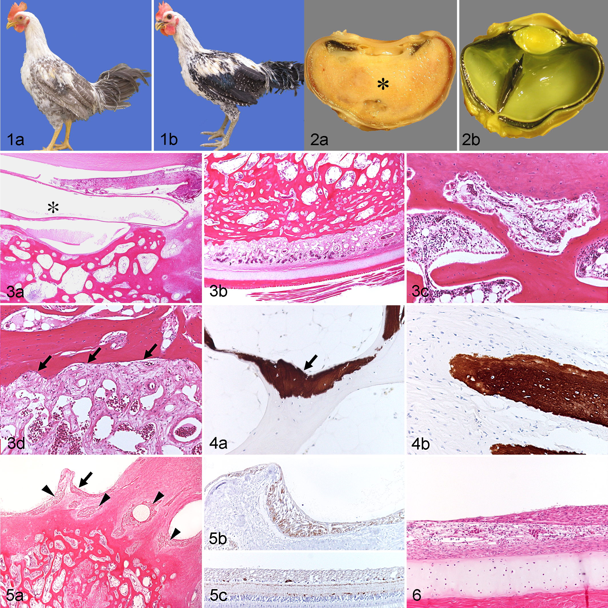
Chickens. (a) GSP/pe male, No. 1. Plumage color is diluted. (b) GSP male, No. 5. Plumage color is normal.
Grossly, the eyes of GSP/pe chickens (Fig. 2a) were larger than those of GSP chickens (Fig. 2b), and amorphous hard material filled both eyes of 2 males and the right eye of both of the 2 females of the GSP/pe strain (Fig 2a). This material was mature osseous tissue that occupied a large portion of the posterior chamber, replacing the vitreous (Fig. 3a,b). The osseous tissue resembled cancellous bone and formed trabecula with osteocyte-containing lacunae and fatty bone marrow lined by osteoblasts and occasional osteoclasts (Fig. 3c). The bone marrow contained hematopoietic cells along with vascular sinuses within a loose fibrous stroma (Fig. 3c). Osseous tissue involved both the pecten and retina (Fig. 3b), which were difficult to identify. Osseous tissue was not present in the choroid or any anterior portions of the eyes, including lens, anterior chamber, or cornea (Fig. 3a). Fibrous and cartilaginous foci were scattered in some internal and surface portions of the osseous tissue. Retinal pigment epithelium (RPE) was not present between or within osseous areas. The choroid was thick and fibrous (Fig. 3d). A few areas of osseous tissue were stained by the von Kossa method despite having been decalcified (Fig. 4). Small segments of degenerate neural retina were scattered in the osseous tissue (Fig. 5a). The neural retina was positive for β III tubulin (Fig. 5b). The ciliary body and iris were deformed by the osseous tissue and contained mild inflammatory cell infiltrates (Fig. 3a). The lens was mostly liquefied, sparing the capsule, thus resembling a hypermature cataract (Fig. 3a). In the 2 eyes without osseous tissue (GSP/pe females), the retina was completely detached and markedly degenerate. RPE and Bruch’s membrane remained on the surface of the choroid, and in several segments, RPE cells were spindloid and arranged in fibrous nodules (Fig. 6).
In birds, membranous bone structures (scleral ossicles) are present in the peripheral sclera. 6 In the cases described herein, intraocular osseous tissues were not connected to the scleral ossicles or the scleral cartilage and did not invade the choroid. The pecten and vitreous were involved in the metaplastic process, but there was no proliferation or transition of these tissues into bone. In addition, the ciliary body, iris, and lens, although deformed, degenerate, and inflamed, were not displaced. These results suggest that osseous tissues were not derived from the sclera, choroid, pecten, vitreous, ciliary body, iris, or lens. A few reports have described intraocular osseous tissues originating from the ciliary body in dogs 8 and guinea pigs 3 and from the lens 7 and choroid 11 in humans.
Recently, RPE has been suggested to retain the capability of epithelial to mesenchymal transdifferentiation. 4,14 The intraocular bone incorporated in PVR of 2 human patients was thought to result from metaplasia of RPE. 16 In young humans, ocular trauma has been presumed to be the most common cause of intraocular ossification and to induce osteoblastic transformation of the RPE to form compact bone. 12 In the current study, a change of RPE from an epithelial phenotype to a spindle phenotype was present in GSP/pe hens unaffected by ossification. In addition, ocular osseous tissue contained fibrous and cartilaginous foci. In 1 study, cultured RPE cells derived from PVR were both epithelioid and fibroblast/fusiform and simultaneously expressed cytokeratin 19, vimentin, and glial fibrillary acidic protein, suggesting dedifferentiation of RPE cells with epithelial-mesenchymal transition. 2 During serial passage, cultured porcine RPE cells had decreased expression of cytokeratin 18, rearrangement of their actin cytoskeleton, and increased expression of α–smooth muscle actin, suggesting transdifferentiation of RPE from an epithelial to a mesenchymal phenotype. 4 It has been suggested that in inflamed eyes, transforming growth factor β1 (TGFβ1) initiates fibrous metaplasia of RPE, and bone morphogenic protein 7 (BMP-7) could promote transdifferentiation of metaplastic RPE to osteoblasts, and then growth differentiation factor 5 (GDF-5) could stimulate osseous metaplasia. 14 In the present cases, RPE cells were not present between the choroid and osseous tissue or in any inner portions of the osseous tissue, although fragmented neuronal retina was scattered in the metaplastic bone. RPE with a spindloid appearance and Bruch’s membrane were present in the upper surfaces of the choroid of eyes without osseous formation, although the retina was detached. From these data, it is likely that osseous tissue may be derived from RPE cells, and spindle cell transformation and proliferation of RPE may be an early stage of osteogenesis. The presence of mature cancellous bone with normal marrow indicates that the intraocular osseous tissue of GSP/pe eyes is different from neoplastic proliferation such as primary ocular osteosarcomas of dogs. 5 In older birds of the SJ strain, metaplastic bone and cartilage filled the vitreous chamber, which is similar to our cases of aged GSP/pe chickens, although the original breed of these strains is different. 13 In ovo study of intraocular osteogenesis is possible in birds. Thus, the GSP/pe chicken may be a suitable animal model for investigating the potential of epithelial-mesenchymal transdifferentiation of RPE cells.
Footnotes
Acknowledgements
We thank Kazuo Sato, Fumitoshi Umeda, and Hideo Suzaki for their technical assistance.
Declaration of Conflicting Interests
The author(s) received no potential conflicts of interest with respect to the research, authorship, and/or publication of this article.
Funding
The author(s) received no financial support for the research, authorship, and/or publication of this article.