
Abstract
Renal pigmentation due to the administration of exogenous compounds is an uncommon finding in most species. This report describes renal pigmentation and intranuclear inclusions of the proximal convoluted tubules due to chronic bismuth administration in a rhesus macaque. An 11-year-old Indian-origin rhesus macaque with a medical history of chronic intermittent vomiting had been treated with bismuth subsalicylate, famotidine, and omeprazole singly or in combination over the course of 8 years. At necropsy, the renal cortices were diffusely dark green to black. Light and electron microscopy revealed intranuclear inclusions within the majority of renal proximal tubular epithelial cells. These inclusions appeared magenta to brown when stained with hematoxylin and eosin and were negative by the Ziehl-Neelsen acid-fast stain. Elemental analysis performed on frozen kidney measured bismuth levels to be markedly elevated at 110.6 ppm, approximately 500 to 1000 times acceptable limits. To our knowledge, this is the first report of renal bismuth deposition in a rhesus macaque resulting in renal pigmentation and intranuclear inclusions.
Bismuth compounds are a rarely reported cause of both tissue pigmentation and intranuclear inclusions in humans and laboratory animals. Historically, parenteral bismuth administration was used as a common treatment for syphilis. Histologic examination of renal tissue from deceased patients routinely revealed intranuclear and intracytoplasmic bismuth deposition in epithelial cells of the convoluted tubules. 4,10 Oral bismuth preparations, a mainstay of treatment for diarrhea, can result in transient pigmentation of the tongue and fecal material within the colon through reaction of bismuth salts with hydrogen sulfide produced in the oral cavity or within the colon, creating an insoluble black salt. 2
Bismuth subsalicylate (BSS) is the primary ingredient in Pepto-Bismol (Proctor & Gamble, Cincinnati, OH). It is estimated that roughly 99% of bismuth ingested is excreted in the feces. 2 Bismuth levels in blood measured with atomic absorption spectroscopy increase with chronic administration but are not routinely associated with toxicity. 2 Despite the low levels of bismuth absorbed and the minimal risk of toxicity, the literature discourages prolonged administration. 2 The absorption of bismuth from the gastrointestinal tract may be increased under a variety of situations, including patient variation, the formulation of the bismuth compound or salt, time of gastric emptying, alteration of gastric pH, and coadministration of bismuth with products or foodstuffs that contain thiol groups, cysteine, and/or fruit juice. 1,6,7,8 Bismuth is deposited in multiple organs but is retained longest in the kidney. 7 Renal bismuth inclusions have been identified 1 to 30 years after parenteral treatment. 7
The purpose of this report is to describe the gross, histologic, histochemical, electron microscopic, and heavy metal analysis findings in a case of a renal bismuth accumulation in a rhesus macaque.
Case History
An 11-year-old intact male Indian-origin rhesus macaque, born at the Oregon National Primate Research Center (ONPRC), was housed indoors and had an 8-year history of frequent intermittent vomiting of undetermined cause. The diet consisted of standard laboratory diet monkey chow and/or monkey chow soaked in Ensure (Abbott Laboratories, Abbott Park, IL). Miscellaneous fruits and fruit juice were routinely offered as a form of enrichment.
Over the course of 8 years, this animal’s therapeutic regime consisted variably of 262 to 524 mg/d of BSS, 3.5 to 5 mg/d of famotidine, and 6 mg/d of omeprazole administered alone or in combination (Suppl. Table S1).
The animal was euthanized as part of a terminal research protocol. All experimental procedures were approved by the Oregon Health & Science University’s (OHSU’s) and ONPRC’s Institutional Animal Care and Use Committee (IACUC). Research was conducted in compliance with the Animal Welfare Act and other federal statutes and regulations relating to animals and experiments involving animals, and it adhered to the principles stated in the 1996 and 2011 editions of the National Research Council’s Guide for the Care and Use of Laboratory Animals. The facility where this research was conducted is fully accredited by the Association for Assessment and Accreditation of Laboratory Animal Care International.
At necropsy, the animal weighed 10.3 kg and had a body condition score of 3.5 of 5. Significant gross findings included renal cortices that were bilaterally and uniformly dark green to black, with dark red medullas (Fig. 1). The urinary bladder contained approximately 15 ml of clear, pale yellow urine. The transverse colon had several diverticula containing firm to inspissated black pigmented stool. Representative samples of major organs were fixed in 10% neutral buffered formalin. Tissues were embedded in paraffin, routinely processed, sectioned at 4 μm, and stained with hematoxylin and eosin (HE).
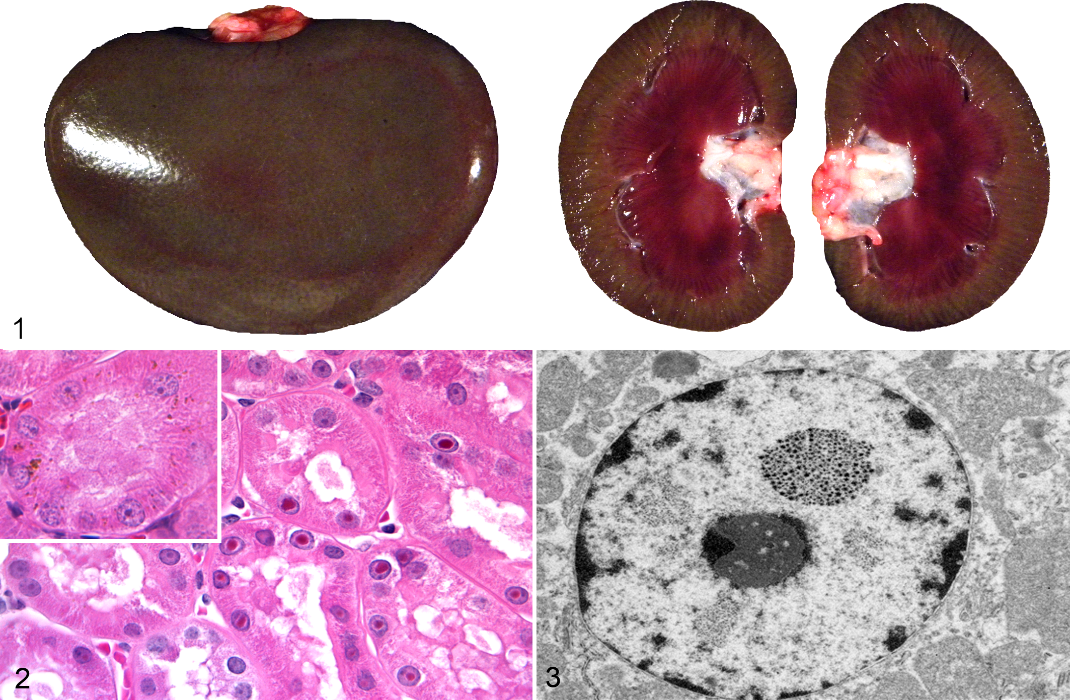
Kidney; rhesus macaque (Macaca mulatta). Renal cortices were uniformly dark green to black, with dark red medullas.
Light microscopy of HE-stained sections revealed numerous proximal renal tubular epithelial cells that contained a single, large, discrete, well-demarcated magenta to brown intranuclear inclusion (Fig. 2). Fewer tubular epithelial cells contained granular brown intracytoplasmic material. The inclusions were negative when stained with Ziehl-Neelsen acid-fast stain (Suppl. Fig. S1) and appeared pale gray (negative) with Perl’s iron, blue to gray (negative) with periodic acid–Schiff (PAS), light to dark brown (negative) with Alizarin red stain for calcium (Suppl. Fig. S2), and light gray (negative) with reticulin stain. The inclusions stained black with Fontana Masson (Suppl. Fig. S3) and Churukian-Schenk stains. Unstained tissues had brown inclusions; when unstained tissue sections were treated with a 10-second application of 30% hydrogen peroxide, the inclusions were no longer visible. With all these stains, the granular cytoplasmic material displayed the same staining characteristics as the intranuclear inclusions.
Samples were prepared for electron microscopy with 1% osmium tetroxide and 0.8% potassium ferrocyanide stains in 0.1 M sodium cacodylate buffer for 2 hours, rinsed with water, stained en bloc with 1% uranyl acetate for 30 minutes, and then dehydrated with a graded series of acetone (50%, 70%, 90%, and 100% for 20 minutes each). Samples were then infiltrated with a 1:1 mix of acetone and Epon 812 (cat. 14120; EMS, Hatfield, PA) overnight with rotation. After this incubation step, the 1:1 mix was replaced with 100% Epon 812 and allotted time to polymerize overnight at 60°C. Thin sections (90 nm) were imaged at 120 kV on a FEI Tecnai Spirit TEM system (FEI, Hillsboro, OR). Images were collected as a 2048 × 2048–pixel, 16-bit gray scale using FEI’s TEM Imaging & Analysis (TIA) interface on an Eagle 2 KCCD multiscan camera (FEI, Hillsboro, OR) at the OHSU Biomedical Engineering Multi-scale Microscopy Core (Portland, OR). Electron microscopy imaging revealed numerous renal cortical tubular epithelial cells that contained a single, variably sized intranuclear inclusion. The inclusions were composed of densely grouped, coaggregated, electron-dense crystalline material that appeared variably star or asterisk shaped (Fig. 3). The inclusions did not peripheralize or displace the nucleolus, and the remaining nuclear structures appeared normal.
Toxic element screening, including bismuth levels, was performed on frozen kidney tissue with inductively coupled plasma atomic emission spectrometry at Michigan State University’s Diagnostic Center for Population and Animal Health. Bismuth levels were 110.6 ppm. Lead levels were unremarkable at <0.50 ppm.
Discussion
The diagnosis of renal bismuth pigmentation in this macaque was based on the combination of a history of chronic BSS administration, elemental screening, gross and histologic findings, histochemical staining, and electron microscopy.
Toxic element screening confirmed not only the presence of bismuth (110.6 ppm) but that the levels were measured at 500 to 1000 times higher than normal acceptable bismuth levels. Normal levels of bismuth are <0.1 ppm in the liver and muscle of wild game birds and <0.2 ppm in the kidneys of rats. 1,5 The renal levels of lead in this macaque were considered within normal limits, disproving lead as the source of the intranuclear inclusions. Although nephrotoxicity due to bismuth has been reported in other species, in this case, pigmentation due to bismuth accumulation was not accompanied by clinical or microscopic evidence of toxicity. 7
The dark green to black renal cortical pigmentation was consistent with pigmented bismuth compound accumulation. Since the kidney is the primary target organ for bismuth accumulation, this was the most likely organ to exhibit pigmentation. 7 There was no gross or histologic evidence of this pigment in any other organs. Other causes of grossly visible green black renal pigment such as hemozoin and melanin were disproved through microscopic appearance of intranuclear localization and patient history. The animal had lived in a nonendemic area for malaria-causing Plasmodium sp throughout its lifetime, and the animal had not received any blood or tissue transplants that could have resulted in iatrogenic transmission. Furthermore, there was no histologic evidence of malaria, effectively ruling out hemozoin pigment.
A variety of histochemical stains were employed to help correlate the appearance of the intranuclear inclusions and intracytoplasmic granules observed in the renal tubular epithelium with the elemental screening results. With the Ziehl-Neelsen acid-fast stain, lead inclusions stain positively on paraffin-embedded tissue, but the reaction is less intense than that in fresh tissue. 9 In contrast, bismuth inclusions stain acid-fast positive with frozen tissue and negative with paraffin-embedded tissue. 3 The results with Perl’s and Alizarin red stains for ferric iron and calcium were also negative. To demonstrate that the material was intrinsically pigmented, unstained tissues were examined, revealing brown inclusions. When unstained tissue sections were treated with hydrogen peroxide, the inclusions were no longer visible. The presumptive mechanism for decolorization with hydrogen peroxide is that the hydrogen peroxide oxidizes the bismuth sulfide, resulting in formation of colorless bismuth sulfate. 10 Melanin is resistant to decolorization with this short-term hydrogen peroxide application. 10 Fontana Masson and Churukian-Schenk silver-based stains, used for the nonspecific demonstration of melanin and argyrophilic granules, stained the inclusions brown-black. As the hydrogen peroxide treatment suggested that the material was not actually melanin, it was presumed that the bismuth salt inclusions possessed the ability to bind silver from a silver solution and reduce it to visible metallic silver with and without a reducing agent, thus exhibiting both argentaffin and argyrophilic properties.
Electron microscopy confirmed the presence of an electron-dense material consistent with heavy metal or mineral within the nucleus of proximal renal tubule epithelial cells. The appearance of bismuth inclusions depends on the method of fixation; sections fixed with glutaraldehyde exhibit a homogeneous electron-dense appearance. 4 However, inclusions in tissue fixed with osmium have a more granular and fibrillar appearance. 4 The latter is more consistent with the inclusions we identified in our osmium fixed samples.
Increased absorption of bismuth in this macaque is thought to be related to a variety of factors, including chronic vomiting, which likely altered the time of gastric emptying. In addition, famotidine and omeprazole were frequently administered with the BSS for this animal’s gastrointestinal disturbance. Famotidine is an H2 receptor antagonist. Work performed in humans with other H2 receptor antagonists (ranitidine) has demonstrated an increased systemic absorption of tripotassium dicitrato bismuthate. 6 Omeprazole, a proton pump inhibitor, has also been linked to increased absorption of tripotassium dicitrato bismuthate. 8 Although this animal was administered BSS, not tripotassium dicitrato bismuthate, this still suggests an additional potential for increased absorption. Furthermore, this animal’s diet of routine monkey chow contained normal dietary levels of cysteine, and as enrichment, this animal routinely received whole fruits and fruit juices. Both cysteine because of its thiol group and ascorbic acid derived from fruits and fruit juices have been linked to the formation of soluble bismuth in vivo and in vitro. 7 The prolonged administration of BSS by itself or in conjunction with drugs that alter gastric pH, as well as routine administration of foodstuffs that contain thiol group compounds and ascorbic acid, could have singly or in combination been responsible for increased systemic absorption of bismuth and consequently this animal’s gross and histologic findings.
Footnotes
Acknowledgements
We thank Drs Claudia Lopez, Andreas Lehner, and Dana Scott for their gracious support with electron microscopy, toxic element screening, and electron microscopy interpretation, respectively.
Declaration of Conflicting Interests
The author(s) declared no potential conflicts of interest with respect to the research, authorship, and/or publication of this article.
Funding
The author(s) disclosed receipt of the following financial support for the research, authorship, and/or publication of this article: This work was supported by grant P51OD011092 from the National Institutes of Health.
References
Supplementary Material
Please find the following supplemental material available below.
For Open Access articles published under a Creative Commons License, all supplemental material carries the same license as the article it is associated with.
For non-Open Access articles published, all supplemental material carries a non-exclusive license, and permission requests for re-use of supplemental material or any part of supplemental material shall be sent directly to the copyright owner as specified in the copyright notice associated with the article.