
Abstract
An adult male domestic pigeon (Columba livia) was presented for necropsy following natural death after a period of chronic weight loss and severe intestinal ascariasis. Histopathologic examination of the liver found moderate to marked, multifocal necrotizing hepatitis with large, basophilic intranuclear inclusion bodies. Transmission electron microscopy of affected hepatocytes demonstrated numerous intra- and perinuclear icosahedral virions arranged in a lattice structure, consistent with adenoviral infection.
History and Gross Findings
Tissues from an adult male domestic pigeon in the Columbus Zoo and Aquarium collection were submitted to The Ohio State University applied pathology service following the animal’s natural death. The pigeon flock had experienced chronic intestinal ascariasis and weight loss, for which the birds received fenbendazole and ivermectin per os. Three days after treatment, this pigeon was found dead in its enclosure. Complete gross postmortem examination performed by attending zoo veterinarians revealed that the bird was in poor body condition and was heavily parasitized by intestinal nematodes (Ascaridia columbae), which filled the small intestinal lumen. No other significant gross lesions were documented. The heart, lung, kidney, liver, and intestines were collected, fixed in 10% neutral buffered formalin, and submitted for histopathology. Other diagnostic tests were not performed.
Microscopic Findings
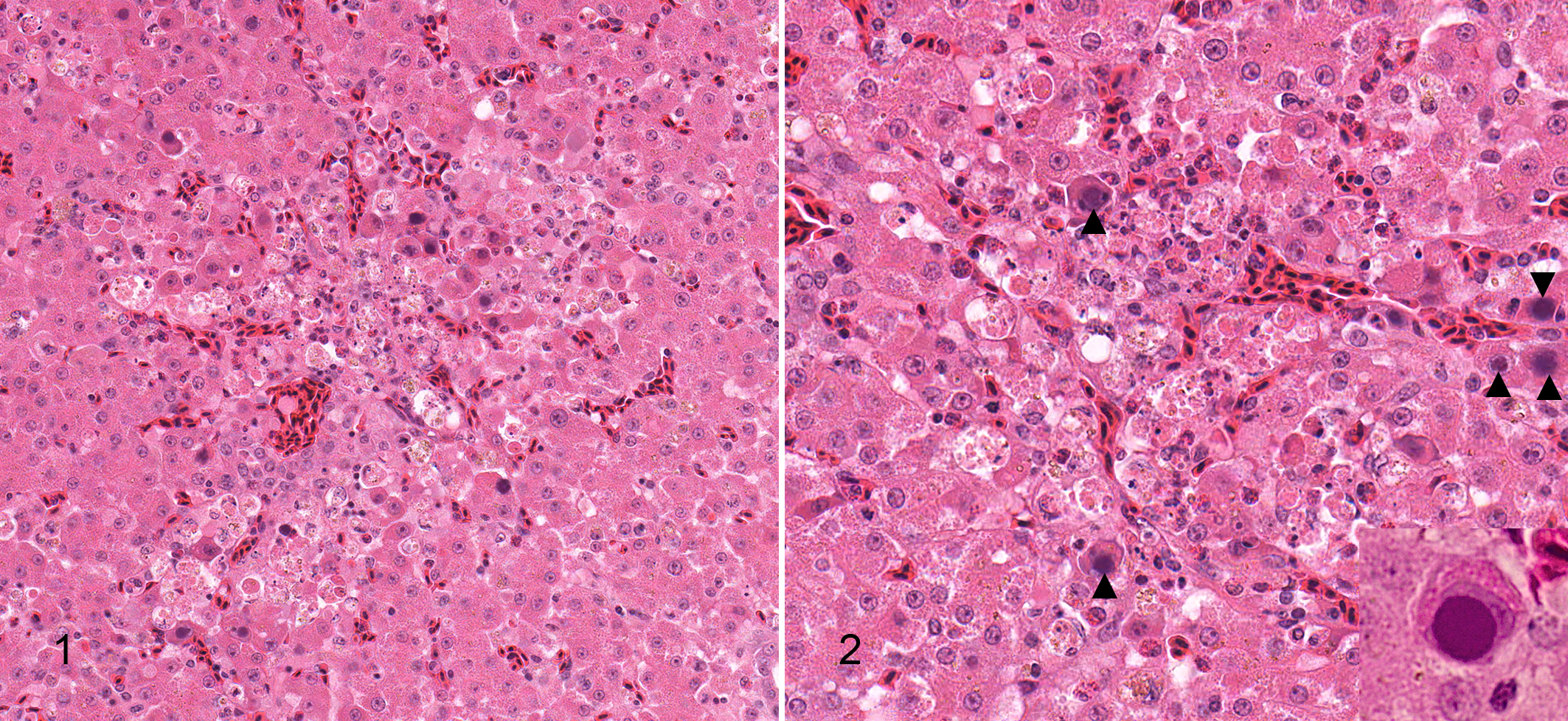
Formalin-fixed tissues were processed routinely and stained with hematoxylin and eosin (HE). Significant histologic lesions in this bird were confined to the liver. Throughout the liver, there were multifocal to coalescing, random areas of hepatocellular necrosis, characterized by loss of parenchymal architecture with individualization of hepatocytes, cytoplasmic hypereosinophilia, and mild infiltrates of heterophils (Fig. 1). Karyomegaly of many necrotic hepatocytes was observed, attributable to single, large, basophilic, round intranuclear inclusion bodies, occasionally with margination of chromatin (Fig. 2). Some viable hepatocytes adjacent to foci of necrosis also contained these intranuclear inclusion bodies.
Differential Diagnoses
Differential diagnoses for necrotizing hepatitis include toxic, ischemic, or infectious causes. Given the random localization of hepatocellular necrosis and the presence of intranuclear inclusion bodies, a viral etiology was favored. Based on the histologic characteristics of the intranuclear inclusion bodies, differentials ranked from most to least likely included pigeon adenovirus (PiAV), fowl adenovirus (FAV), avian polyomavirus, or pigeon herpesvirus 1.
Transmission Electron Microscopy
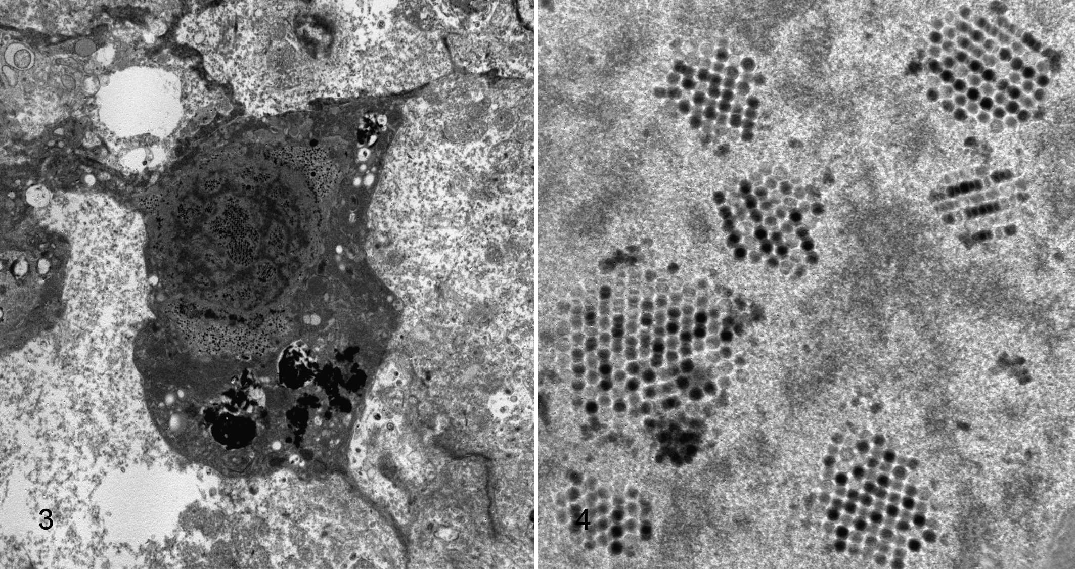
Formalin-fixed liver tissue was processed routinely for transmission electron microscopy (TEM). Optimal electron microscopic assessment of the liver was somewhat impaired by formalin fixation and storage at room temperature. TEM demonstrated the presence of numerous icosahedral, nonenveloped viral particles measuring 75 to 85 nm in diameter. These virions were most concentrated inside the nucleus of hepatocytes but were also occasionally observed outside the perimeter of a disrupted nuclear membrane (Fig. 3). In many nuclei, virions were arranged in a crystalline lattice (Fig. 4). The structure, size, and lattice formation by these viral particles support the diagnosis of an adenoviral infection. 1,7,10,11
Diagnosis
Based on the histologic lesions observed by light microscopy and further characterization of the intranuclear inclusion bodies by TEM, the diagnosis was necrotizing hepatitis with intranuclear adenoviral inclusion bodies.
Discussion
Liver lesions in this pigeon are consistent with adenoviral hepatitis. Adenoviral diseases of poultry are found worldwide and include marble spleen disease of pheasants, hemorrhagic enteritis of turkeys, egg drop syndrome in chickens, quail bronchitis virus, and inclusion body hepatitis (IBH) of chickens. 3,4,7 Avian adenoviruses are categorized into 3 serogroups, and multiple serotypes exist within each group. 3 To date, 7 different serotypes of group I FAV have been identified in pigeons, and in addition to these FAV serotypes, specific PiAV strains have been found. 2,3,6 There are 2 clinical syndromes of PiAV infection reported: “classical adenovirosis” is classified as type 1 and “necrotizing hepatitis” is designated as type 2, which is a distinct entity from IBH of other fowl. 3 Microscopic liver disease characterized in this bird is consistent with the latter clinical classification. Type 2 pigeon adenovirus infection can affect pigeons of any age, and sudden death of infected animals occurs within 24 to 48 hours, frequently without showing clinical signs. 3,9 In flocks where acute deaths are attributable to adenoviral necrotizing hepatitis, pigeons that survive remain clinically normal. Gross lesions of type 2 PiAV, if recognized, have been reported as a slightly swollen, yellow-tinged liver or multiple, depressed, red, pinpoint foci throughout the hepatic parenchyma. 3,7 No gross lesions of the liver were identified by zoo veterinarians performing the postmortem examination on this bird. Histologic demonstration of extensive hepatic necrosis with characteristic intranuclear inclusion bodies in hepatocytes is considered highly suggestive of pigeon adenoviral infection. 2,3 Although polymerase chain reaction and in situ hybridization assays for some passerine and psittacine adenoviruses are now being used for viral detection, 5,8 pigeon strain-specific viral diagnostics are not widely available. In this report, TEM was used to ultrastructurally characterize the intranuclear inclusion bodies, which corroborates the diagnosis of adenoviral necrotizing hepatitis.
Footnotes
Acknowledgement
The authors would like to thank Mamoru Yamaguchi for his tireless efforts in performing electron microscopy.
Declaration of Conflicting Interests
The author(s) declared no potential conflicts of interest with respect to the research, authorship, and/or publication of this article.
Funding
The author(s) received no financial support for the research, authorship, and/or publication of this article.