
Abstract
The cadherin family of adhesion molecules regulates cell-cell interactions. N-cadherin is expressed by neural and fibroblast cells but not by normal epithelial cells. In human medicine, the role of N-cadherin in breast cancer remains controversial, but some studies have described the switch from E-cadherin to N-cadherin as a critical step in the malignant progression of neoplastic cells. The present study was carried out on 160 feline mammary tumors (21 adenomas and 139 carcinomas). The relationship between the immunohistochemical expression of N-cadherin in neoplastic epithelial cells and 2 established prognostic factors such as regional metastasis and tumor grade was examined. The results of the study showed a statistically significant relation between the expression of N-cadherin and the 2 prognostic factors, and also a reduced expression of E-cadherin in tumors that expressed N-cadherin.
Cadherins are a family of transmembrane proteins that, together with their associated intracellular catenins, have important functions in cell-cell adhesion. Different cell types express different members of the cadherin family. 2 Epithelial (E)-cadherin and neural (N)-cadherin are the most extensively studied members of the cadherin family, but while they share many structural and functional features, they are expressed in an almost mutually exclusive manner. 9,12 E-cadherin is a key component of adherent junctions in epithelial cells and functions as a suppressor of tumor growth and invasion. Perturbation of its function is associated with an invasive phenotype in many tumors. N-cadherin is expressed by neural and fibroblast cells, where it mediates a less stable and more dynamic form of cell-cell adhesion. N-cadherin is not expressed in normal epithelial cells, but its expression has been demonstrated in several types of carcinomas. 1 Because of the important role played by cadherins in cell recognition, adhesion, and signaling, modulation of their function and expression has significant implications for the progression of tumors. 10 For instance, in a series of human breast carcinomas, it has been demonstrated that a switch from E-cadherin to N-cadherin expression contributes to increased tumor cell migration, invasion, and metastasis. 1,12
In the field of veterinary pathology, studies in canine mammary carcinomas have pointed to a relationship between the loss of E-cadherin and malignancy. 5,6 The role of E-cadherin has also been studied recently in feline mammary carcinomas in which preservation of this protein was associated with maintaining tissue architecture and inhibiting tumor cell invasiveness. 11,14 However, no studies have been published in veterinary science about cadherin switching involving the loss of E-cadherin and the expression of N-cadherin in mammary carcinomas. The aim of the present study is to analyze the expression of N-cadherin using a series of feline mammary adenomas and carcinomas (with and without regional metastasis) whose World Health Organization (WHO) classification, grading, and immunohistochemical expression of E-cadherin had previously been published. 11
The immunohistochemical expression of N-cadherin in feline mammary tumors was analyzed in a panel of adenomas (n = 21) and carcinomas (n = 139). Among the carcinomas, 73 had regional lymph node metastasis at the moment of diagnosis. In the present study, cases of animals with distant metastases, multiple mammary tumors, or recidivant neoplasms were discarded. Lymph node metastases were assessed by the evaluation of hematoxylin and eosin–stained slides as well as immunohistochemistry using a pancytokeratin monoclonal antibody, as described previously. 11 Tumors with regional micrometastases were classified as metastatic carcinomas. Carcinomas had been classified 11 according to the WHO as tubulopapillar (46/139), cribiform (62/139), or solid (31/139). The grading of the carcinomas had been obtained 11 by the modification of the Nottingham method described by Castagnaro et al. 4 The grading revealed 37 grade I, 66 grade II, and 36 grade III carcinomas. The expression of E-cadherin had been analyzed in a previous study, 11 following the method described by Brunetti et al. 3 As previously published, 11 all the adenomas (n = 21) showed normal membrane labeling of E-cadherin, while only 72 of the 139 carcinomas exhibited this pattern of expression. The other 67 carcinomas showed a reduced membrane expression of E-cadherin; in most cases, this reduced E-cadherin expression consisted of a combination of reduced labeling in the membrane and cytoplasmatic expression, and only 6 carcinomas showed exclusively cytoplasmic expression of the protein.
Immunolabeling of N-cadherin was carried out by the avidin-biotin-peroxidase complex method (ABC) using a monoclonal antibody (clone 6G11; Dako, Carpinteria, CA) following a pretreatment heat (121°C in citrate buffer, pH 6; 8 minutes). Counterstaining involved Mayer’s hematoxylin. Normal feline brain served as a positive control for each immunohistochemical series. Negative controls were carried out by replacing the primary antibody with an isotype-matched irrelevant antibody.
The presence of more than 5% of the neoplastic mammary epithelial cells showing cytoplasmic and/or membranous labeling of N-cadherin was interpreted as a gain of expression of this cadherin.
The association between the expression of N-cadherin and the presence of metastasis or the expression of E-cadherin was examined using the Pearson χ2 test. For each association, the odds ratio with a confidence interval (CI) of 95% was calculated. The association between N-cadherin expression and WHO histological classification or tumor grade was examined using a Kruskal-Wallis test. Analyses were performed using SPSS 19.0 software (SPSS, Inc, an IBM Company, Chicago, IL). A P value of <.05 was considered statistically significant.
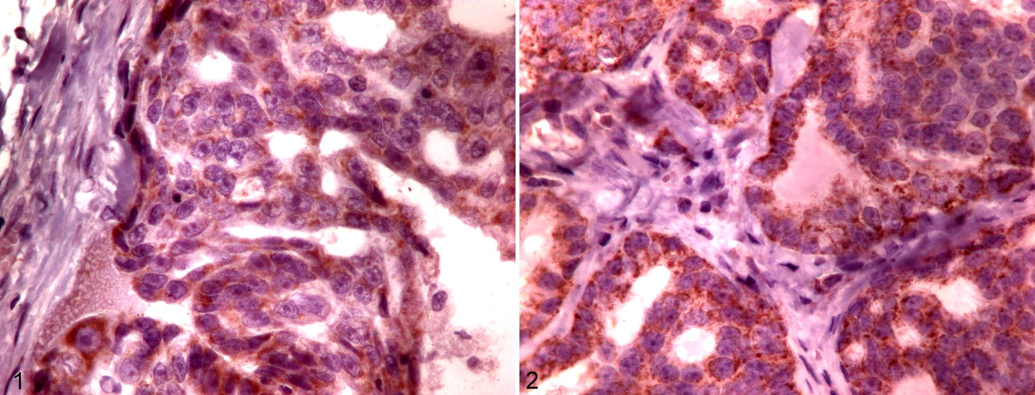
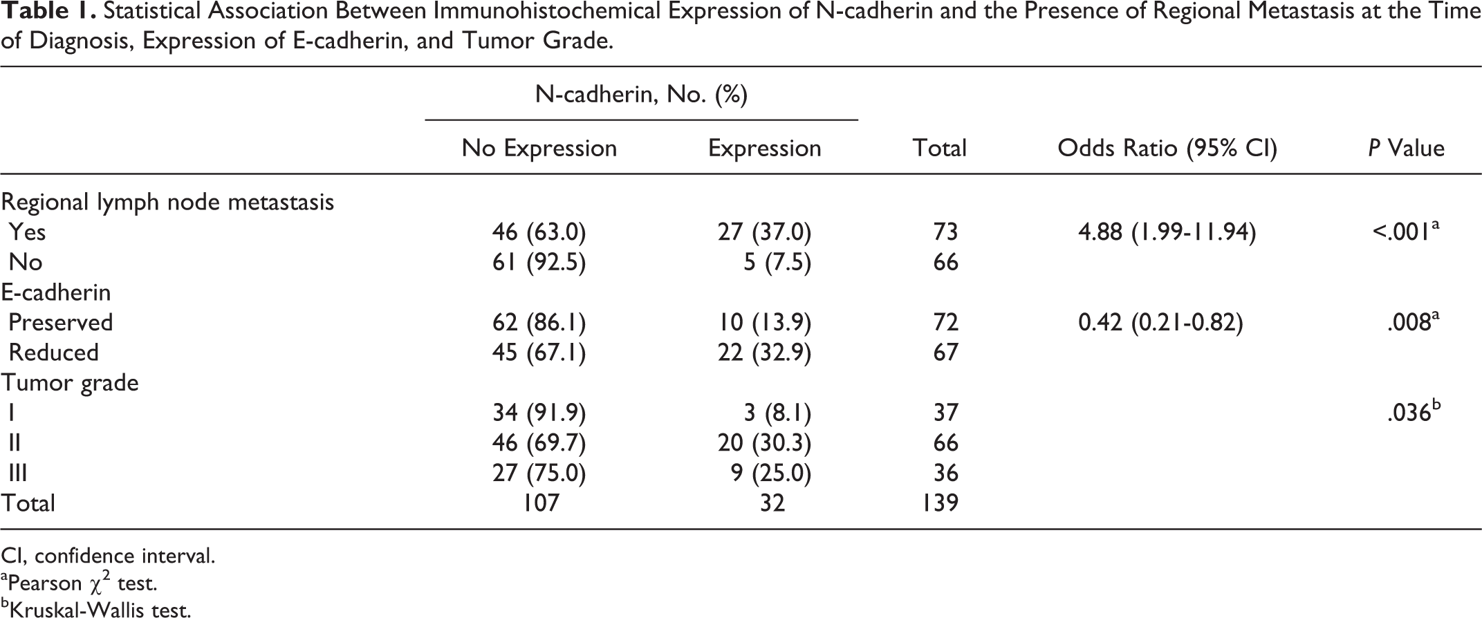
No N-cadherin expression could be found in the neoplastic cells of the 21 studied adenomas, while the immunohistochemical expression of N-cadherin was detected in 32 of the 139 analyzed carcinomas. The positive immunohistochemical expression of the molecule was predominatly cytoplasmic (Fig. 1), but staining of the membrane was also seen in 4 cases (Fig. 2), although membranous staining alone was never seen. When the relationship between the expression of N-cadherin and the morphology of carcinomas was examined, no significant differences were found. In contrast, when the relationship between the expression of N-cadherin and the grade of the carcinomas was studied (Table 1), a statistically significant relationship (P = .036) could be found. The expression of N-cadherin was lower in grade I carcinomas (3/37; 8.1%) than in grade II (20/66; 30.3%) or grade III (9/36; 25.0%) carcinomas. In fact, when a χ 2 test was applied to the association between N-cadherin expression and grade I or grade II/III, a significant difference was found (P = .012), although there were no significant differences between grade II and III tumors.
Statistical Association Between Immunohistochemical Expression of N-cadherin and the Presence of Regional Metastasis at the Time of Diagnosis, Expression of E-cadherin, and Tumor Grade.
CI, confidence interval.
aPearson χ2 test.
bKruskal-Wallis test.
The immunohistochemical expression of N-cadherin was also related (P < .001) to the presence of regional metastasis (Table 1), since most (84%) of the N-cadherin–positive tumors had metastases in the regional lymph nodes at the moment of diagnosis. The expression of N-cadherin also showed a negative relationship (P = .008) with the expression of E-cadherin (Table 1): only 10 of the 32 carcinomas expressing N-cadherin showed preserved expression of E-cadherin in the membrane of the neoplastic cells.
The results of the present study confirm the relationship between the immunohistochemical expression of N-cadherin in feline mammary carcinomas and 2 well-established prognostic factors 7,13 such as the presence of regional metastasis and tumor grade. Thus, the expression of this molecule could be considered a sign of malignancy. The association of the expression of the molecule with a loss of E-cadherin seems to point in the same direction. The mechanism related to the effect of N-cadherin in the progression of epithelial tumors has been studied in human medicine, where in vitro studies demonstrated that N-cadherin promotes a state of dynamic adhesion that allows both attachment and detachment of individual cells from the primary tumor and selective association with tissues such as the stroma or the endothelium. 8 Also, the formation of the extracellular complex involving N-cadherin and the fibroblast growth factor receptor has been described, resulting in a higher expression of metalloproteinase 9 and increases in cellular invasiveness. 1
Although this article is not a prognostic study, the results described indicate that the expression of N-cadherin may be a promising marker to be taken into account in future prognostic studies.
Footnotes
Declaration of Conflicting Interests
The author(s) declared no potential conflicts of interest with respect to the research, authorship, and/or publication of this article.
Funding
The author(s) disclosed receipt of the following financial support for the research, authorship, and/or publication of this article: C. Peñafiel-Verdu was the recipient of a predoctoral grant from the Fundacion Seneca (Regional Govern of Murcia).