
Abstract
The hearts of 30 dogs naturally infected with Leishmania infantum chagasi were evaluated histologically and immunohistochemically. Myocardial lesions were detected in all dogs, including lymphoplasmacytic myocarditis (27/30), myonecrosis (24/30), increased interstitial collagen (22/30), lepromatous-type granulomatous myocarditis (7/30), fibrinoid vascular change (3/30), and vasculitis (1/30). The parasite was detected in the hearts of 20 of 30 dogs. The number of parasitized cells correlated with the intensity of the inflammation and with the number of granulomas. The results indicate that cardiac lesions are prevalent in dogs with naturally occurring leishmaniasis even in the absence of clinical signs of cardiac disease.
Canine leishmaniasis may be subclinical or may manifest as a systemic disease with weight loss, muscular atrophy, anemia, hepatosplenomegaly, and cutaneous, renal, or ocular lesions. 2 Although clinical signs of cardiac disease are rarely reported in canine leishmaniasis, 3,10 myocardial lesions occur, 1,5,8 and the parasite can be found in cardiac tissue. 3,5,8,10 The aim of the present study was to document by histology and immunohistochemistry lesions and parasites in 4 regions of the heart in dogs naturally infected with Leishmania infantum chagasi.
Materials and Methods
Thirty dogs, naturally infected with L. infantum chagasi, were selected from Araçatuba, an endemic area in São Paulo State, Southern Brazil. The diagnosis was based on serology and confirmed by cytologic examination of bone marrow and lymph nodes. Serologic tests for Ehrlichia canis infections were negative in all dogs. Because Araçatuba is in a nonendemic area for Trypanosoma cruzi, Chagas disease has not been reported. For the same reason, the dogs were also considered negative for T. cruzi infection. All dogs had physical examinations, including blood pressure evaluation and electrocardiograms. Serum urea nitrogen and serum creatinine concentrations were determined to evaluate renal function. Seventeen dogs were female and 13 were male, with an age range of 6 months to 12 years (mean [SD], 3.29 [2.30] years). The study and its methods were approved by the Ethics Committee for Animal Experimentation (protocol number 2447/2011).
At necropsy, longitudinal and transverse sections of myocardium, about 1 cm3, were collected from the middle of the right atrial free wall, right and left ventricular free walls, and the interventricular septum. Tissue samples were fixed in formalin, embedded in paraffin, sectioned at 5 μm, and stained with hematoxylin and eosin (HE) and Masson’s trichrome. The myocardium of dogs with leishmaniasis was compared with that of healthy dogs and lesions semi-quantitatively scored for myocardial injury on a scale of 0 to 3: 0 = absent, 1 = mild (small focal lesions), 2 = moderate (larger, multifocal or focally extensive lesions), and 3 = severe (affecting most areas, with coalescence). Anti-Leishmania immunohistochemistry was performed as described. 9
Statistical analysis was performed using a commercially available software program (InStat; GraphPad Software, La Jolla, CA). Results were correlated using the Spearman test, whereas lesion intensity in the different cardiac areas was compared using the Friedman test. A P value <.05 was considered statistically significant.
Results
Although all dogs had clinical disease, none had clinical signs of cardiac disease or arrhythmias. One dog had a systolic arterial blood pressure of 186 mm Hg, above normal limits of 160 mm Hg. Three dogs had uremic syndrome. All dogs had histologic lesions in at least 1 of the 4 cardiac regions. The prevalence and severity of the main histologic changes are summarized in Supplemental Table S1. Amastigotes of Leishmania sp were observed in the myocardium of 20 of 30 dogs, mainly within a few macrophages (Fig. 1).
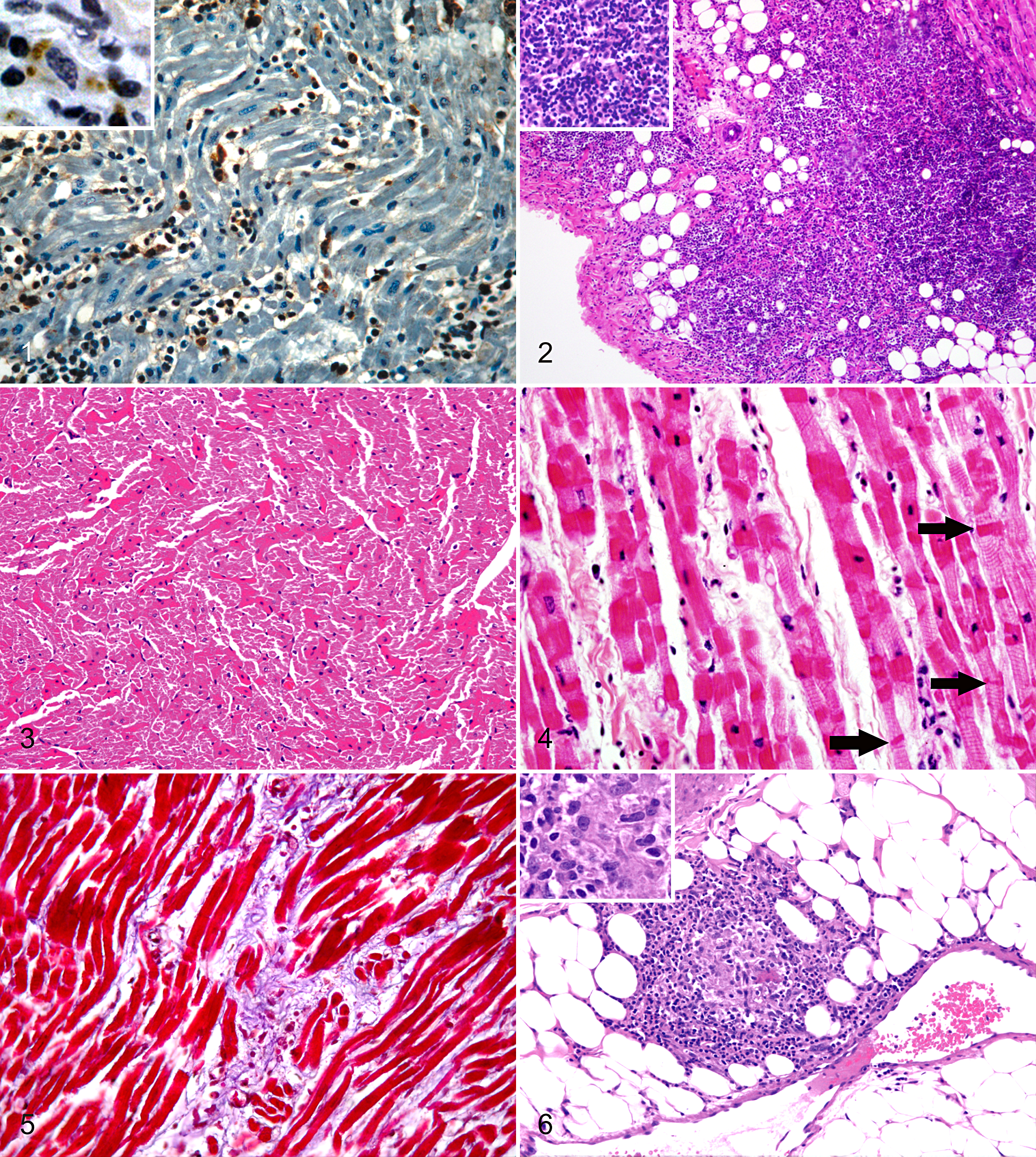
The number of parasitized cells correlated positively with the severity of myocardial inflammation in the right atrium (P = .039) and right ventricle (P = .018). The most common cardiac injury was lymphoplasmacytic myocarditis (Fig. 2), which was observed in 27 of 30 dogs. Leukocytes were concentrated in the subendocardial and subepicardial regions, especially in the right atrium, which was significantly different in comparison to the left ventricle (P = .031).
Coagulative necrosis of cardiomyocytes (Fig. 3) or contraction band necrosis (Fig. 4) was observed in 24 of 30 dogs, mainly in the right atrium and right ventricle. An increase in interstitial collagen was observed in 22 of 30 dogs (Fig. 5). Poorly defined, lepromatous-type granulomas, consisting of an inflammatory infiltrate composed mainly of macrophages with few lymphocytes and plasma cells, were observed in 7 of 30 dogs (Fig. 6). The number of granulomas correlated positively with the number of infected cells in the right atrium (P = .046) and interventricular septum (P = .022). Fibrinoid change was observed in the small arteries of the right atrium and right ventricle in 3 of 30 dogs, whereas vasculitis was observed in only 1 dog.
Discussion
Despite the low number of parasites detected in macrophages within heart tissue, with the exception of 1 dog that had several parasitized cells, 20 of 30 dogs had parasites in at least one of the areas evaluated, in contrast to a previous study, which found parasites in the myocardium of only 1 of 12 (8.4%) dogs. 1 The positive correlation between the number of parasitized cells and the severity of inflammation could suggest a direct role of the parasite in cardiac inflammation. Although the prevalence of myocarditis was similar, the severity of inflammation was more severe in this study than in a previous one. 1 The right atrium was most severely affected by lymphoplasmacytic myocarditis in this study, as in a previous case report. 8 Cardiomyocyte necrosis was more common in this study than previously reported. 1 The degree of cardiomyocyte necrosis was generally mild to moderate; only 1 dog had extensive necrosis. Myocardial necrosis was usually coagulative; however, contraction band necrosis was also observed. Although 22 of 30 dogs had an increase in interstitial collagen, myocardial fibrosis was generally mild and mostly associated with areas of inflammation. The association between fibrosis and inflammation may be explained by the role of leukocytes in fibrogenesis, which is mediated by cytokines and growth factors released during the healing phase in tissue injury. 7 The presence of granulomas, which have been described in the myocardium of dogs with leishmaniasis, 8 may reflect a chronic inflammatory response to uncontrolled parasitic infection rather than a protective response, because the number of granulomas was positively correlated with the number of infected cells. Fibrinoid change in myocardial arteries, which was observed in 3 dogs, may be a consequence of injury to the tunica intima and tunica media, which has been described in immune-mediated vasculitis 4 and is often observed in dogs with uremic syndrome and systemic arterial hypertension. 6 The 3 dogs with fibrinoid necrosis had uremic syndrome due to leishmaniasis, and 1 of these dogs also had systemic arterial hypertension. In summary, the results of this study indicate that myocardial lesions may be common and important in dogs with leishmaniasis even in the absence of clinical signs.
Footnotes
Declaration of Conflicting Interests
The author(s) declared no potential conflicts of interest with respect to the research, authorship, and/or publication of this article.
Funding
The author(s) received no financial support for the research, authorship, and/or publication of this article.
References
Supplementary Material
Please find the following supplemental material available below.
For Open Access articles published under a Creative Commons License, all supplemental material carries the same license as the article it is associated with.
For non-Open Access articles published, all supplemental material carries a non-exclusive license, and permission requests for re-use of supplemental material or any part of supplemental material shall be sent directly to the copyright owner as specified in the copyright notice associated with the article.