
Abstract
This article documents an epizootic of inflammation and neoplasia selectively affecting the lateral line system of lake trout (Salvelinus namaycush) in 4 Finger Lakes in New York from 1985 to 1994. We studied more than 100 cases of this disease. Tumors occurred in 8% (5/64) of mature and 21% (3/14) of immature lake trout in the most severely affected lake. Lesions consisted of 1 or more neoplasm(s) in association with lymphocytic inflammation, multifocal erosions, and ulcerations of the epidermis along the lateral line. Lesions progressed from inflammatory to neoplastic, with 2-year-old lake trout showing locally extensive, intense lymphocytic infiltrates; 2- to 3-year-old fish having multiple, variably sized white masses up to 3 mm in diameter; and fish over 5 years old exhibiting 1 or more white, cerebriform masses greater than 1 cm in diameter. Histologic diagnoses of the tumors were predominantly spindle cell sarcomas or benign or malignant peripheral nerve sheath neoplasms, with fewer epitheliomas and carcinomas. Prevalence estimates did not vary significantly between sexes or season. The cause of this epizootic remains unclear. Tumor transmission trials, virus isolation procedures, and ultrastructural study of lesions failed to reveal evidence of a viral etiology. The Finger Lakes in which the disease occurred did not receive substantially more chemical pollution than unaffected lakes in the same chain during the epizootic, making an environmental carcinogen an unlikely primary cause of the epizootic. A hereditary component, however, may have contributed to this syndrome since only fish of the Seneca Lake strain were affected.
From 1985 to 1994, an epizootic of inflammatory and neoplastic lesions affecting the lateral line tissue of lake trout was monitored in the New York Finger Lakes. The Finger Lakes are a series of long, relatively deep, glacially formed lakes located in upstate New York. 9 Recent synoptic water quality and sediment core analyses of the Finger Lakes, undertaken by the New York State Department of Environmental Conservation (NYS DEC), indicate that during the 1970s and continuing today, these lakes have had water conditions of varying quality. 15,28,39,41 Numerous organic and inorganic chemical contaminants were found in sediment cores retrieved from the Finger Lakes in 1997 and 1998, correlating to synoptic lake conditions during the 1970s. Primary organic chemicals detected in these cores included dichlorodiphenyl-trichlorethane (DDT) and polychlorinated biphenyls (PCBs). Primary inorganic chemicals detected included arsenic, nickel, chromium, copper, zinc, lead, and calcium.
The Finger Lakes were traditionally used primarily as summer resort areas, with more year-round residences established in recent years. Small tourism-oriented or college towns and agricultural activity such as viticulture and winemaking surround the lakes, and thus these regions have not been heavily affected by industrial pollutants. These lakes, however, have been variably affected by siltation and eutrophication due to agricultural and residential development in their watersheds. 28,39
Lake trout (Salvelinus namaycush, Walbaum, 1792) and brook trout (Salvelinus fontinalis, Mitchill, 1814) are native to New York state waters. Lake trout typically inhabit deep, cold, well-oxygenated lakes such as the Finger Lakes. Lake trout may live up to 20 years. 9 Unlike the brook, brown, and rainbow trout, lake trout are substrate spawners, scattering their eggs over rocky shoals in the slow-moving shallows of lakes as opposed to the rushing waters of streams.
The fish population in Cayuga Lake is an example of the stocking efforts undertaken by the NYS DEC throughout the Finger Lakes. Trout present in Cayuga include lake trout, rainbow trout (Oncorhynchus mykiss, Walbaum, 1792), and brown trout (Salmo trutta, Linnaeus, 1758). Their main forage base is alewives (Alosa pseudoharengus, Wilson, 1811), smelt (Osmerus mordax mordax, Mitchill, 1814), yellow perch (Perca flavescens,
Lesions of lateral line are rarely noticed or reported by fishermen, biologists, or scientists studying feral fish. Neoplasia of this organ system is extremely rare in both experimental carcinogenesis studies and in field epidemiology studies of neoplasia in fish. 3,34,36 Therefore, this epizootic presented a remarkable biological and pathological finding worthy of intensive investigation.
The lateral line system is a complex intradermal mechanosensitive organ system found on the head and trunk of fish and amphibians. The basic unit of the lateral line system is the neuromast, a complex receptor organ derived from the embryological neural crest and epidermal placode, composed of sensory hair cells, support cells, cupula mantle cells, periderm, and afferent fibers. 16,18,24,25,57
Hair cells of the neuromast are clustered and surrounded by slender supporting cells. Cupula mantle cells secrete the gelatinous cupula that covers the projections of the hair cells, causing them to deflect in unison when impinged upon by vibrating water. Such deflection results in the mechanical opening of ion channels, likely transient receptor potential subfamily channels, causing a shift in the membrane potential and ultimately leading to the stimulation of afferent nerves. 32 Neuromasts exist in 3 various configurations along the lateral line system: free, skin covered, or bony scale encased. Free neuromasts are located on the head, trunk, and fins of fish. Skin-covered neuromasts are found mainly on the head and are of various branching patterns and form the anterior lateral line. The bony scale encased neuromasts run bilaterally along the flanks of the trunk and form the posterior lateral line.
The closest homologous structure to lateral line neuromasts in terrestrial mammals is the hair cell of the inner ear. Recently, the lateral line system has been recognized as a useful fish model system for the study of mechanisms of drug toxicity to neurosensory cells of the inner ear. 14,42 Fish also possess an inner ear tuned to high-frequency vibrations, but the lateral line system detects distant low-frequency hydrodynamics as well as movements of objects at close range, such as schoolmates, prey, or predators. 25
Materials and Methods
Morphologic Studies
Diagnostic submissions were contributed by anglers. Additional specimens were obtained by gill net. Live fish were euthanized with a lethal dose of phosphate-buffered tricaine methane sulfonate (MS-222; Argent, Redmond, WA). All experiments with fish used for field epidemiology or experimental disease studies were approved by Cornell University’s Institutional Animal Care and Use Committee and were conducted according to guidelines provided in the Guide for the Care and Use of Laboratory Animals. Complete postmortem examinations were conducted on each case. In the initial 30 cases, histopathologic examination was performed on all major organs and gross lesions. In subsequent cases, histologic examination was limited to lateral line lesions. Tissues were fixed in neutral buffered 10% formalin for light microscopy or in Karnovsky’s fixative (1:4.5 concentrated fixative in cacodylate buffer, pH 7.2; Electron Microscopy Sciences, Hatfield, PA) for electron microscopy. For routine light microscopic study, tissues were embedded in paraffin, cut at 6 μm, and stained with hematoxylin and eosin (HE). Selected sections of neoplasms were stained with Masson’s trichrome. Step sections of early lateral line lesions were prepared with tissue embedded in glycol methacrylate (GMA; HistoResin, Leica Microsystems, Wetzlar, Germany), cut at 4-μm intervals, and stained with HE. For transmission electron microscopy (TEM), tissue (n = 13 cases, 5–6 blocks per case) was postfixed in 1% osmium tetroxide in 0.1 M cacodylate buffer, then dehydrated by passing through a graded series of ethanols followed by propylene oxide. Tissue was embedded in Epon/araldite, and toluidine blue–stained 1-μm sections were examined to select the best 3 to 4 blocks per case. Ultra-thin sections were prepared using a diamond knife and stained with uranyl acetate and lead citrate. TEM was conducted on a Philips EM-301 (Philips, Andover, MA) or Zeiss 902 (Carl Zeiss, Oberkochen, Germany).
Immunohistochemistry Procedures Applied to Neoplasms
Five tumors from 5 fish were evaluated using an immunohistochemical staining panel including antibodies directed against S100 protein (S100), neuron-specific enolase (NSE), or pan-cytokeratin (CK). The slides were heated in a 60°C oven for 20 minutes and then deparaffinized using xylene (Fisher Scientific, Waltham, MA) and rehydrated using a graded series of ethanol (Pharmco, Brookfield, CT) baths and then distilled H2O. For NSE, heat-induced epitope retrieval was performed by microwaving slides immersed in EDTA (pH 8.0) for two 10-minute cycles. Slides were placed on the Autostainer Plus (Dako, Glostrup, Denmark), rinsed with buffer, and then immersed in a 3% hydrogen peroxide solution for 5 minutes at room temperature to block endogenous peroxidase activity. For CK, slides were treated with pepsin for 20 to 45 minutes. Nonspecific protein adhesion was blocked using Zymed (San Francisco, CA) goat anti-rabbit kit (S100, NSE) or Zymed goat anti-mouse kit (CK) for 5 minutes at room temperature. The following primary antibodies were applied at room temperature with an incubation time of 30 to 90 minutes: polyclonal rabbit–anti-human S100, 1:400 dilution (Dako); polyclonal rabbit–anti-human NSE, 1:3000 dilution (Dako); and monoclonal mouse–anti-human AE1/AE3 CK 1:100 dilution (Dako). The secondary antibody, Zymed biotinylated goat anti-rabbit (S100 and NSE) or Zymed biotinylated goat anti-mouse (CK), was applied and the slides were incubated for 10 to 20 minutes at room temperature. A streptavidin-peroxidase conjugate, Zymed goat anti-mouse kit or Zymed goat anti-rabbit kit, was applied for 10 minutes at room temperature. Chromogen, 3,3′-diaminobenzidine-tetrahydrochloride (DAB from DakoCytomation, Carpenteria, CA), was applied to the slides for 1 minute at room temperature. Slides were counterstained using hematoxylin (DakoCytomation) for 2 minutes and then washed in distilled water. Slides were dehydrated using ethyl alcohol and cleared with xylene and ProPar (Anatech Ltd, Battle Creek, MI). Finished slides were coverslipped using Permount mounting media (Fisher Scientific).
Virus Isolation Procedures
Virus isolation was attempted using methods established for isolation of major salmonid viruses. 50,54,55 Briefly, cell-free preparations were evaluated from anterior kidney, spleen, and lateral line lesions/tumors of 4 fish from Owasco Lake and 10 fish from Cayuga Lake. Tissue homogenates, 1/10 w/v in sterile phosphate-buffered saline (PBS), pH 7.4, with 100 U/ml penicillin, 100 μg/ml streptomycin, and 25 μg/ml Fungizone (PSF), were prepared in sterile stainless steel blenders. Homogenates were centrifuged 5 minutes at 1000 × g. The fat-free supernatant fluid was filtered through a low-binding 0.45-μm filter (Millex GV; Millipore, Billerica, MA). Serial dilutions of the filtered homogenate (1:100, 1:1000) were prepared in Leibovitz medium (Gibco, Grand Island, NY) with 5% fetal bovine serum (FBS) and PSF (Leibovitz-5%). Using monolayers of 4 fish cell lines (rainbow trout gonad—RTG-2, Chinook salmon embryo—CHSE-214, epithelioma papulosum cyprini—EPC, and lake trout fry—CB3) grown in 24-well plates (2 cm2 per well), 3 wells each were inoculated with 0.1 ml of 1:10, 1:100, and 1:1000 dilutions of each tissue homogenate. 13,22,54 Appropriate triplicate uninoculated control and positive control (infectious pancreatic necrosis virus) wells were prepared on each tissue culture plate. Homogenate aliquots were allowed to adsorb to monolayers for 1 hour at 15°C, and then an additional 1 ml of Leibovitz-5% was added to each well. For each cell line, duplicate plates were sealed in plastic bags. One plate was incubated at 10°C and the other at 15°C for 1 month. At this time, plates were frozen at –80°C and thawed, and then 0.1-ml aliquots of each well were used to inoculate fresh monolayers for a second passage that was incubated for 1 month with replicates at both 10°C and 15°C.
To check for the presence of highly cell-associated viruses, whole live cells from anterior kidney, spleen, or lateral line neoplasms were cocultivated with monolayers of selected fish cell lines. 10 Single-cell suspensions were prepared from the tissues by teasing them through a 60-μm mesh sterile screen (Tetko, Inc, Elmsford, NY). Suspensions of these cells were prepared in Leibovitz-5%, and cell concentration was determined using a hemocytometer (AO Scientific Instruments, Buffalo, NY). Cell viability was assessed using trypan blue exclusion. 54 Monolayers of the cell lines in 24-well plates were inoculated with 10 4 or 10 5 live cells. This cocultivation procedure was conducted on 1 fish from Cayuga Lake using CHSE and RTG cell lines incubated at 15°C and on 1 fish from Seneca Lake using the CB3 cell line incubated at both 10°C and 15°C. Inoculated cultures were incubated for 1 month. Second passages of these cultures were prepared as with the conventional virus isolations. Cells from selected wells were prepared for TEM by trypsinization of monolayers, fixation of cells in 1:4.5 Karnovsky’s fixative, and processing as described above.
Tumor Cell Cultures
To determine whether viral particles might be more readily shed in cultures of neoplastic cells removed from the trout’s immune system, we initiated primary cultures of neoplasms from the heads of Cayuga, Owasco, and Seneca Lake fish. The surface of the neoplasms was disinfected with 70% ethanol. The superficial 2 mm of the neoplasms was removed to minimize microbial contamination and fixed for histology. The remainder of the neoplasms was excised aseptically, rinsed in sterile PBS, and placed in PBS with PSF. Neoplastic tissue was minced and placed in a 1:10 w/v sterile trypsin-versene (TV) solution containing 0.05% trypsin and 0.025% versene in a PBS base. The tissue was trypsinized with gentle mixing on a stir plate for 2 to 6 hours at 4°C. Freed cells were harvested and the tissue residue was again trypsinized for up to 8 hours. Trypsin was inactivated by addition of 1:10 volume of FBS to harvested cell suspensions. Cell suspensions were counted using a hemocytometer and viability was determined with trypan blue. Cell suspensions were centrifuged 10 minutes at 200 × g and the cells resuspended in Leibovitz medium containing 20% FBS and PSF (Leibovitz-20%). Aliquots containing 6 × 10 6 viable cells were seeded per 25-cm2 flask in Leibovitz-20% and incubated at 10°C, 15°C, or 20°C. For long-term preservation, confluent cell cultures were trypsinized and cells resuspended in freeze medium composed of Leibovitz-20% containing 7% dimethylsulfoxide. Aliquots of cells in freeze medium were frozen at –80°C or in liquid nitrogen.
Supernatant fluid from the 10th passage of the 15°C neoplastic cell culture from a Cayuga Lake fish was negatively stained for TEM. Copper grids (300 × 75 mesh) previously coated with Formvar (Fullam, Schenectady, NY) were dipped in a mixture of 4 parts 100 μg/ml bacitracin (Sigma, St Louis, MO), 1 part 3% phosphotungstic acid, and 1 part supernatant fluid or 1 part concentrated supernatant fluid. For the concentrated supernatant fluid, 50 ml of supernatant fluid was centrifuged 30 minutes at 10 000 × g to remove debris, and then clarified supernatant fluid was centrifuged 1 hour at 100 000 × g. The pellet was resuspended in 1 ml distilled water. Cells from the cell culture were also trypsinized, fixed in Karnovsky’s fixative, and processed as described above for TEM.
Disease Transmission Trials
To determine whether the lateral line syndrome was transmissible, lateral line lesion material and kidney and spleen cells from affected fish were injected into hatchery-reared lake trout fry. Two strains of lake trout were used: Seneca Lake (male) × Lake Ontario (female) fry from the Tunison Laboratory of Aquatic Science (United States Geological Survey: USGS), Cortland, New York, and Seneca Lake fry from the NYS DEC Bath Fish Hatchery, Bath, New York. Both strains were raised in 9°C flowing water. The mean weights of the fry were 7.3 g from Tunison and 6.6 g from Bath. Groups of fry were identified by pelvic (right/left), adipose, and no fin clips. The 2 strains of fry were housed in separate tanks and acclimated for 2 weeks. Fish were randomly assigned to 1 of 5 treatment groups comprising 20 to 27 fish each: (1) no injection (anesthesia only); (2) media only (Leibovitz with 2× PSF + gentamycin); (3) homogenized lateral line, kidney, and spleen; (4) whole live cells from the lateral line, kidney, and spleen; and (5) whole live cells from the lateral line. For injections, fish were anesthetized in phosphate-buffered MS-222. Control fish (group 2) received two 0.1-ml injections, one subcutaneously, on the left side just caudal to the trailing edge of the operculum on the lateral line, and the second intraperitoneally, on the left ventral trunk caudal to the pelvic fin. Lateral line preparations were injected subcutaneously into the lateral line on the left side of the trunk. Kidney/spleen preparations were injected intraperitoneally. Fry were housed in flowing, dechlorinated water in aerated 10-gallon aquaria set in Living Streams (Frigid Units, Toledo, OH). Living Streams were maintained at 14 ± 2°C for the first 2 months of the study and at 10°C to 15°C for the fall/winter. A 12-hour light/dark cycle was imposed using incandescent white lights. Detritus was siphoned as needed, water quality was measured and recorded weekly (total ammonia, pH, nitrite, and hardness), and dissolved oxygen and residual chlorine were monitored monthly. Fry were initially fed 2% body weight but were reduced to 1% body weight on day 32 due to water fouling concerns.
Collection of Material for Injection
Lake trout showing the lateral line syndrome were collected from Cayuga Lake during the summer of 1991 by NYS DEC biologists using gill nets. Ninety percent of the fish collected were immature. Affected fish were placed on ice and transported to Cornell University for dissection. Lateral line lesions were excised and placed in Leibovitz with 2× PSF + gentamycin, and kidney and spleen samples were passed through a sterile screen (Tetko, Inc) to create single-cell suspensions in Leibovitz with 2× PSF + gentamycin. All samples were held at 4°C until injection.
Lateral Line Lesion Preparations
Lateral line lesions were prepared as both homogenates and whole live-cell preparations. Homogenates were prepared from explanted tissue at 10% w/v in Leibovitz with 2× PSF + gentamycin, sonicated twice at 70% maximum for 30seconds, centrifuged at 500 × g for 10 minutes, and passed through a 0.45-μm FBS-soaked filter. Whole live-cell preparations began with explanted tissue trypsinized in TV (10× by volume) at 8°C for 1.5 hours, and then a second harvest was collected with additional TV added (10× by volume) to remaining tissue and harvested 1 hour later. FBS was added to make 10% of the volume of TV during each harvest. Cells in TV/FBS were centrifuged at 500 × g for 10 minutes and resuspended in Leibovitz with 2× PSF + gentamycin. Cells were counted using a hemocytometer and viability determined by trypan blue exclusion. Cells were diluted to 105 live cells/0.1 ml.
Preparation of Kidney/Spleen Suspensions or Homogenates
Whole live-cell suspensions were counted using a hemocytometer and trypan blue exclusion. Suspensions were centrifuged at 500 × g for 10 minutes and resuspended in Leibovitz with 2× PSF + gentamycin for a final concentration of 106 live cells per 0.1 ml. Homogenates were prepared starting with 106 live cells per 0.1 ml that were homogenized, sonicated twice at 70% maximum for 30seconds, and centrifuged at 500 × g for 10 minutes, and the fat-free supernatant was passed through an FBS-soaked 0.45-μm filter.
Evaluation of Fish
Fish were monitored daily, and any dead or moribund fish were necropsied. Every 2 months, fish were anesthetized and examined for evidence of lateral line lesions. Since 0 to 8 fish per treatment group died within 1 day of injection from stress of anesthesia or injection, at the first evaluation time point 2 months postinjection, up to 10 fish were sampled for histology from each treatment group to equalize the number of fish per tank, so that growth rates would be comparable in each treatment group. Fish were maintained in laboratory tanks for 16 months. At the termination of the experiment, fish were anesthetized in phosphate-buffered MS-222, weighed, examined for any gross lesions, and necropsied. Major tissues were fixed in neutral buffered 10% formalin.
Evaluation of Lateral Line Lesions for Molecular Evidence of Papillomavirus or Walleye Dermal Sarcoma Virus
Using methods previously reported, molecular probes to cottontail rabbit and bovine papillomaviruses or walleye dermal sarcoma retrovirus were applied to Southern blots of DNA extracts from lateral line lesions of 10 different fish, including those with large neoplastic masses, early small neoplasms, and preneoplastic lesions. 43 Negative controls were normal skin from an unaffected fish or buffer.
Assessment of Lateral Line Lesions for Evidence of RNA-Dependent DNA Polymerase Activity
Tissue from the same 10 fish evaluated with molecular probes as well as control fish skin was assessed for RNA-dependent DNA polymerase activity using published methods. 43 As with the brown bullhead tissue assessed previously, the RNA-dependent DNA polymerase activity of lake trout tissue was expressed most strongly in the presence of Mn2+ rather than Mg2+. Tissue extracts were centrifuged on sucrose gradients to determine whether RNA-dependent DNA polymerase activity was elevated in the fraction with density of retroviral particles.
Results
Epizootiology
The lateral line syndrome has been observed in lake trout from Cayuga, Seneca, Owasco, and Canandaigua Lakes, with 66, 11, 16, and 7 of the initial 100 cases being from each of these lakes, respectively. Interestingly, the syndrome has not been observed from the 2 Finger Lakes, Keuka and Skaneateles, in which natural recruitment of lake trout is sufficient and stocking has not occurred in recent decades. Biologic surveys conducted by gill net resulted in prevalence estimates of 1% in adult lake trout in Cayuga (500 mature fish examined) and Seneca Lakes (1500 mature fish examined) in 1989–1990 and 5% (28/600) in immature lake trout in Cayuga Lake in 1989–1991. The prevalence estimates from a survey of Owasco Lake in 1989 were 8% (5/64) in mature and 21% (3/14) in immature lake trout. Biologists and fishermen report that the disease was not observed prior to the early 1980s. Affected fish ranged from 2 to 11 years old. Neoplasms occurring on 2-year-olds were less than 3 mm in diameter, and many were microscopic. Young of the year that were examined in summer were not affected, having been stocked 3 to 4 months earlier. The severity of the disease did not vary seasonally. Male and female fish were equally affected, exhibiting similar lesions (31 males and 31 females among cases with sex identified). The Aquatic Animal Health Program at Cornell University has not received any additional cases of the lateral line syndrome during the past 15 years, and biologists report that they no longer observe these lesions in lake trout from the Finger Lakes.
To help determine whether the lateral line syndrome was due to genetic or environmental influences, we examined 53 anesthetized 10-year-old Seneca Lake strain broodstock of the 1978 year class that had been raised and maintained in the Allegheny National Fish Hatchery, Warren, Pennsylvania. We did not observe any abnormalities of the lateral line system. Photographs of typical early lesions and large neoplasms in affected Finger Lakes fish were shown to the hatchery manager, who testified that he had not observed such lesions in any of the year classes of Seneca Lake strain broodstock maintained at that hatchery, including 300 fish of the 1981 year class.
Gross Evaluation
We have studied more than 100 cases of the lateral line syndrome in lake trout from New York’s Finger Lakes, examining abnormal tissues from 75 of these cases at the light microscopic level. A typical case consisted of 1 or more neoplasms in association with multifocal erosions and ulcerations of epidermis along the lateral line. Forty-eight of the initial 100 cases had 1 or more neoplasm(s) on the head, with two-thirds of these cases having multiple neoplasms. Twenty-nine cases had 1 or more neoplasm(s) on the trunk. Eight cases had neoplasms on both head and trunk. Eighty percent of the cases had segments of trunk lateral line that were eroded, ulcerated, or scarred; most lesions were multifocal and bilateral. Neoplasms were soft, white, ulcerated, usually cerebriform masses up to 4 cm in diameter (Figs. 1–3).
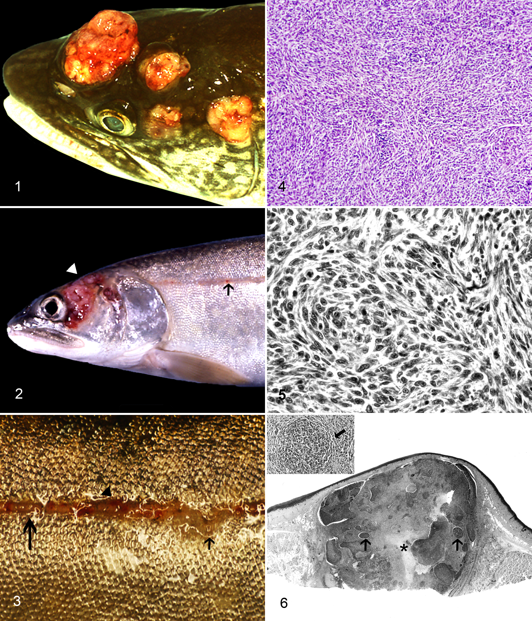
Head; 8-year-old lake trout from Seneca Lake, case No. 1. Spindle cell sarcomas of the lateral line canal system.
Light Microscopic Lesions
Table 1 reports the range of morphologic diagnoses and anatomic location of lateral line neoplasms. Neoplasms were similar grossly, except for epitheliomas, which differed from most malignant neoplasms in having a diameter of 2 mm or less. Because malignant neoplasms were typically highly anaplastic, differences of opinion regarding classification occurred among consulted pathologists. Four board-certified veterinary pathologists, 2 neuropathologists, and 3 fish pathologists examined representative neoplasms. Among these pathologists, diagnoses of high-grade astrocytoma, amelanotic melanoma, and malignant fibrous histiocytoma were given for anaplastic neoplasms in addition to diagnoses reported in Table 1. We classified malignant neoplasms based on the dominant histologic feature. Thus, neoplasms comprised principally of highly anaplastic cells, with a prominent spindle cell component, were classified as spindle cell sarcomas.
Neoplastic Lesions Affecting the Lateral Line of Lake Trout From New York’s Finger Lakes.
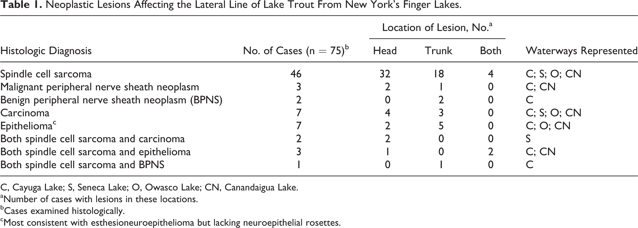
C, Cayuga Lake; S, Seneca Lake; O, Owasco Lake; CN, Canandaigua Lake.
aNumber of cases with lesions in these locations.
bCases examined histologically.
cMost consistent with esthesioneuroepithelioma but lacking neuroepithelial rosettes.
Three-quarters of the head neoplasms and two-thirds of the trunk neoplasms were spindle cell sarcomas. Most neoplasms over 2 mm in diameter were spindle cell sarcomas, regardless of location. These neoplasms contained pleomorphic spindle to stellate cells arranged in interlacing bundles and whorls within a scant fibrovascular stroma. A secondary component of these neoplasms was clusters and small sheets of pleomorphic round to oval cells with scant to abundant homogeneous eosinophilic cytoplasm. A small percentage of these round cells formed syncytia with 3 to 20 nuclei or bizarre cytomegalic and/or karyomegalic cells. Nuclear features of spindle and oval cells varied widely, with some having finely stippled or coarsely granular chromatin and others being large and vesicular with prominent, sometimes multiple, nucleoli. Spindle cell sarcomas over 5 mm in diameter exhibited areas of Antoni type A differentiation typical of nerve sheath neoplasms and identified as dense aggregates of spindle cells organized into interlacing parallel bundles with nuclear palisades (Figs. 4, 5). Trichrome stain revealed minimal collagen in regions of the neoplasms resembling nerve sheath. Small islands of highly anaplastic basophilic round cells with scant cytoplasm were often embedded within a field of more differentiated spindle cells (Figs. 6, 7).
Neoplasms over a few millimeters in diameter often had central areas of coagulative necrosis and hemorrhage. Pseudopalisading of nuclei surrounding these necrotic foci resembled patterns occurring in high-grade astrocytomas of mammals. In such neoplasms (Figs. 8–11), plump stellate tumor cells with abundant eosinophilic cytoplasm resembled gemistocytic astrocytes. 20,31 Malignant neoplasms frequently invaded subjacent connective tissue, skeletal muscle, bone, and blood vessels. However, neither gross nor microscopic evidence of metastasis to distant sites was observed.
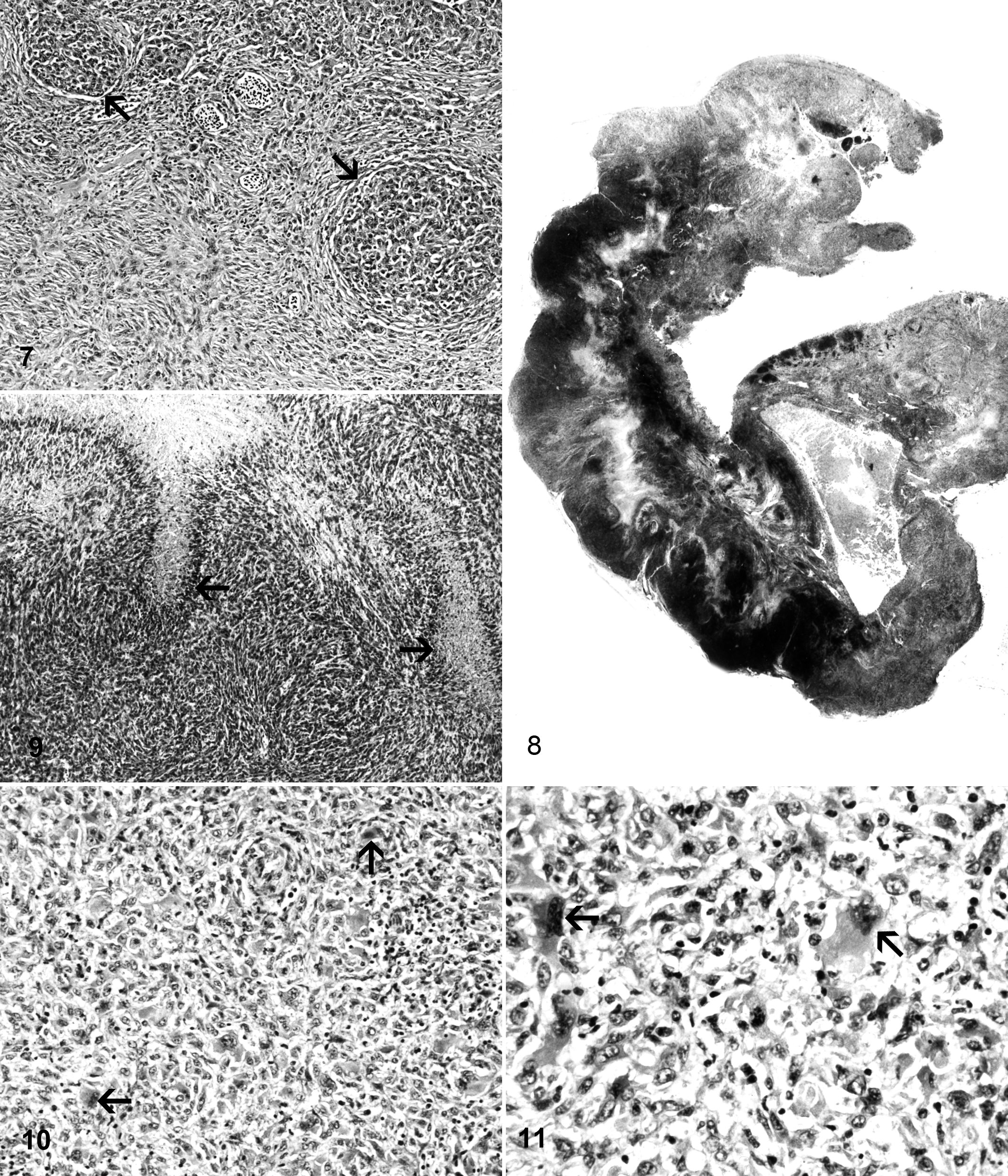
Head; adult lake trout from Seneca Lake, case No. 6. Islands of highly anaplastic basophilic round cells (arrows) set in a sheet of more differentiated spindle cells. Hematoxylin and eosin (HE).
Abnormal segments of lateral line without neoplasia nearly always exhibited mild to severe lymphocytic inflammation of the lateral line canal, with moderate to marked osteoclasia of the scale surrounding the canal (Figs. 12–14). In cases of severe chronic inflammation, complete resorption of the bone of the lateral line canal occurred (Figs. 15, 16). Erosions and ulceration of the epidermis adjacent to the lateral line ostea were common in the case of moderate or severe canal inflammation. Hemorrhage often accompanied inflammation of the lateral line tissue. Grossly abnormal segments of lateral line that lacked active inflammation were occasionally surrounded by several concentric layers of compact collagen (fibrosis).
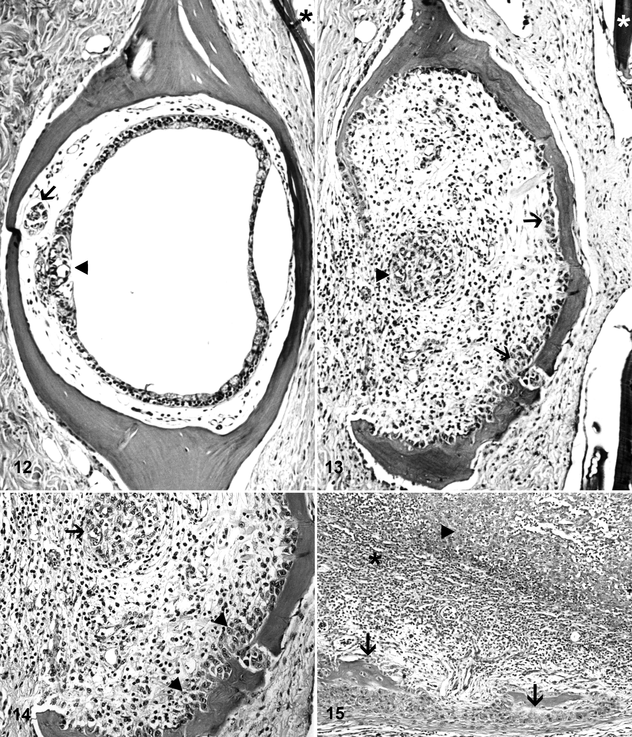
Trunk; normal adult lake trout. Normal lateral line canal containing neuromast (arrowhead) innervated by lateral line nerve (arrow). Scale near skin surface (*). Unaffected adult lake trout from Owasco Lake. Hematoxylin and eosin (HE).
Step sections revealed foci of hyperplasia and dysplasia of the epithelium lining the lateral line canal in several cases with severe, chronic inflammation of the canal. In hyperplasia, well-differentiated epithelium of the canal was mildly to markedly thickened up to 20 cells deep compared with the normal 3 to 5 cells (Figs. 15, 16). In dysplasia, the orderly sequence of epithelial differentiation was disrupted by clusters of pleomorphic epithelial cells bearing large, deeply basophilic nuclei and cytoplasmic vacuoles, particularly within the deeper strata.
Unfortunately, the intense lymphocytic inflammation that preceded neoplasia tended to obliterate the sensory neuroepithelium of the lateral line canal. Thus, despite extensive study of step sections of GMA-embedded early lesions, we were unable to definitively discern whether inflammation or early neoplasms first occurred in the neuromast tissue or elsewhere in the lateral line canal system. Occasionally, step sections of chronic inflammatory lesions near the neurosensory epithelium contained a single nonneoplastic neuroepithelial rosette, which indicated reparative responses of the neuroepithelium to chronic damage. However, neuroepithelial rosettes were not observed forming in neoplasms.
Those neoplasms comprised of well-differentiated or more anaplastic spindle cells in which the nerve sheath pattern predominated were designated benign or malignant peripheral nerve sheath neoplasms, respectively. The smallest microscopic neoplasms were designated simply epitheliomas (Fig. 17) because the cell of origin was uncertain. These neoplasms occluded the lateral line canal and consisted of sheets of uniform, small, closely packed oval to polygonal epithelial cells, with indistinct cytoplasmic borders and scant eosinophilic cytoplasm. Nuclei of epitheliomas were of uniform size, with finely stippled to coarsely granular chromatin. Nucleoli were single and inconspicuous. Carcinomas (Figs. 18–21) consisted of sheets of polygonal to round cells, with scant to abundant eosinophilic cytoplasm. Carcinomas often contained bizarre syncytial or giant neoplastic cells.
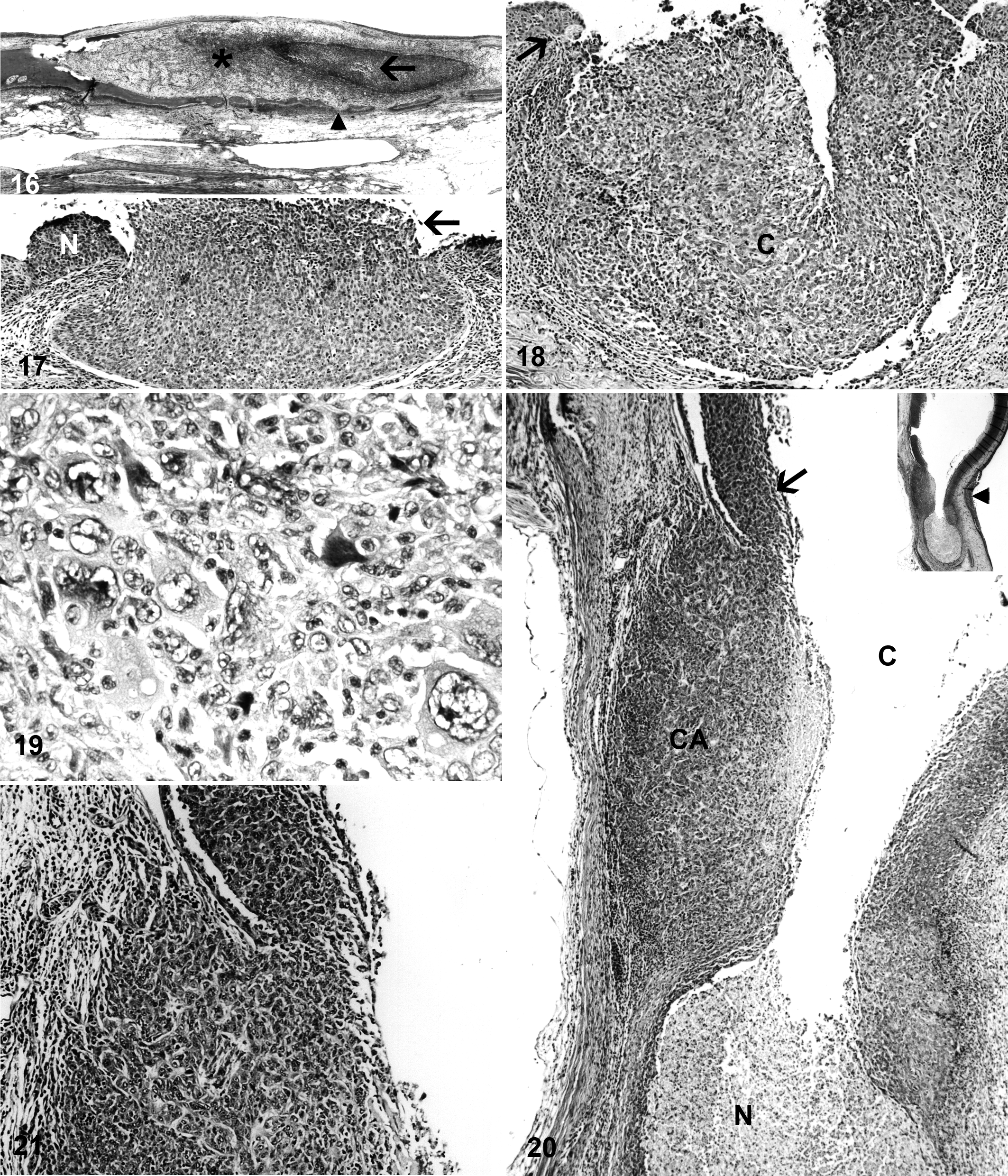
Head; adult lake trout from Owasco Lake, case No. 9. Intense lymphocytic inflammation (*). Hyperplasia of epithelium of canal (arrow). Osteoclasia of bone of canal (arrowhead). Hematoxylin and eosin (HE).
Microscopic neoplasms containing epithelioma tissue also frequently contained 1 or more other types of neoplastic tissue, including spindle cell sarcoma, benign peripheral nerve sheath neoplasm, or carcinoma. Small neoplasms remained within the epithelium of the lateral line canal, spreading laterally, obliterating the canal lumen, and often forming intraepithelial nests resembling those seen in malignant melanomas. As neoplasms evolved to become more anaplastic, invasion through the basement membrane inevitably occurred (Figs. 20, 21).
Ultrastructural Studies
In TEM studies, tissues from 13 cases were examined, including fish from Seneca, Owasco, and Cayuga Lakes. We studied spindle cell sarcomas from 10 cases, carcinomas from 3 cases, 1 epithelioma, and inflammatory lesions from 3 cases. Viral agents were not observed in any of the tissues examined. Ultrastructurally, more differentiated spindle cell sarcomas were similar to the epithelioma. Both had tightly adherent cells with scant to moderate amounts of cytoplasm and moderately convoluted nuclei containing primarily euchromatin. The cytoplasm contained abundant free ribosomes and granular endoplasmic reticulum, moderate numbers of mitochondria, occasional Golgi complexes with abundant vesicles nearby, and occasional bundles of cytoplasmic intermediate filaments. Well-differentiated desmosomes with small arrays of associated tonofilaments occurred at up to 3 per cell (Fig. 22). Anaplastic spindle cell sarcomas and carcinomas were somewhat similar to more differentiated neoplasms but with wider intercellular spaces, almost no heterochromatin, even more free ribosomes, and fewer mitochondria.
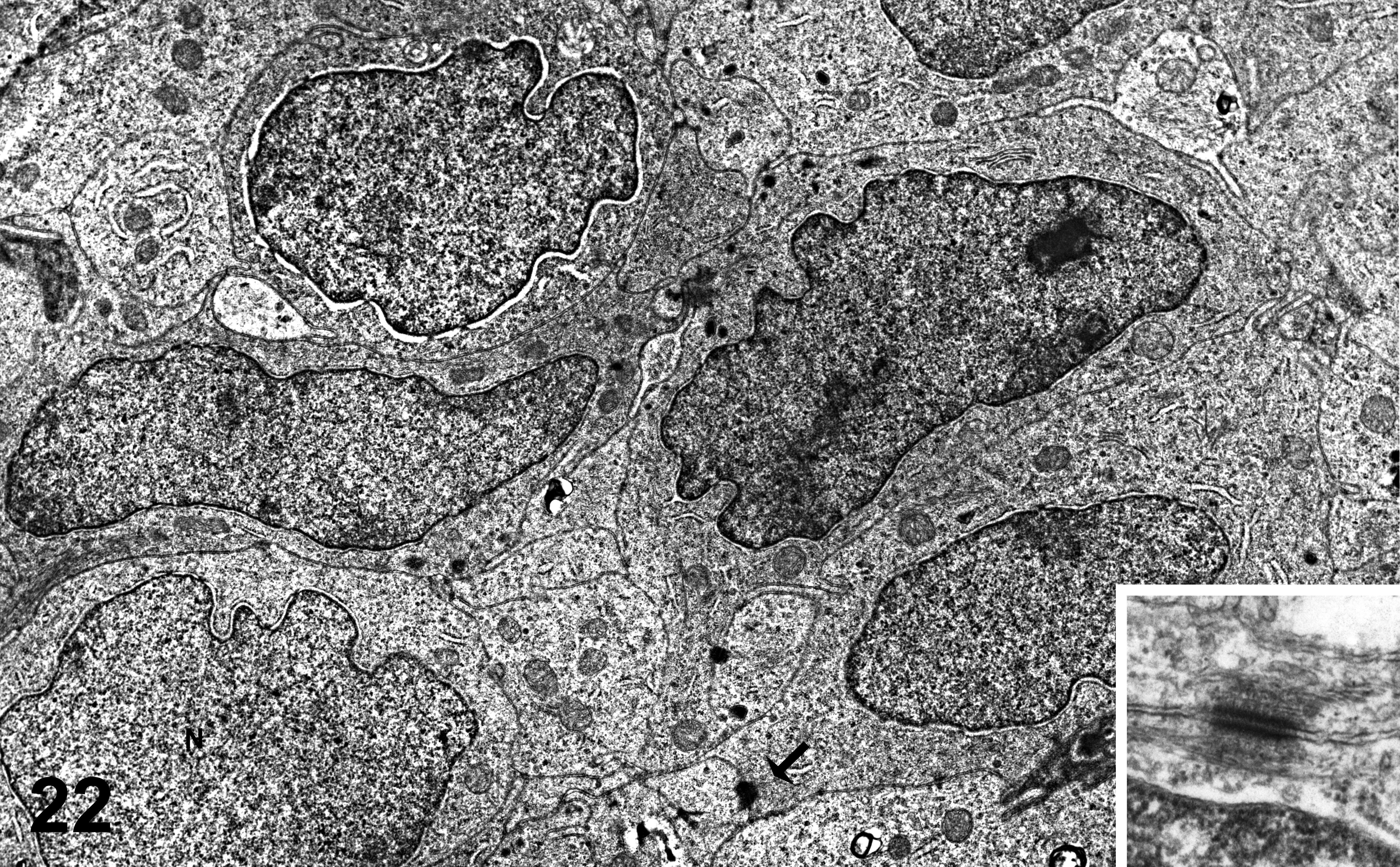
Trunk; 3-year-old lake trout from Owasco Lake, case No. 3. Spindle cell sarcoma mass protruding from pit of lateral line canal. Tightly packed neoplastic cells containing large moderately convoluted nuclei (N), with primarily euchromatin. Desmosomes (arrow) are present multifocally on intercellular junctions. Inset: desmosome.
Immunohistochemistry Studies of Neoplasms
Findings from immunohistochemistry studies of various histologic categories of neoplasia confirmed the diverse cell lineages forming these neoplasms. 2,11,52 A malignant peripheral nerve sheath neoplasm stained strongly with S100 and moderately strongly with NSE (Fig. 23, case No. 4) but negative for cytokeratin, consistent with peripheral nerve tissue. Anaplastic sarcoma tissue (Figs. 24 and 25, case Nos. 6 and 2) stained strongly positive with S100 in both cases, lightly positive for NSE in 1 of 2 cases, and negative for cytokeratin, consistent with a neural origin of these poorly differentiated tumors. Two carcinomas (Figs. 26 and 27, case Nos. 10 and 12) stained moderately or strongly positive for cytokeratin and negative for S100 and NSE, consistent with an epithelial origin of these neoplasms. Unfortunately, the glial fibrillary acidic protein antibody to mammalian tissue used in these immunohistochemistry studies did not stain normal glial tissue of lake trout, so we were unable to evaluate the possible contribution of glial lineages to the neoplasms.
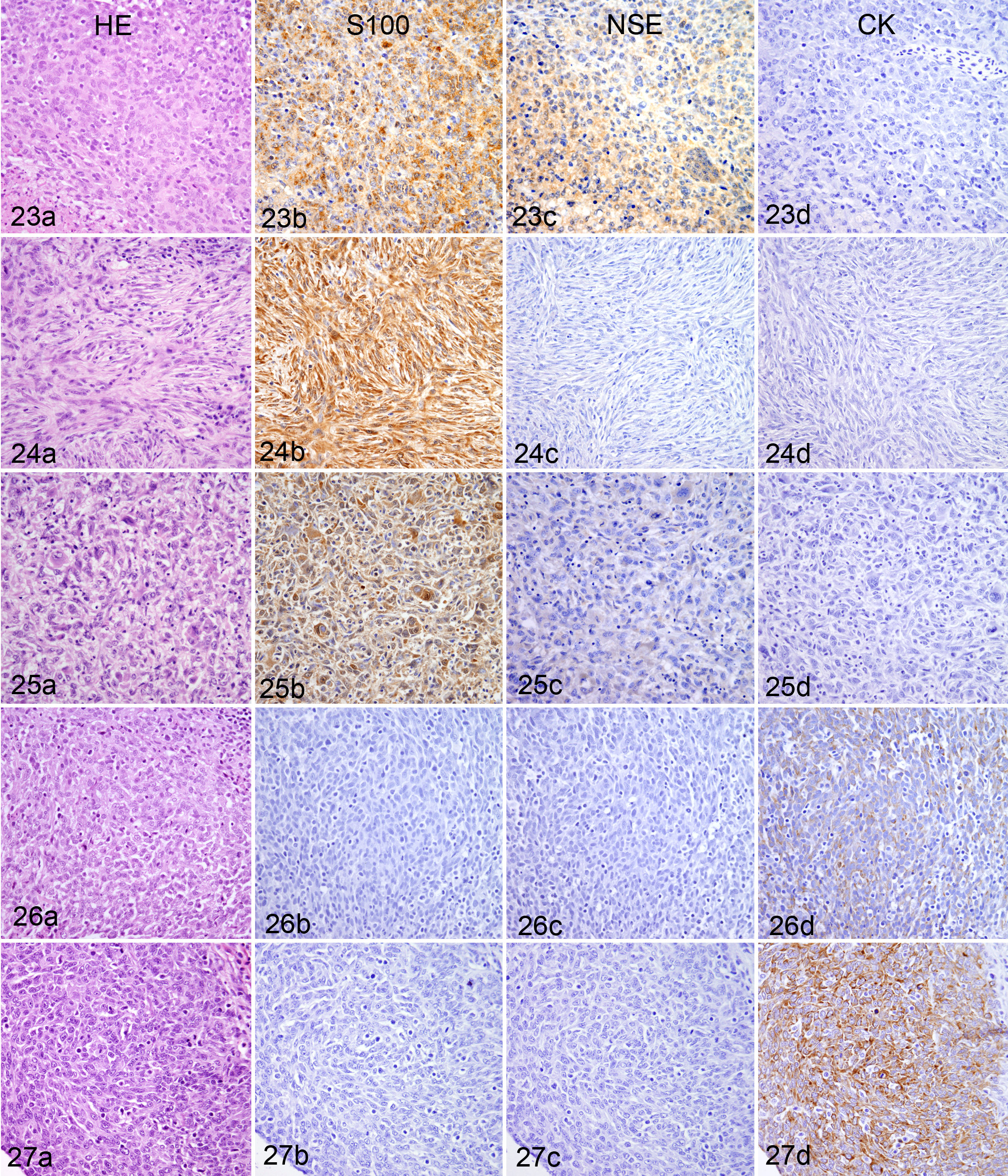
Head; adult lake trout from Seneca Lake, case No. 4. Immunohistochemistry studies of anaplastic region of malignant peripheral nerve sheath neoplasm using streptavidin-peroxidase, 3,3′-diaminobenzidine-tetrahydrochloride (DAB) as chromogen, and hematoxylin as counterstain. Column a, hematoxylin and eosin (HE). Column b, S100 strongly positive. Column c, neuron-specific enolase (NSE) moderately positive. Column d, cytokeratin (CK) negative.
Virus Isolation
Evidence of viral pathogens was not observed in cell cultures. Viral agents were not detected in TEM preparations of cell cultures.
Cell Culture of Neoplasms
Cell cultures of neoplasms grew best at 10°C or 15°C rather than at 20°C. The most vigorous culture, which was derived from a Cayuga Lake fish, has undergone 41 passages and is composed of stellate cells. These cells grow well in tissue culture flasks, forming clumps, small irregular sheets, and syncytia with intervening open spaces, but never forming confluent monolayers of polygonal or spindle cells typical of epithelial or fibroblast cell lines, respectively. Unlike cell lines of normal lake trout cells that we have developed, cultures of these neoplastic cells readily survive freeze-thaws when protected in freeze medium, even at a relatively low passage number such as 16. 13 Neither negatively stained preparations of culture supernatant fluid nor cultured neoplastic cells revealed viral agents when examined by TEM.
Tumor Transmission Trial
We did not observe gross or microscopic lesions of the lateral line syndrome in control or tumor-injected fish of either of the 2 strains of fish that were studied. Additional fish examined at earlier sampling times also showed no lesions of the lateral line syndrome.
Southern Blots Probed With Markers for Selected Viruses
Southern blots probed with markers for cottontail rabbit and bovine papillomaviruses or walleye dermal sarcoma retrovirus revealed no evidence of these viruses.
Assays of RNA-Dependent DNA Polymerase Activity in Abnormal Lateral Line Tissues
RNA-dependent DNA polymerase activity in tissues from abnormal lateral line of 10 fish varied from 2230 to 139 410 counts per minute (cpm) with Mn2+, polyriboadenylic acid, and oligo-deoxythymidylic acid, with just 2 of the samples showing activity less than 40 000 cpm. The highest activity in abnormal lateral line tissue occurred in a sample of a small early spindle cell sarcoma from the trunk. The positive control sample was supernatant fluid from a culture of ovine fibroblasts infected with the retrovirus ovine progressive pneumonia virus with activity of 124 827 cpm. Negative control cell culture fluid, fresh cell culture media, and normal fish skin had activities of 2403, 4720, and 3842 cpm, respectively. Fractions of abnormal lateral line collected from positions in sucrose gradients with the density of retroviruses did not show higher RNA-dependent DNA polymerase activity than other fractions.
Discussion
We describe here a unique epizootic of neoplasia affecting the lateral line of lake trout in specific Finger Lakes in New York state. Thus far, we have been unable to determine the cause of the disease. The evidence generated to date most strongly supports the hypothesis that these neoplasms are hereditary, as with familial neurofibromatosis in humans. Familial neurofibromatosis of humans is most often an autosomal dominant disorder caused by mutation in the tumor suppressor genes NF1 or NF2. 30,47 In the lateral line syndrome of lake trout, all affected individuals have been of the Seneca Lake strain, originating in one of several fish hatcheries. Those Finger Lakes (Keuka and Skaneateles) with sufficient natural recruitment have not been stocked with lake trout in recent years and do not have the lateral line syndrome in lake trout. Seneca Lake strain fish raised in the Allegheny National Fish Hatchery might not show the lateral line syndrome if this syndrome is caused by a gene occurring relatively rarely in the population. By chance, the samples of broodstock selected for the Allegheny hatchery may have lacked the gene. Or the mutation may have occurred as fish of the Seneca Lake strain were raised in another hatchery. Extensive molecular genetic studies will be required to clarify the possible role of genetic factors in a species such as lake trout, having a generation time of 5 years or more. Despite the lack of evidence for causes other than a genetic abnormality, the pronounced lymphocytic inflammation of the lateral line canal that inevitably precedes neoplasia is relatively uncommon in the case of hereditary neoplasia. 48 However, recent studies of neurofibromatosis I in humans reveal a reciprocal paracrine growth factor relationship between mast cells and neoplastic Schwann cells, with mediators secreted by neoplastic cells stimulating local proliferation of mast cells and vice versa. Pharmacologic agents blocking mast cell secretion reduce neoplastic Schwann cell growth. 17,53,56 Inflammation is also commonly a feature of vestibular schwannomas in neurofibromatosis caused by mutation in NF2. 49 If the lateral line neoplasia is hereditary, it may still require an environmental component for expression, such as a tumor promoter, which might occur in natural dietary items or in certain natural waters or sediments.
Virus isolation attempts, tumor transmission trials, Southern blots probed with markers for known viruses, assays of RNA-dependent DNA polymerase conducted on fractions from sucrose gradients at the density of retroviruses, and ultrastructural studies of affected lateral line tissues did not provide evidence of a viral etiology. At the time that this research was conducted, telomerase was not yet recognized as a reverse transcriptase. Thus, we were rather perplexed by the high levels of RNA-dependent DNA polymerase observed in the lateral line tumors as well as in the other fish neoplasms that we evaluated such as orocutaneous papillomas of brown bullhead when we were unable to identify retroviruses in the tissues. 43 We now realize that telomerase was markedly upregulated in these tumors as well as in preneoplastic lesions in lake trout in comparison to activity levels in normal skin of these fish species. Telomerase reverse transcriptase has recently been studied extensively in the model fish species Xiphophorus and found to be upregulated following ultraviolet light exposure. 19
A number of toxicants such as asbestos, copper, petroleum hydrocarbons, and pesticides can cause degeneration and inflammation of the lateral line tissue of fish. 3,5,46 However, we believe that the lateral line syndrome in lake trout is not likely due to exposure to 1 or more environmental carcinogen(s) because the disease is remarkably tissue and species specific. Several other salmonid species, including rainbow trout, brown trout, and landlocked Atlantic salmon, inhabit affected waters but showed no lesions of the lateral line. In lake trout affected by the lateral line syndrome, no other tissues were frequently or consistently abnormal.
Unfortunately, ultrastructural studies failed to clarify the cell type(s) of origin of the lateral line neoplasia. Neither epitheliomas nor carcinomas displayed the characteristics of epidermal filament (Malphigian) cells of normal or papillomatous fish skin, including intercellular bridges, ample cytoplasm containing abundant cytoplasmic intermediate filaments typically comprising more than 50% of total cytoplasmic volume, abundant desmosomes (10 or more per cell), or long arrays of tonofilaments associated with desmosomes. 44 Nor did these epithelial neoplasms exhibit cilia, microvilli, or tight junctions characteristic of neuromast epithelium. Ultrastructural features of all neoplasms examined most closely resembled embryonal neuroepithelial tissue. 26 Since a variety of fish cell types in addition to epithelial tissue can exhibit occasional desmosomes, including cardiac myocytes, perisinusoidal Ito cells of liver, astrocytes, and neural cells, the presence of small numbers of desmosomes in neoplasms of the lateral line does not indicate the cell type of origin. 20
A limited scanning electron microscopic study of early lesions in the lateral line of 10 affected fish failed to reveal the site of origin of neoplasms within the lateral line, so details of this work are not reported. Immunoperoxidase studies conducted with antibodies to mammalian S100, neuron-specific enolase, and cytokeratin corroborated the morphologic diagnoses assigned to the different histologic categories of neoplasia occurring in lake trout lateral line.
Two neoplasms affecting the lateral line of fish are cataloged in the Registry of Tumors in Lower Animals (RTLA) at the Smithsonian Institution. Both neoplasms were classified by Dr Harshbarger of the RTLA as esthesioneuroepitheliomas. One of these neoplasms occurred in a sheepshead from the Chesapeake Bay (RTLA 3102), and the other was in a goldfish from Big Bear Lake, San Bernardino, California (RTLA 1931). Each of these neoplasms comprised 2 distinctive components: sheets of densely packed basophilic round cells forming neuroepithelial rosettes and other sheets resembling a well-differentiated nerve sheath neoplasm, with interlacing bundles of spindloid cells showing nuclear palisading and storiform arrangements.
Five olfactory neuroepithelial neoplasms, 4 benign and 1 malignant, are reported in various bony and cartilaginous fish in the RTLA (RTLA 1707, 2051, 2566, 3132, and 3409). 29 Like the neuroepithelial neoplasms reported in the lateral line in the RTLA and unlike the neoplasms of the lateral line that we report in lake trout, the olfactory neuroepithelial neoplasms exhibited characteristic neuroepithelial rosettes.
The less well-differentiated lateral line neoplasms of lake trout strongly resembled malignant schwannomas of rats and mice. In murine malignant schwannomas and in lake trout neoplasms, sheets of more differentiated Antoni type A tissue were admixed with islands of highly anaplastic tissue composed of large ovoid mononuclear cells, often exhibiting cytomegaly, karyomegaly, multilobulated nuclei, or syncytial cell formation. 27,37,38 Most of the lateral line neoplasms of lake trout were highly malignant histologically, yet, unlike the malignant schwannomas of murine species that often metastasized widely, lake trout neoplasms did not metastasize. Despite the histologic resemblance of lake trout neoplasms to nerve sheath neoplasms, no inflammation or other abnormalities were observed in lateral line nerves or their branches except immediately adjacent to severely inflamed neuromast organs.
Careful study of the various lesions in this large group of affected fish suggests the following sequence for development of neoplasms in lake trout affected with the lateral line syndrome: mild lymphocytic inflammation of the lateral line canal may regress, leaving localized fibrosis, or may progress to intense lymphocytic inflammation accompanied by hyperplasia and dysplasia of epithelium lining the lateral line canal. Small well-differentiated epithelial neoplasms arise multifocally in the lateral line. Some of these neoplasms evolve into large pleomorphic, highly anaplastic neoplasms with central necrosis. Whether a single pluripotential cell type gives rise to the various neoplasms or whether multiple cell types can undergo neoplastic transformation in affected fish remains uncertain.
The evolution of the lateral line neoplasms from small benign epithelial lesions to large, invasive spindle cell neoplasms is similar to the epithelial to mesenchymal transition that occurs during the evolution of many neoplasms in mammals, including carcinomas and melanomas, as they become invasive and metastasize. 8 Neural stem cells of mammals may undergo an epithelial to mesenchymal transition to display mesodermal phenotypes. 6 Certain zebrafish mutant lines show an epithelial to mesenchymal transition during the evolution of neoplasia in the fish. 45 Recent drug discovery studies using zebrafish have targeted the epithelial to mesenchymal transition. 23 Although neoplasms that are highly malignant morphologically rarely metastasize in most fish species, these species still share many morphological and molecular features with mammalian cancer as the tumors evolve to become more anaplastic and invasive. 33,50
The lateral line syndrome does not appear to have a devastating effect either on lake trout populations in affected lakes or on the health of individual fish. Less than 10% of spawning lake trout in affected Finger Lakes exhibited lesions of the disease. The presence of smaller, often preneoplastic lesions in most affected fish younger than 4 years suggests that the disease progresses slowly. Affected fish, both immature and adult, even fish with large or multiple neoplasms, were in good nutritional status. This indicates that the morphologically normal segments of lateral line in these fish functioned appropriately, allowing the fish to locate prey and avoid predation. Once fish biologists became aware of the lateral line syndrome, they selected against affected individuals when collecting eggs for the lake trout stocking program. The recent disappearance of the lateral line syndrome due to management changes further supports a genetic basis for the disease.
Fish models used in the laboratory and in field epidemiology have provided important insights into risks from environmental contaminants as well as basic mechanisms of carcinogenesis. 34,35,36 Genetically tractable model fish species such as zebrafish, medaka, and Xiphophorus have allowed careful study of the roles of specific genes and signaling pathways in the development of hereditary neoplasia. Zebrafish genetic models for neuroblastoma and peripheral nerve sheath neoplasia are now available. 1,4,21 Basic understanding of molecular mechanisms of tumorigenesis gleaned from model fish species may guide scientists in clarifying the role of genetic factors in naturally occurring epizootics. Sophisticated new techniques such as the Virochip, a microarray-based platform for detecting and genotyping viral pathogens, will allow better detection of viral genetic material in neoplasms and should prove helpful in future studies of epizootic neoplasia in fish. 12
In summary, we describe a syndrome of chronic inflammatory lesions and neoplasms specifically affecting the lateral line system of lake trout in certain Finger Lakes in New York state. The cause of this syndrome remains uncertain; however, electron microscopy, virus isolation, and experimental transmission studies have failed to reveal evidence of a viral etiology. Since all affected fish are of a single strain, the Seneca Lake strain, a genetic basis for the lesions is possible.
Footnotes
Acknowledgements
We thank biologists from the New York State Department of Environmental Conservation for assistance in fish collections. We thank Ms Teena Smith for excellent technical advice and assistance in immunoperoxidase assays. Dean L’Amoreaux, Derek Currie, and Sujuan Zhang provided outstanding support in cell culture and electron microscopy studies. We appreciate the expertise of Alexis Wenski-Roberts in photomicrograph preparation. Initial cases of this disease were presented at the Fish Health Section of the American Fisheries Society/Eastern Fish Health Workshop, Annapolis, Maryland, 1989.
Author Note
The field and laboratory components of this research were conducted while Dr Spitsbergen served on the faculty at Cornell University in the Department of Avian and Aquatic Animal Medicine, which has been incorporated into the Aquatic Animal Health Program in the Department of Microbiology and Immunology.
Declaration of Conflicting Interests
The author(s) declared no potential conflicts of interest with respect to the research, authorship, and/or publication of this article.
Funding
The author(s) disclosed receipt of the following financial support for the research, authorship, and/or publication of this article: This project was funded by the “Return a Gift to Wildlife” program of the New York State Department of Environmental Conservation.