
Abstract
Thirteen proliferative diseases in fish have been associated in the literature with 1 or more retroviruses. Typically, these occur as seasonal epizootics affecting farmed and wild fish, and most lesions resolve spontaneously. Spontaneous resolution and lifelong resistance to reinfection are 2 features of some piscine retrovirus–induced tumors that have stimulated research interest in this field. The purpose of this review is to present the reader with the epidemiological and morphological features of proliferative diseases in fish that have been associated with retroviruses by 1 or more of the following methods: detection of C-type retrovirus-like particles or reverse transcriptase activity in tumor tissues; successful tumor transmission trials using well-characterized, tumor-derived, cell-free inocula; or molecular characterization of the virus from spontaneous and experimentally induced tumors. Two of the diseases included in this review, European smelt spawning papillomatosis and bicolor damselfish neurofibromatosis, at one time were attributed to a retroviral etiology, but both are now believed to involve additional viral agents based on more recent investigations. We include the latter 2 entities to update the reader about these developments.
Members of several virus families have been implicated in the etiology of tumors in fish. The literature contains numerous publications in which members of virus families Herpesviridae, Papillomaviridae, and Retroviridae are associated with the development of tumors in fish. In this report, we review the epidemiological, gross, microscopic, and ultrastructural features of 13 piscine tumors reported in the literature for which there is evidence of a retroviral involvement. In some instances, the evidence supporting a retroviral association is compelling and includes viral sequencing and successful transmission trials using a well-characterized inoculum. In other instances, a retroviral association is based on detection of retrovirus-like particles or reverse transcriptase activity in tissues from proliferative lesions. These viruses have been the subject of several reviews. 4,48,49 For this review, we have accessed archival materials primarily from the archived Registry of Tumors in Lower Animals as well as cases from the Aquatic Animal Health Program at the College of Veterinary Medicine, Cornell University.
Before evaluating a lesion, any case of a fish tumor must be considered in light of the fish as an ectotherm. Much of the biology of fish is affected by water temperature and seasonality of various physiological processes. It is well known that the immune response in fish is modified by changes in water temperature and other physiological stressors, such as spawning activity. 39 A review of the case records of most fish disease diagnostic laboratories will show the largest number of neoplastic cases to occur in the spring, a time of changing water temperature and a time when many, but not all, fish spawn. Water temperature may also affect the replication of a virus. In the pathogenesis of walleye dermal sarcoma, spring tumors had more infectious virus than those lesions evaluated in the fall. 14 It is not uncommon to observe seasonality in virally caused tumors in fish. 24
Proving a retroviral etiology in the pathogenesis of tumors is notoriously challenging. Due to the paucity of fish cell culture lines, most fish retroviruses are amplified by polymerase chain reaction (PCR) and sequenced using degenerate PCR approaches, rather than cell culture. Only 6 tumorigenic piscine retroviruses have been fully or partially sequenced. These include walleye dermal sarcoma virus, walleye epidermal hyperplasia viruses 1 and 2, perch epidermal hyperplasia viruses 1 and 2, and salmon swim bladder sarcoma virus. 49
Techniques to incriminate retroviruses in tumorigenesis have evolved significantly over time. Initial associations were made by the detection of retrovirus-like particles identified by transmission electron microscopy (TEM) in the lesion. Detection of retrovirus-like particles by TEM, however, may be due to coincidental expression or selective replicative advantage of mitotically active cells. 18 The discovery of reverse transcriptase, an enzyme essential for retrovirus replication, was considered a watershed event in the study of these diseases. The assays used in the detection of reverse transcriptase in affected tissues are complicated and may produce false positives in addition to detecting endogenous reverse transcriptase. 18 Currently, we rely on a variety of advanced molecular techniques to establish a definitive relationship between a retroviral agent and tumorigenesis. 18,48,52
In addition to tumors associated with retroviruses, we include 2 entities previously associated with retroviruses, European smelt spawning papillomatosis and bicolor damselfish neurofibromatosis, but for which viral associations have become complicated by the discovery of additional viral agents within lesions.
Walleye Dermal Sarcoma
Dermal sarcoma in the walleye (Sander vitreus) was first described in fish collected from Oneida Lake, New York, by Walker. 65 Walleye dermal sarcoma disease is characterized as single to multifocal, benign, cutaneous tumors on adult walleye. The lesions are seasonal and first arise in the late fall and remain on the fish through the winter until the spring, when they are observed during the spawning season. The lesions regress in the late spring to summer. 7,8,26 In the Great Lakes Basin, prevalence estimates in the spring range between 20% and 30% and are highest in young adults. There is evidence that a walleye found to be tumor positive during any given year will not develop walleye dermal sarcoma in the subsequent year, suggesting that tumor resolution results in lifelong resistance to the disease. 25 –27
Spontaneous dermal sarcomas in walleye are multifocal to coalescing, white to pale pink dermal nodules, up to 1 cm in diameter, with a sessile base and a smooth to cobblestone surface (Fig. 1). Nodules can occur anywhere on scaled skin. Histologically, the skin is elevated by a well-demarcated, pseudoencapsulated neoplasm that arises within the dermal stratum spongiosum (scale bed) and thus often envelops a central scale (Fig. 2). Tumors are composed of spindle cells organized into streams that contour and vaguely whorl around the central scale and are supported by a dense collagenous matrix containing variable foci of osseous metaplasia. Spindle cells have moderate amounts of pale, vacuolated cytoplasm and indistinct borders. Nuclei are oval with vesicular chromatin and a central nucleolus. Mitoses are infrequent. 40 Reports of invasive tumors are rare and are limited to experimental trials in fingerling walleyes challenged by injection and 3 wild adult walleyes with natural disease. 9,12,20 Tumor regression is characterized by a progressively intense mononuclear cell infiltration and epidermal ulceration followed by tumor necrosis and invoution. 8
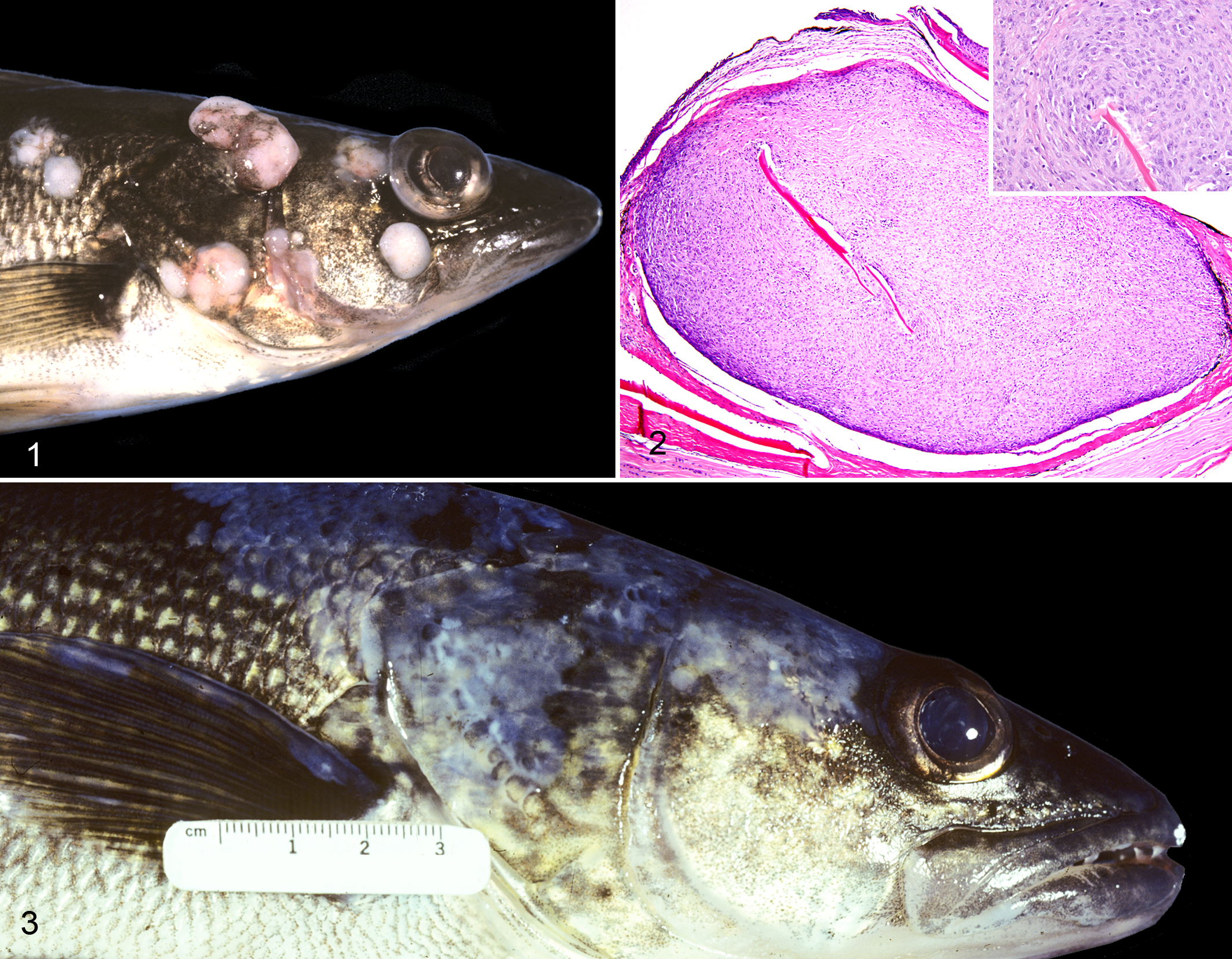
Walleye dermal sarcoma virus has been sequenced and identified as a complex piscine retrovirus. 29 The viral gene orfA, which encodes a cyclin-C–like homologue, termed retrovirus-cyclin (RV-cyclin), has been identified in the viral genome, suggesting that viral oncogenesis results from perturbations in the cell cycle. 36,52 The virus has been identified in neoplastic and inflammatory cells in proliferative lesions by in situ hybridization and immunohistochemistry, and viral RNA levels decrease with tumor regression. 46 The lesion has been experimentally transmitted in year 1 walleyes by intramuscular injection, feeding, and topical application of cell-free filtrates of tumor homogenates. 40 Transmission has also been achieved in walleye by cohabitation. 11 Successful experimental infection trials in sauger (Sander canadensis) and yellow perch expanded the potential host range of this virus. 13,30
Walleye Discrete Epidermal Hyperplasia
Discrete epidermal hyperplasia in walleye is a benign seasonal disease that is observed most commonly in spring spawning season on adult fish. Lesions typically regress by late summer. 7,28 The disease is termed walleye discrete epidermal hyperplasia to differentiate it from walleye diffuse epidermal hyperplasia, the latter being characterized by epidermal plaques with indistinct borders. A herpesvirus has been isolated from walleye diffuse epidermal hyperplasia. 32 In contrast to the other known retrovirus-associated skin tumor affecting walleye, walleye dermal sarcoma, prevalence estimates for discrete epidermal hyperplasia are comparatively low and increase with age, affecting up to 20% of year 8+ fish. 28
Walleye discrete epidermal hyperplasia is characterized by expansive to coalescing, translucent, flat plaques with lobate to stellate, sharply delimited margins. Lesions are widely distributed on the skin of the head, trunk, and fins (Fig. 3). Microscopically, the epithelium is abruptly expanded up to 5 times in thickness by proliferating Malpighian cells with abundant mitotic figures. The subjacent basement membrane and dermis are unaffected. There is goblet cell hyperplasia at the interface with the normal dermis. 45,69
Two retroviruses, walleye epidermal hyperplasia retrovirus 1 and 2, have been characterized from spontaneous lesions and fully sequenced. 37,52 Transmission trials using cell-free filtrates derived from spontaneous lesions known to harbor both viruses were successful. The presence of both viruses was subsequently confirmed in experimentally induced lesions via PCR assay. 10
Atlantic Salmon Swim Bladder Leiomyosarcoma
Leiomyosarcoma of the swim bladder of Atlantic salmon (Salmo salar) was first reported in 1978 in 2 separate studies as leiomyosarcoma and fibrosarcoma in aquacultured fish in Scotland. 19,38 A relatively low (4.6%) incidence of the disease was reported. Swim bladder leiomyosarcoma in Atlantic salmon again occurred in 1996–1997, when it was found in juvenile (year 1–2) Atlantic salmon from the Pleasant River, Maine, held at a fish hatchery in Massachusetts as brood fish in an Atlantic salmon restoration effort. 5 The tumors developed to such a degree that a persistent, low-level mortality was observed. Affected fish exhibited signs of lethargy, inappetance, and hemorrhages on the fins and body surfaces.
Grossly, coalescing, smooth, firm, pale-tan masses, up to 2 cm in diameter, expand the wall of the swim bladder and impinge on the luminal space (Figs. 4, 5). Microscopically, well-demarcated, unencapsulated, multinodular masses expand within the swim bladder wall and are contained within the luminal and serosal basal laminae. Tumors are composed of neoplastic spindle cells organized into long interlacing streams subdivided by thin bands of collagen. Neoplastic cells have moderate anisocytosis and variable nuclear pleomorphism with abundant mitotic figures and frequent pyknotic nuclei (Fig. 6). The neoplastic cells stain magenta with Masson’s trichrome (Fig. 7) and are variably immunopositive for α–smooth muscle actin (Fig. 8) and strongly immunopositive for desmin (Fig. 9). The tumor morphology and histochemical and immunohistochemical staining patterns are consistent with the diagnosis of leiomyosarcoma. 5
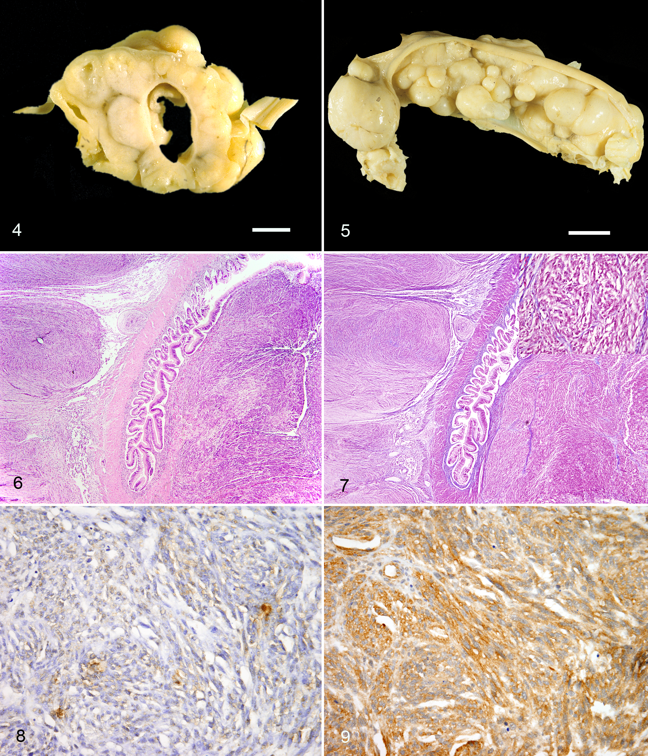
An exogenous retrovirus associated with the leiomyosarcoma was first identified by degenerate PCR amplification of the pol gene and then sequenced. 44 Tumor transmission trials have not been attempted.
Yellow Perch Discrete Epidermal Hyperplasia
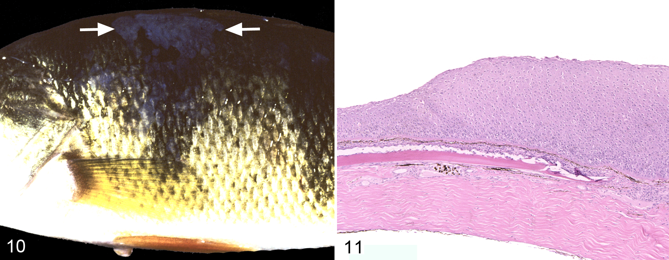
A single case of discrete epidermal hyperplasia of yellow perch (Perca flavescens) was observed in an adult fish during the spring spawning season. The lesion was grossly and microscopically identical to walleye discrete epidermal hyperplasia (Figs. 10, 11). As with the lesion in the walleye, 2 retroviruses were isolated from yellow perch discrete epidermal hyperplasia and have been termed perch discrete epidermal hyperplasia retrovirus 1 and 2 (P. R. Bowser and J. W. Casey, unpublished data). Segments of the highly conserved polymerase gene, pol, were sequenced from both perch viruses and were unique compared with other known piscine retroviruses. 49 Although experimental transmission efforts have not been successful, the limited amount of tissue and virus concentration made available for the referenced study may have been insufficient to replicate the disease experimentally. 13
Chinook Salmon Plasmacytoid Leukemia
Plasmacytoid leukemia of Chinook salmon (Oncorhynchus tshawytscha), also known as “marine anemia,” is a disease of seawater and freshwater salmonids on the Pacific Coast of the United States and Canada and in salmonids reared in net-pens in Chile. 34 The disease has caused significant (up to 50%) mortality in both market-size fish in the marine environment and in young fish raised in the freshwater environment. The most prominent clinical signs are pale gills (especially in young fish), lethargy, and a darker than normal skin color. Occasionally, fish present with bilateral exophthalmos due to dilation of retrobulbar vessels. 35
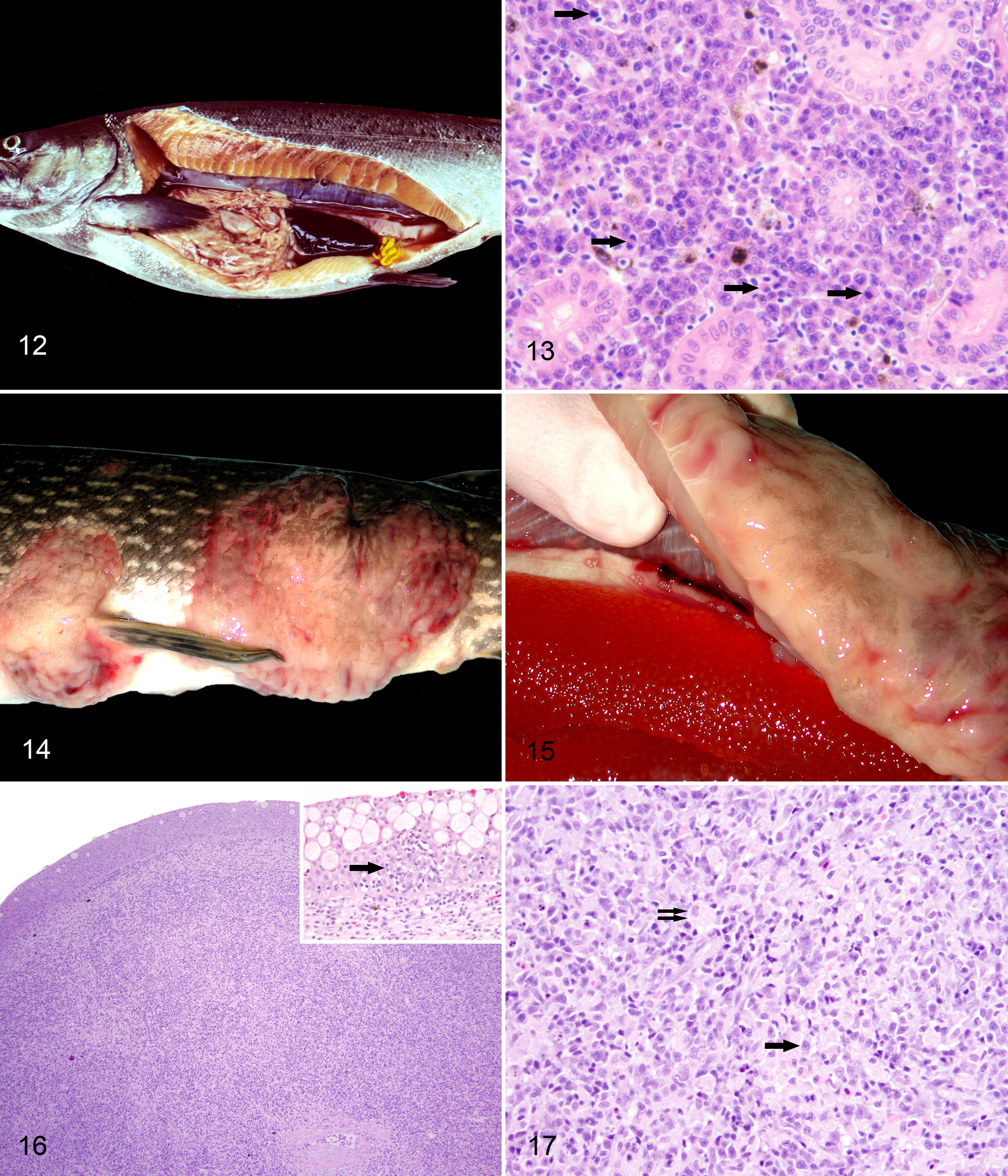
Internally, the most common signs are serosanguinous ascites and renosplenomegaly (Fig. 12). Cytologic impression smears of affected organs are often diagnostic and reveal high numbers of discohesive round to ovoid cells with pink to gray, finely granular cytoplasm and eccentric round to deeply cleaved nuclei bearing prominent nucleoli. Giemsa-stained imprints reportedly accentuate the perinuclear halo. 35 On microscopic evaluation of paraffin-embedded tissues, the renal interstitium (Fig. 13), splenic red pulp, lamina propria of the distal intestine, and vascular-rich areas of the pancreas, liver, pericardium, and retrobulbar tissues (ie, rete mirabile) are variably infiltrated by sheets of immature plasmablasts that replace normal structures. Plasmablasts range from 10 to 20 μm in diameter and are slightly ovoid with deeply basophilic, finely granular cytoplasm and distinct borders. Nuclei are round and eccentric with the typical perinuclear halo. Mitoses are generally abundant (Fig. 13). 33,35
Plasmacytoid leukemia has been associated with a retrovirus, but coinfection with Enterocytozoon salmonis, a microsporidian parasite, and Renibacterium salmoninarum, the causative agent of bacterial kidney disease, is disproportionately common among infected fish. 33 Evaluation of renal and periocular tissues from Chinook salmon with natural and experimentally induced diseases revealed 110 nm diameter retrovirus-like particles by TEM as well as reverse transcriptase activity. The virus has been isolated in cell culture and is tentatively termed salmon leukemia virus. 21 Experimental transmission trials using cell-free, ultra-filtrated inoculum from kidneys and spleens of fish with clinical plasmacytoid leukemia that had received antiprotozoal treatment resulted in successful transmission of the disease. 33 Together, these studies suggest that plasmacytoid leukemia in Chinook salmon is caused by a retrovirus, but the role of protozoal and bacterial agents as possible cofactors in the etiopathogenesis of this disease remains undetermined at this time. 34
Muskellunge and Northern Pike Lymphosarcoma
Esocid lymphosarcoma or lymphoma is a disease of adult northern pike (Esox lucius) in Europe and North America and muskellunge (Esox masquinongy) in North America. 67 The diseases manifest as cutaneous round cell tumors with a low prevalence in the summer but increased prevalence in the fall, winter, and spring. It is hypothesized that horizontal transmission of esocid lymphosarcoma occurs in the spring during the spawning season of esocid fish. Although the histogenesis of these tumors is unknown, immunohistochemical and ultrastructure studies suggest that this neoplasm is T lymphocytic or histiomonocytic in origin. 63,64
Grossly, cutaneous lesions are characterized by expansive, soft, pale-tan, knobby plaques with irregular margins (Fig. 14). Cutaneous masses often coalesce, reaching several centimeters in diameter, and may become extensively ulcerated. On section, there is infiltration of the subjacent skeletal muscle and cut surfaces bulge (Fig. 15).
Microscopically, the dermis and epidermis are infiltrated by a poorly demarcated, unencapsultated, variably ulcerated neoplasm composed of pleomorphic round cells organized into sheets in the dermis and forming clusters of up to 12 cells in the Malpighian and basal strata, effacing the normal epidermal architecture (Fig. 16). Neoplastic round cells have moderate amounts of vacuolated, eosinophilic cytoplasm and indistinct borders. Nuclei are up to 10 μm in diameter and are generally round with fine granular chromatin and a central nucleolus. Occasionally, nuclei are larger (up to 12 μm in diameter) and a reniform with vesicular to coarsely clumped chromatin and prominent nucleolus. There is moderate anisocytosis and anisokaryosis, and mitoses average 10 in 10 high-magnification (×400) fields. Intermingled throughout the neoplastic population are high numbers of small, 8-μm diameter lymphocytes and individual necrotic cells (Fig. 17).
C-type viral particles and reverse transcriptase activity have been detected in cell-free homogenates prepared from tumor tissues, but efforts to culture the retrovirus have been unsuccessful. 42,43 Experimental transmission trials using cell-free filtrates have resulted in successful replication of the disease in both northern pike and northern pike × muskellunge offspring. 6,41
Atlantic Salmon Epidermal Papillomatosis
Epidermal papillomatosis of Atlantic salmon (S. salar) is a benign, proliferative, multicentric, epizootic disease reported in cultured and wild Atlantic salmon inhabiting both the freshwater and marine environments. 15,17 The lesions are seasonal, developing in the summer and resolving spontaneously in the fall and winter. The disease is reported to cause minimal morbidity and mortality, but associated lesions may reduce carcass value. The highest prevalence estimates recorded, up to 55%, were from freshwater hatchery-reared parr (freshwater salmon in their second summer). 17
Grossly, 2 subtypes of epidermal masses, plaque-like or exophytic and verrucous, grow to 4 cm in diameter and are distributed on the opercula, fins, and tail. Masses increase in size until later summer and then appear to slough off, occasionally leaving remnant ulcers prone to secondary infections by opportunistic pathogens.
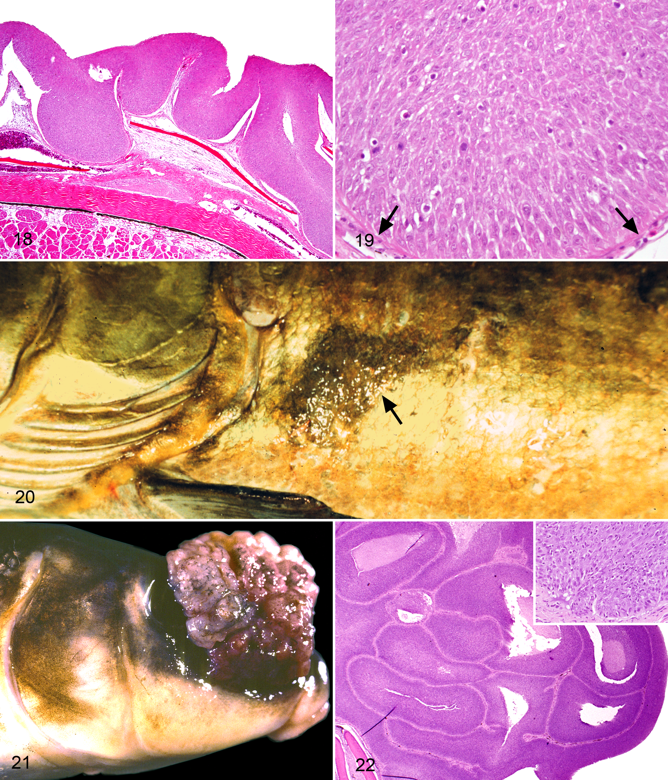
Microscopically, both subtypes are characterized by well-demarcated proliferations of Malpighian cells (nonkeratinizing epithelial cells that comprise the outer layers of the piscine epidermis) that expand the epidermis up to 15 times normal thickness. 17,22 Plaque-like subtypes have abrupt margins and a flat surface. Exophytic, verrucous masses have deep rete pegs with intervening fibrovascular stalks (Fig. 18). Anaplasia, pleomorphism, and bizarre mitotic figures have been reported in both subtypes (Fig. 19), and dermal invasion is not a feature. In a small proportion of masses, proteinaceous, eosinophilic inclusions and rarely intranuclear inclusions have been reported in degenerate cells. 17 The regressive stage is characterized by neovascularization and leukocyte infiltration, followed by necrosis, ulceration, and then rapid epithelialization. Deaths due to concomitant infection by the aquatic oomycete Saprolegnia sp and defective osmoregulation have been reported as a rare complication of the ulcerative stage. 17
Ultrastructure studies of spontaneous lesions revealed cytoplasmic, 125 to 150 nm diameter, C-type viral particles in a small proportion (5/52) of blocks examined. Subsequent virus isolation by cell culture was unsuccessful. 16 Transmission trials have not been attempted. The epizootic and seasonal factors that characterize this syndrome suggest the etiology may be related to infectious, hormonal, or environmental factors. A common environmental exposure, however, seems unlikely based on the widespread nature of epizootics occurring in both freshwater hatcheries and in the wild. Studies in cultured fish have demonstrated increasing prevalence in affected raceways, supporting the role of a transmissible infectious agent. 51 Although the etiology of Atlantic salmon epidermal papillomatosis is unknown, infectious and hormonal factors remain possible cofactors in this disease. 16
Muskellunge and Northern Pike Smooth-Type Epidermal Hyperplasia
Smooth-type epidermal hyperplasia affects muskellunge (E. masquinongy) and northern pike (E. lucius) in Europe and North America. Prevalence estimates up to 7.4% have been reported from tributaries of the Hudson Bay in Canada. 68 This disease is characterized by multifocal skin lesions that resemble glistening, mucoid plaques and must be differentiated from grossly similar lesions caused by esocid herpesvirus-1, termed blue spot disease. The latter is characterized by opaque, pale blue epidermal plaques with a notably “gritty,” granular surface. 66,68
Grossly, areas of smooth-type epidermal hyperplasia are slightly elevated, translucent plaques, up to 3 cm in diameter, and are distributed on the skin of the body and fins. Generally, these plaques have a bluish hue, but some can be dark and red (Fig. 20). Microscopically, the epidermis is expanded by a sharply demarcated proliferation of uniform, basilar epithelial cells organized into sheets. Proliferating basal cells have minimal morphologic features of atypia, but mitotic figures are reported. 68 Proliferative cells replace all of the epidermal strata as well as specialized components, such as goblet cells, alarm cells, and mechanosensory organs, but respect the basement membrane. Occasionally, there is goblet cell hyperplasia at the interface with the adjacent normal epidermis.
Ultrastructure evaluation of these lesions revealed abundant intercellular clusters of C-type viral particles, up to 150 nm in diameter with a 120 nm diameter capsid and an 80 nm diameter electrodense core. Occasionally, viral particles were observed budding from the cytoplasmic membrane of epithelial cells. Attempts to isolate the virus via cell culture were unsuccessful. 68 Experimental transmission has not been accomplished.
Hooknose Cutaneous Fibroma/Fibrosarcoma
A cutaneous fibroma/fibrosarcoma of the hooknose (Agonus cataphractus) has been reported for fish from the German Wadden Sea, an intertidal zone off the coasts of the Netherlands and Germany, by Anders et al. 2 Neither a seasonal pattern nor tumor regression has been reported. 2
The lesions were originally reported as yellow or red-black, well-circumscribed, raised nodules on the skin that were 3 to 9 mm in diameter. Microscopically, dermal masses are composed of nonencapsulated, infiltrative proliferations of spindle cells organized into streams and interlacing bundles. Morphological features of cellular atypia and local invasion have been reported. 2,45
Ultrastructure studies identified C-type viral particles, 86 to 132 nm in diameter, within cytoplasmic vacuoles of tumor-infiltrating lymphocytes. They were composed of double cores and lateral bodies. Morphologically, these particles were interpreted as lentiviruses. 2 Tumor transmission trials, virus isolation, and viral cloning have not been performed.
White Sucker Epidermal Papilloma
Epidermal papillomas of white sucker (Catostomus commersoni) have been reported in mature fish at various locations across the northern United States and Canada. Prevalence estimates vary by region and are as high as 59%, but mortality is rare. 60,62 Originally, lesions were thought to be associated with polluted waters, where prevalence estimates were significantly higher compared with less polluted waters. 60 This association was challenged, however, by laboratory-based studies in which fish were maintained in clean water. Within the latter fish, lesions regressed on some fish, remained unaltered on some, and developed anew on other fish. 61 Although lesions are reported to increase in both size and density throughout life, spontaneous regression has been reported. 61,62
Grossly, masses have a predilection for the lips and head. They appear as white, firm nodules with a smooth to rugose texture and commonly coalesce or directly appose one another (Fig. 21). Less common subtypes include plaque-like mucoid epidermal masses and small areas of discrete epidermal hyperplasia located elsewhere on the body. 60
Microscopically, papillomas are sharply demarcated with broad dermal papillae and are composed of proliferative basilar and Malpighian cells. Areas of affected epidermis are variably infiltrated by leukocytes and contain a notable paucity of goblet cells and mechanoreceptors (Fig. 22).
In early studies performed by Sonstegard, 62 100 nm diameter C-type viral particles were detected budding from cytoplasmic membranes of proliferative epithelial cells, and reverse transcriptase activity was demonstrated in particulate fractions from the lesions. Inoculation of cell cultures failed to induce transformation, and a virus was not isolated. More recent studies have failed to identify the presence of any viral particles in these lesions. 47,59 Transmission trials have also had conflicting results. Early trials using tumor homogenates and cell-free inocula did not reproduce the disease. 62 A more recent trial using cell-free inocula prepared from only proliferating lesions did result in successful transmission of lesions at the site of injection in 50% of inoculated fish. However, the moderate rate of spontaneous papillomas that developed in control and cohabitating fish during this trial hindered the interpretation of this finding. 47
Angelfish Lip Fibroma
A fibroma of the lip of the angelfish (Pterophyllum scalare) has been found on several occasions in populations of this popular freshwater aquarium-maintained fish species. 23 All known cases have been associated with angelfish in captivity, which may suggest stress, density, and environmental factors (including trauma associated with being held in captivity) as cofactors. There are no known reports of wild angelfish with this lesion.
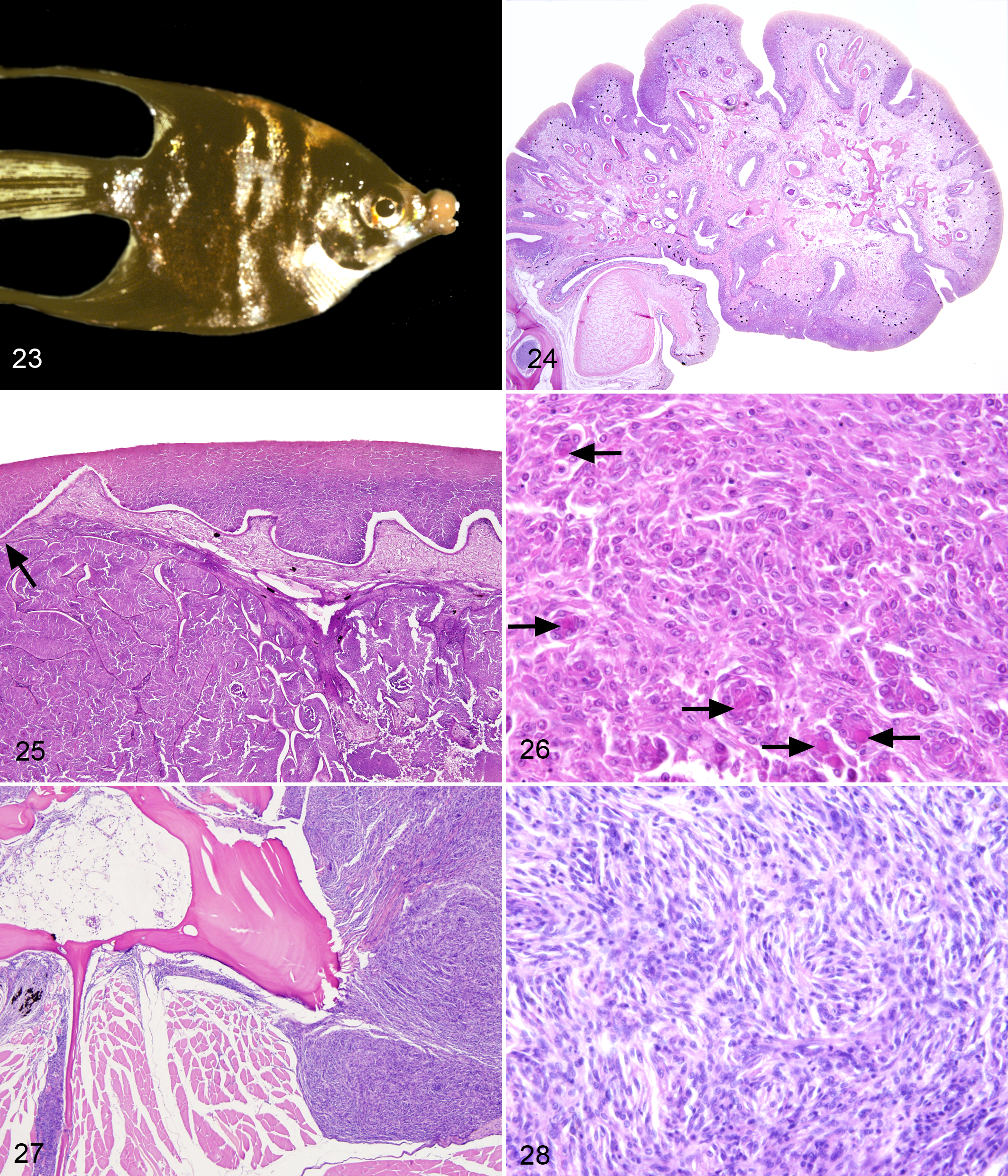
Grossly, tumors arise from the labial mucosa and often develop on both the upper and lower arcades (Fig. 23). Microscopically, pedunculated, dermal masses elevate the labial skin and are composed predominantly of spindle cells organized into interlacing bundles within a loose, edematous fibrovascular stoma. Often the tumor stroma contains entrapped, surface-oriented tooth buds and central areas of osseous metaplasia (Fig. 24).
Ultrastructure studies revealed A-type and C-type viral particles within neoplastic fibroblasts. Transmission trials using cell-free inocula have been unsuccessful. 23 Virus isolation and viral cloning have not been attempted.
European Smelt Spawning Papillomatosis
Spawning papillomatosis of European smelt (Osmerus eperlanus) is considered a benign proliferative epidermal lesion reported in up to 5.5% of sexually mature smelt in the spring during spawning in German estuary waters near the Baltic Sea. This seasonal pattern resulted in the name that included the reference to the time of year when spawning occurs. 3
Grossly, smooth, white, ovoid nodules, up to 0.5 cm in diameter, affect the head and fins. Less commonly, larger, plaque-like masses may be found on the trunk. Fin nodules can be easily dislodged near the end of the spawning season. Microscopically, unencapsulated, multinodular proliferations of epithelial cells expand initially within the stratum compactum of the dermis and abut the overlying epidermis (Fig. 25). Epithelial cells have moderate anisocytosis and anisokaryosis, as well as abundant mitotic figures, and form syncytia containing up to 8 nuclei. Nuclei are enlarged and occasionally have prominent marginated chromatin. Syncytial cells contain irregular, eosinophilic inclusions, up to 15 μm in diameter (Fig. 26). Interestingly, these dermal nodules progressively migrate into the overlying epidermis, where they become pseudoencepsulated by the cutaneous epithelium and eventually undergo degeneration along the basilar margin. At this stage, the nodules are shed, leaving behind remnant ulcers.
Ultrastructure studies performed on nodular lesions of the fin reported 2 sizes of C-type viral particles, 88 to 101 nm and 55 to 76 nm in diameter, as well as numerous herpesvirus-like particles, 95 to 130 nm in diameter. 1,3 More recent ultrastructure evaluation of archived fin lesions from smelt revealed numerous herpesvirus-like intranuclear viral capsids and large, intracytoplasmic comma-shaped virions but did not report observation of C-type viral particles. 31 Subsequent attempts to isolate a herpesvirus in cell culture were unsuccessful, perhaps due to prolonged tissue storage and limited availability of fish cell lines ideal for this technique. Apparently, the concentration of herpervirus-like particles was high enough that its DNA could be detected above background cell DNA. The genomic DNA was analyzed by DNA restriction endonucleases, and the virus was termed O. eperlanus herpesvirus-1 (OEHV1) or Comet herpesvirus of smelt. 31 Experimental transmission has not been accomplished. Based on histopathologic and ultrastructural findings, European smelt spawning papillomatosis may be associated with a herpesvirus-like agent rather than a retrovirus, but critical genomic analysis of OEHV1 and River’s postulates need to be fulfilled.
Bicolor Damselfish Neurofibromatosis
A neurofibromatosis-like disease has been documented in bicolor damselfish (Stegastes partitus), a marine reef fish in southern Florida, and has gained particular interest as a potential model for neurofibromatosis type 1 (NF1) in humans. 58 Prevalence estimates reach 23% of adult fish on some reefs, and disease prevalence is associated with fish density and size class of the population cluster. 54 This disease is progressive and invariably fatal. 58
Grossly, tumors are multicentric and track along cutaneous nerves on the body and fins, as well as spinal nerves, such as the trigeminal and facial nerves, and are sometimes associated with nerves of internal organs in advanced stages of the disease. Two gross subtypes have been identified: (1) hyperpigmented plaque-like epidermal masses that minimally invade the subjacent dermis and bones but often are associated with erosion and distortion of adjacent scales and fin rays and (2) larger, nonpigmented subcutaneous nodules that commonly invade skeletal muscle, internal organs, and bone and eventually erupt through the skin (Fig. 27). Focal hyperpigmented plaques are the first detectable lesion, and these may eventually coalesce into rugose, flat masses that cover a large surface area of the body. Nonpigmented subcutaneous to dermal nodules dominate the later stages of the disease and have been characterized as neurofibromas and malignant peripheral nerve sheath tumors.
Microscopically, nerve sheath tumors are unencapsulated, invasive proliferations of spindle cells and plump pleomorphic cells variably organized into plexiform and storiform patterns (Fig. 28), whorls, and interlacing fascicles. 58 Neoplastic cells are consistently associated with peripheral nerves, which exhibit hyperplastic and degenerate changes such as enlarged myelin sheaths with disruption of axonal fibers. 57
Transmission trials using uncharacterized tumor homogenate and cell-free filtrates as well as tumor cell lines have been successful at reproducing tumors identical to spontaneous disease. 53,57 C-type viral particles, 90 to 100 μm in diameter, and reverse transcriptase activity have been detected in tumor cell lines derived from both spontaneous and experimentally induced lesions. 55 Subsequently, however, multiple retroviral DNA fragments were detected in cell lines derived from lesions and normal tissues, and transmission studies demonstrated that none of these were either necessary or sufficient for tumor transmission. Due to the inconsistent association of a retrovirus with tumors, the authors concluded that a retroviral etiology for bicolor damselfish neurofibromatosis is unlikely. 56 More recent research suggests that this disease may be associated with an extrachromosomal DNA virus-like agent, termed the damselfish virus-like agent, rather than a retrovirus. 50,56
Summary
The vast majority of tumors in fish that are associated with retroviruses are readily visible when the fish is examined. As such, these may constitute a large number of tumors observed by the general public and subsequently brought to the attention of governmental environmental resource agencies. The veterinary pathology community might also reasonably expect that such specimens would eventually be presented to them for evaluation. Our goal in the development of this document was to provide a contemporary summary of the literature that would be of value when these specimens are evaluated in a diagnostic laboratory setting.
Footnotes
Acknowledgements
We thank Dr J. Wolf of Experimental Pathology Laboratories, Sterling, Virginia, for providing access to the archived materials of the Registry of Tumors in Lower Animals. We also thank Dr M. Kent, Oregon State University, for providing materials for our use for description of Chinook salmon plasmacytoid leukemia and Dr M. Schmale, University of Miami, for providing materials for our use for description of bicolor damselfish neurofibromatosis.
Declaration of Conflicting Interests
The author(s) declared no potential conflicts of interest with respect to the research, authorship, and/or publication of this article.
Funding
The author(s) received no financial support for the research, authorship, and/or publication of this article.