
Abstract
Naked mole rats (NMRs; Heterocephalus glaber) are highly adapted, subterranean, eusocial rodents from semiarid regions of the eastern horn of Africa and the longest-living rodent known with a maximum life span of up to 30 years. They are a unique model for aging research due to their physiology, extreme longevity, and, when compared to mice and rats, resistance to cancer. Published surveys of disease in NMRs are sparse. Captive colonies in zoological collections provide an opportunity to monitor spontaneous disease over time in a seminatural environment. This retrospective study describes common lesions of a zoo population over a 15-year period during which 138 adult NMRs were submitted for gross and histologic evaluation. Of these, 61 (44.2%) were male, 77 (55.8%) female, 45 (32.6%) died, and 93 (67.4%) were euthanized. The most frequent cause of death or reason for euthanasia was conspecific trauma (bite wounds) and secondary complications. Some common histologic lesions and their prevalence were renal tubular mineralization (82.6%), hepatic hemosiderosis (64.5%), bite wounds (63.8%), chronic progressive nephropathy (52.9%), and calcinosis cutis (10.1%). In sum, 104 (75.4%) NMRs had more than one of the most prevalent histologic lesions. No malignant neoplasms were noted; however, there was a case of renal tubular adenomatous hyperplasia with nuclear atypia and compression that in rats is considered a preneoplastic lesion. This retrospective study confirms the NMR’s relative resistance to cancer in spite of development of other degenerative diseases and highlights the utility of zoological databases for baseline pathological data on nontraditional animal models.
Heterocephalus glaber, also known as the sand puppy or, more commonly, the naked mole rat (NMR), is a mouse-sized (up to 35 g at adult weight) hairless and eusocial, long-lived rodent that lives in subterranean burrows in arid and semiarid regions of Kenya, Somalia, and Ethiopia. 7,8,13,21,92 The NMR is highly adapted to harsh conditions, thriving in dark, poorly ventilated, underground burrow systems up to 2 m below the surface with low oxygen and high carbon dioxide levels. 57,62,92 NMRs have a large, broad head and procumbent incisors for digging and short yet agile legs allowing quick movement both forward and backward. Other adaptations to living underground include a low metabolic rate, thermoregulation (poikilothermic), minimally developed lungs, and high circulating hemoglobin and myoglobin concentrations. NMRs are also resistant to cerebral hypoxia and have decreased cutaneous pain sensation, attributed to lack of Substance P, which is thought to be a protective mechanism against acid buildup in tissues due to high environmental carbon dioxide. 14,64,69,76 NMRs have virtually no exposure to sunlight and have small eyes with reduced visual capacity that likely serve a circadian function and as sensors for incursions of light into the burrow system. 14,21 In addition, like other mole rats, they do not have an environmental source of cholecalciferol (vitamin D3), nor do they obtain it in the diet. 23,83,84 Despite undetectable serum levels of the principal circulating metabolite and perpetual vitamin D3 deficiency, NMRs do not develop the typical associated pathologies and are able to maintain mineral homeostasis. 20,23,99 Likewise, vitamin D supplementation in captive colonies has resulted in intoxication and soft tissue mineralization. 20,21 NMRs are herbivorous, coprophagic, hindgut (cecal) fermenters and obtain all of their water requirements from their food. 7,24 In the wild, they feed upon geophytes, which are plants that possess perennating (storage) organs below the soil surface (eg, corm, bulb, tuber, rhizome). 7,92 Captive diets aim to provide high levels of fiber and include a variety of fresh vegetables, the staple food being sweet potato, and small amounts of fruit and baby cereal. 7,21,73,92 Cecotrophes are soft, partially digested fecal pellets that are reingested to provide water, nourishment, and protein in young NMRs and to replenish cecal protozoa and bacteria in older individuals. Maintenance of the cecal microbial population is necessary for the production of essential volatile fatty acids, amino acids, and vitamins that support basal metabolic needs. 21,24
NMRs are 1 of 2 eusocial mammal species (the other is the Damaraland mole rat, Cryptomys damarensis), both of which have been studied extensively. Similar to social structures seen in various insects such as ants, bees, and wasps, NMRs live in large colonies typically ranging from 20 to 300 (average of 75) individuals with a division of labor into reproducers (breeders) and subordinate workers. 7,13,57 Colonies have a single breeding female known as the queen, who is the largest individual of the colony, and 1 to 3 breeding males. 7,9,13,29 Workers are typically smaller and functionally sterile and serve to forage for food, extend the burrow system, defend the colony, and help the queen care for the offspring. 57,92 In free-ranging colonies, NMR workers reportedly have shorter life spans than breeders as they are subject to different predation risks. 16,36 Male and female workers exhibit no sexual dimorphism and are reproductively suppressed by social cues. 30,43,61,91 Worker males have low levels of luteinizing hormone and testosterone with small, intra-abdominal testes, abnormal sperm production, poorly developed epididymi, and impaired fertility. 30,38,41 Additionally, the testes of nonbreeding males have a large volume of interstitial (Leydig) cells with sparsely scattered seminiferous tubules that exhibit disorganized, disorderly spermatogenesis. 38,44,45,75 Subordinate females are anovulatory due to inhibition of hypothalamic gonadotrophin-releasing hormone secretion, which leads to very low levels of sex steroids. 41,42 Females over 6 months old are capable of becoming reproductively active and will fight to establish dominance if the queen dies. 29,31,57 During these upheavals, subordinate female and male NMRs sustain serious injuries and may die in attempts to become breeders. 29,31,57 Furthermore, NMRs are highly xenophobic and will kill individuals from foreign colonies or conspecifics at the command of their queen. 21,31
NMRs are valuable models for aging research due to their reported excellent anti-aging defenses resulting in an exceptionally long life span (9 times longer than laboratory mice), sustained good health and reproductive potential, and minimal age-related changes in body composition, physiology, and molecular function. 15,16,36,51 Buffenstein and colleagues have championed the NMR model and have described the unique biology and physiology that may have implications for healthy human aging. 15,17,18,21
These unique rodents have been used to test theories of aging, including the oxidative stress theory, among others. 2,3,32,51,74,97 Despite exhibiting higher levels of lipid peroxidation, protein carbonylation, and DNA oxidative damage than mice, NMRs age at a much slower rate and appear to have increased resilience to oxidative stress and mitochondrial injury. 4,5,10,36,86 These may be important determinants of healthy aging, in particular in regard to protein and membrane phospholipid stability. 56,70,80 NMRs are also thought to exhibit negligible cellular senescence: a state in which most normal somatic cells are no longer capable of replication. 6,16,46,74 Although older NMRs show changes in morphology, many physiologic functions and fertility (in breeders) are maintained until death. 16 Examples of these virtually unchanged parameters include basal metabolic rate; vascular health and relaxation; bone and cartilage integrity and mineral density; ROS production, antioxidant activity, and oxidative damage; glucose tolerance; and glycated hemoglobin. 3,19,22,32,60,74,82,100 Current studies are examining the stability of the genome and proteins with age. 58,101 For example, sustained high levels of neuregulin-1 were documented in aged NMRs, suggesting that one mechanism of longevity is stability of neuregulin signaling and sustained brain function. 35 Other studies have demonstrated that NMR cells are highly resistant to cell injury and stress, the latter of which is regulated through the nuclear factor erythroid 2-related factor 2 signaling pathway, resulting in a relatively higher expression of cytoprotective genes in NMRs than wild-type mice. 65,88 It is possible that despite some decline in organ function, NMRs resist progression to overt disease. For example, a recent study noted cardiac diastolic dysfunction in female NMRs; however, no apparent impact on health span has yet been noted. 16,52
Unlike aged mice and rats, there is a paucity of data on common histologic lesions and natural disease processes in NMRs. The lack of well-documented comprehensive necropsy and histopathologic findings is likely multifactorial including husbandry and funding. NMRs are housed at 28°C to 30°C with 50% to 60% humidity. 21,92 Autolysis is accelerated at higher temperatures, and if dead animals are not necropsied or tissues fixed in a timely manner, definitive diagnoses cannot be made. Comprehensive histology is relatively expensive, and research grants to study specific aspects of NMR physiology or molecular biology often lack sufficient funds for full comprehensive histopathology by a pathologist. While zoological institutions do routinely perform complete necropsies and histopathology, if autolysis is not advanced, the results are rarely published in widely read journals. To date, Buffenstein and colleagues have not documented a single incidence of cancer as noted either clinically in colony animals (n = 800) 16 or at necropsy (n = 2000) 52 ; however, the condition of the carcasses and the number of cases subjected to histologic examination were not disclosed. 15,16,52 There is one recent report on the histologic findings from 2 extremely old individuals, documenting the presence of nonneoplastic lesions associated with aging in humans and rodents but lack of neoplasia or preneoplastic lesions. 36 Additional personal observation statements regarding disease spectrums in research and some zoological NMR colonies have been made and also suggest a remarkable resistance to cancer. 16,21 This collective experience has driven investigations and intriguing insight into the molecular basis of the NMR’s cancer resistance. 16,36,58,66,90
The objective of this retrospective study was to address the relative dearth of published disease profiles of NMRs by characterizing spontaneous histologic lesions, in particular those classically associated with aging in rodents, in a large zoological population. While these data are specific to one captive population over a 15-year period, it is a comprehensive review of spontaneous histologic lesions in this species and a valuable addition to the current literature. 21,36
Materials and Methods
Gross necropsy and histopathology reports from 138 adult NMRs that died or were euthanized by intracardiac injection of pentobarbital between May 1996 and July 2011 were reviewed. NMRs were euthanized in cases of poor prognosis of return to normal function within the colony either from severe medical conditions or impending prolonged removal from the colony for treatment (ie, greater than 4 days), as in both instances, peaceful reintroduction to the colony would be impossible due to NMR’s eusocial nature. Select archival slides were examined to confirm the original diagnoses. Submitted NMRs were from 8 variably sized colonies maintained at a zoological institution. These colonies were originally derived from a single large colony composed of 19 males and 10 females established in 1987 from a wild-caught male (Tsavo West Kenya) and female (Levata Water Hole North of Arche’s post in Northern Kenya). During the study period, the zoo housed 6 separate colonies of varying size (as low as 3 and up to 40 individuals). All NMR colonies were fed the same diet, which consisted of daily vegetables such as leafy greens, sweet and white potato, turnip, and carrot; smaller amounts of fruit (apple and pear); and twice-weekly supplements of rodent chow and baby cereal. Water requirements were met through the diet. Colonies were maintained in artificial burrow systems in a heated room with an ambient temperature range of 84°F to 86°F and 70% humidity. The colonies on exhibit were highlighted with small incandescent light bulbs in different areas of the artificial burrow system for public observation. The off-exhibit colonies had no access to light unless the caretakers were cleaning, feeding, and servicing the habitat under ceiling-mounted fluorescent light illumination. Signalment and history of each individual were provided by the clinical veterinarian or animal caretaker staff upon submission of the necropsy specimen. Since breeding records and individual animal identifications were not maintained in detail, actual ages in months or years and specific genetic lineage could not be assessed.
Necropsies were performed on all submitted NMRs, excluding specimens that were cannibalized or exhibited advanced autolysis, characterized by discolored, friable, and indiscernible viscera, precluding meaningful interpretation. Samples of all major organ systems were immersion fixed in 10% neutral buffered formalin. Tissues sampled typically included but were not limited to brain, eye, tongue, salivary gland, trachea, thyroid, thymus, esophagus, lung, heart, stomach, small and large intestine, pancreas, mesenteric/tracheobronchial/peripheral lymph nodes, liver, spleen, kidney, adrenal, gonad and reproductive tract, urinary bladder, skin, skeletal muscle, sciatic (or other peripheral) nerve, bone, and bone marrow. Fixed tissues were routinely processed into paraffin, sectioned at 4 to 5 μm, and stained with hematoxylin and eosin. Prussian blue stain was employed in additional sections for evaluation of hemosiderin. Various other stains were used to characterize potential pathogens in selected cases, including Modified Brown and Hopps, Ziehl-Neelsen, Gomoro’s Methamine Silver, and Steiner’s (silver) stain among others.
Results
Over a 15-year period (May 1996 to July 2011) approximately 1147 NMRs died or were euthanized. According to the colony managers, the majority of these NMRs were neonates under 10 days old, considered cases of colony neglect, and typically not necropsied. Approximately 363 individuals were necropsied over this period, and based on body size and approximate birth dates, 225 were neonates or juveniles and excluded from this analysis. In sum, 138 adult NMRs that died (n = 45; 32.6%) or were euthanized (n = 93; 67.4%) were submitted for necropsy and comprehensive histopathology. There were 61 (44.2%) males and 77 (55.8%) females. Lesions and their prevalence are listed in Table 1 and described below categorized by body system. In sum, 104 (75.4%) NMRs had more than one of these lesions. Specific ages were not known for almost all of the NMRs reported herein. For a brief period in 1997, transponders were used for animal identification. This resulted in significant morbidity and mortality (discussed further in Husbandry-Related Lesions), and the practice was discontinued. Other methods of identification have been attempted including tattooing and nail clipping but were overall considered unsuccessful due to inability to visualize individual markings within the habitats. Only animals listed as adult upon submission to the diagnostic laboratory were considered in this analysis.
Histologic findings in captive Naked Mole-Rats (Heterocephalus glaber)
^Total animal (n = 138)
*Multiple tissues affected from different systems
Integumentary and Musculoskeletal Lesions
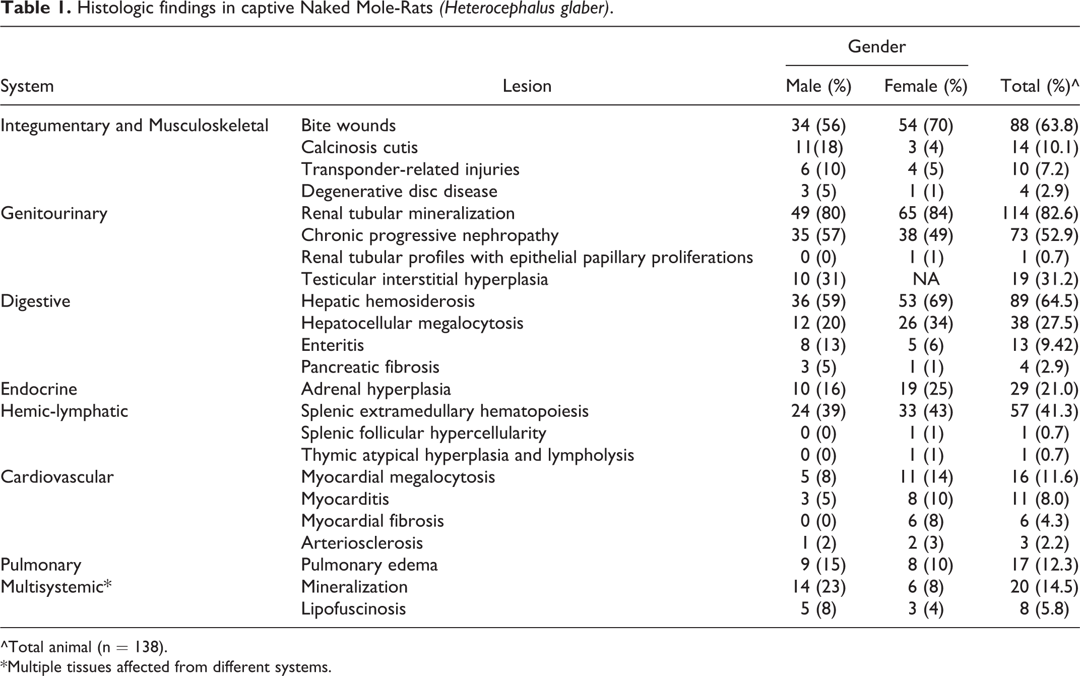
The major cause of death was euthanasia due to severe conspecific trauma; traumatic lesions including bite wounds were found in 88 (63.8%) submitted NMRs. Bite wounds ranged in severity but were generally characterized by short to long, linear lacerations with cutaneous ulceration and crusting over the muzzle, head, trunk, and extremities with associated hemorrhage (Fig. 1). Many traumatized individuals survived initial attacks; however, the extent of their injuries led to gradual debilitation (dehydration, emaciation) and ultimately resulted in euthanasia. Deep bite wounds often led to secondary bacterial infections with cellulitis and myositis and, in few cases, extended through the body wall leading to peritonitis and hemo-, pyo-, or pneumothorax. Conspecific trauma also resulted in fractures of the legs, ribs, or, rarely, vertebrae with subsequent osteomyelitis and, infrequently, digit or distal limb amputations. Bacterial sepsis was suspected in several cases but was confirmed in only a few cases by supportive histologic lesions and positive bacterial culture.
Calcinosis cutis/circumscripta was noted in several cases throughout the 15-year span and was considered clinically significant in many of these individuals resulting in decreased mobility and lethargy. Lesions ranged in severity and consisted of small to large (up to 2-cm diameter), smooth, white to tan nodular subcutaneous swellings along the trunk and flank (cutis) (Fig. 2), and/or surrounding the extremities and digits (circumscripta) (Fig. 3), though similar masses were noted in other tissues (eg, tongue). 20,23 On cut surface, nodules contained white, opaque, pasty to gritty material. Histologically, this material was faintly to deeply basophilic and granular to angular, consistent with mineral (Fig. 4). Mineral aggregates raised the overlying epidermis and compressed and displaced dermal collagen bundles; some mineral was surrounded by small to moderate numbers of foamy macrophages and multinucleated giant cells with fewer lymphocytes, plasma cells, and rare neutrophils.
Secondary infection from microchip transponder implantation resulted in spontaneous death (n = 4) or euthanasia (n = 6) of 10 submitted NMRs over the course of 3 months in 1997. Grossly, affected NMRs had locally extensive subcutaneous edema, hemorrhage, and purulent exudate surrounding the transponder and tracking from the implantation site, over the right lateral abdomen or flank. When examined microscopically (n = 6), there was necrotizing cellulitis and abscess formation with abundant, typically mixed populations (cocci and slender bacilli) of intralesional bacteria. Subjacent skeletal muscle had acute necrosis and hemorrhage with variable degrees of myositis.
Degenerative disk disease, both thoracic and lumbar, was seen in 4 (2.9%) NMRs, with varying degrees of spondylosis and scoliosis, intervertebral arthritis and fibrosis, disk degeneration, collapse, and extrusion, and apparent spinal cord compression.
Genitourinary Lesions
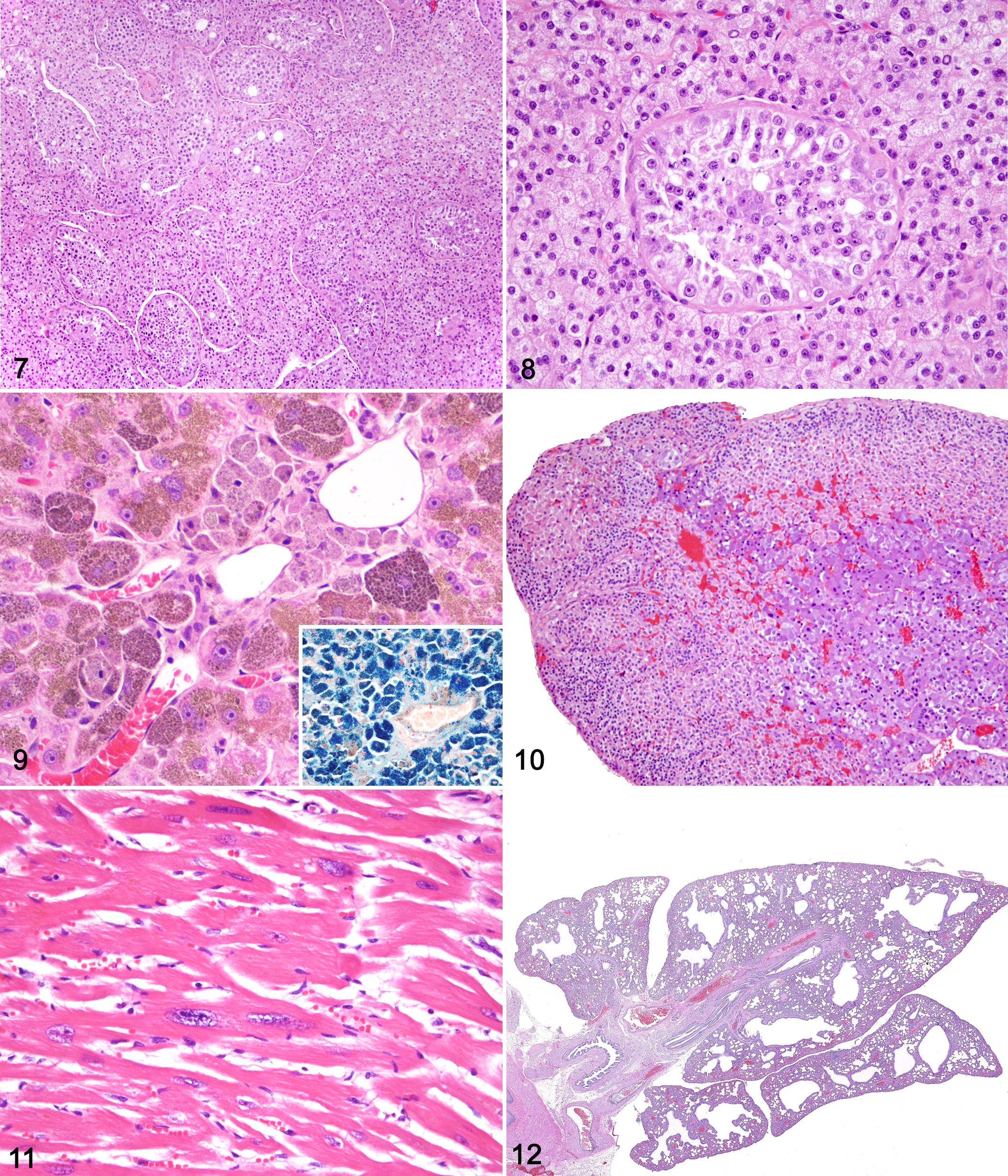
Renal lesions were very common in this NMR population. Over 80% of the examined NMRs had at least rare renal tubular mineralization, characterized by luminal accumulations of deeply basophilic, granular, or angular material that often obscured tubular epithelium with maintenance of the basement membrane. Tubular mineralization was noted in NMRs lacking additional significant renal disease. As noted in a previous report, 36 mild to severe chronic renal disease was frequently observed (52.9%) with a constellation of histologic changes that were considered related. These changes were thought to be progressive stages of a single disease process similar to that seen in aged laboratory rats, known as chronic progressive nephropathy (CPN) and to a less distinguished nephropathy in mice. 78,79 Grossly, kidneys of affected NMRs were diffusely pale tan and either slightly enlarged with multiple pinpoint to 2-mm-diameter cystic structures (microcysts) or shrunken with an irregular knobby to granular cortical surface. In general, kidneys had a range of histologic changes, including membranous glomerular change with synechiae and sclerosis; tubular ectasia and proteinosis with cyst formation; tubular degeneration, necrosis, and regeneration; thickened basement membranes of glomerular corpuscles and tubules; interstitial mixed accumulations of lymphocytes, plasma cells and lesser macrophages; and interstitial fibrosis (Figs. 5, 6). Several cases had lesions compatible with “end stage” kidneys, in which significant portions of the functional parenchyma was lost and replaced by inflammation and fibrous tissue. Many severely affected individuals had evidence of chronic infarction with well-demarcated, wedge-shaped regions of interstitial fibrosis with loss of nephrons. One kidney with mild chronic nephropathy contained a small nodule composed of multiple papillary proliferations of haphazardly arranged, basophilic epithelial cells that extended slightly beyond normal tubule borders, with no prominent basement membrane, mild atypia, and slight compression of adjacent parenchyma (Figs. 13, 14).
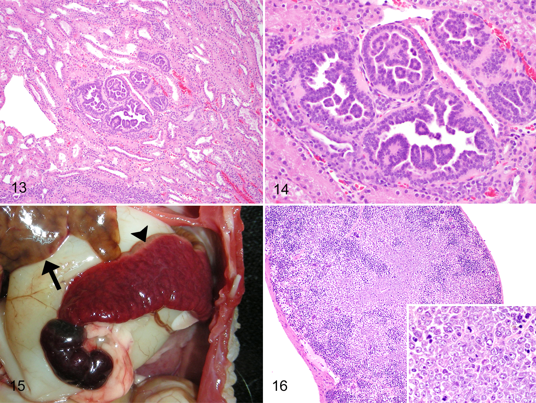
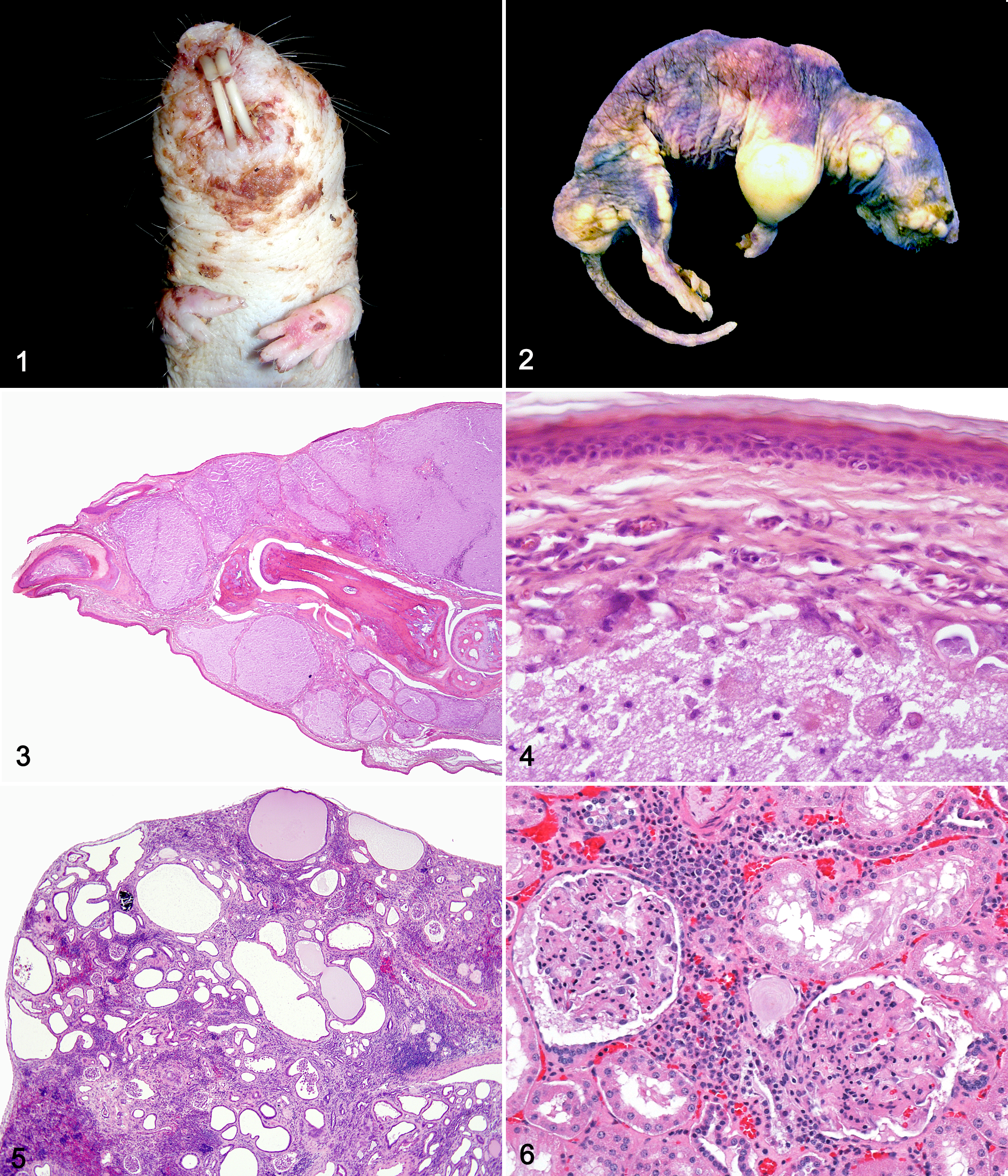
Physiologic testicular interstitial hyperplasia was found in over 30% of male NMRs, all of which were considered subordinates (nonbreeders). Lesions seen in these males were histologically similar to those previously described with abundant polygonal to polyhedral epithelial cells with microvesicular cytoplasm that widely separate seminiferous tubules (Figs. 7, 8). 38,75 Many seminiferous tubules exhibited disorganized spermatogenesis and contained increased numbers of degenerate spermatocytes with rare mature spermatids (Fig. 8).
Digestive Lesions
Hepatic hemosiderosis was seen in 89 (64.5%) of examined NMRs. Hemosiderosis was not typically diagnosed at gross necropsy and best appreciated on microscopic evaluation, though in some cases livers were described as diffusely bronze. Histologically, affected individuals had zonal to diffuse hepatocellular accumulations of brown granular to globular, slightly refractile pigments, confirmed as hemosiderin by Prussian blue stain (Fig. 9 and inset). Additionally, Kupffer cells throughout the sinusoids contained large amounts of similar pigments. Notably, periportal hepatocytes contained more pigments than did midzonal and centrilobular hepatocytes. Hepatocellular megalocytosis was characterized by enlarged hepatocytes with abundant granular cytoplasm and one to multiple, large, occasionally bizarre nuclei compatible with polyploidy. These hepatocytes were typically found in periportal regions and often contained similar hemosiderin pigments (Fig. 9). To a lesser extent, scattered hepatocytes contained tan to gray granular, Prussian blue–negative pigments interpreted as lipofuscin.
Enteritis, in combination with either typhlitis and/or gastritis, was diagnosed in 13 (9.42%) NMRs clustered over several months in 2007 and resulted in subsequent dehydration, endotoxemia, and death in many severely affected individuals. Grossly, affected individuals had gas and/or fluid distention of the intestine and cecum and, to a lesser extent, the stomach. Gastrointestinal (GI) contents, when present, were brown, opaque, watery, and malodorous. Cytologic preparations of GI contents typically contained large numbers of mixed bacilli with lesser numbers of cocci with a predominance of Gram-negative bacteria and numerous commensal protozoa, both ciliates and flagellates. In general, bacterial and protozoal numbers were considered elevated, suggestive of dysbiosis. Some cytologic samples also contained individual or rafts of sloughed mucosal epithelial cells, degenerate neutrophils, erythrocytes, and cellular debris. In some individuals, intestinal and cecal walls had multiple transmural reddened (hemorrhagic) regions, and fewer also had gastric mucosal erosions. Histologically, small intestinal, cecal, and colonic mucosa had epithelial attenuation, hyperplasia, and hypertrophy with some evident dysplasia; scattered crypt necrosis and loss (ulceration); and villous blunting (small intestine). There were also multifocal crypt abscesses with large numbers of bacteria; mucosal and submucosal edema and hemorrhage; lamina proprial accumulations of mixed inflammatory cells, including neutrophils, macrophages, lymphocytes, and plasma cells; and luminal hemorrhage with abundant bacterial and protozoal colonies as noted cytologically. Throughout affected regions, some macrophages contained hemosiderin pigments and/or phagocytosed erythrocytes, indicative of ongoing hemorrhage. In one individual, rare crypt epithelial cells contained smudged basophilic intranuclear inclusions with marginalization of chromatin; however, multiple primer sets targeting rodent parvovirus failed to amplify viral DNA in intestinal scrapings from this and several other affected NMRs. Aerobic and anaerobic bacterial cultures yielded varied results and a single etiology was not confirmed. The physiology of the NMR digestive system relies on the hindgut fermentation of feedstuffs by diverse population of protozoa, bacteria, and fungi, and perturbations in the microbiota rather than infection with one pathogenic agent likely are the underlying cause for intestinal disease outbreaks in NMRs. 23
Pancreatic fibrosis was seen in 4 (2.9%) individuals and was defined as increases in interstitial fibrous tissue with variable isolation of exocrine acini.
Endocrine Lesions
Adrenal cortical hyperplasia, seen in 29 (21.0%) NMRs, was characterized by one to multiple small nodules within the superficial cortex or capsule, composed of densely arranged polygonal (cortical) cells with eosinophilic microvesicular cytoplasm (Fig. 10). Few individuals also had an increased corticomedullary ratio (> 2:1) and evidence of lipofuscin pigments with cortical cells (Fig. 10).
Hemolymphatic Lesions
Splenic extramedullary hematopoiesis (EMH) was prevalent, described in 57 (41.3%) NMRs, and characterized by scattered, occasionally coalescing aggregates of hematopoietic cells including myeloid and erythroid precursors and megakaryocytes. Hepatic and adrenal EMH was also documented in a few other individuals.
In one subordinate female NMR, the spleen was grossly enlarged with multiple raised pale foci (presumptive follicles) and a moderate subacute torsion of the caudal pole (Fig. 15). Histologically, there was marked expansion of the red pulp by EMH, and the normal follicular architecture was obscured by abundant pale staining morphologically mixed cells consistent with centrocytes and centroblasts (Fig. 16). These cells had scant to moderate cytoplasm and large, round to occasionally cleaved nuclei with 1 to 3 nucleoli, moderate anisokaryosis, and 1 to 2 mitoses per 400× field. Renal vessels contained numerous intravascular large leukocytes; however, no foci of parenchymal infiltration were present in the sections. These histologic findings were descriptively diagnosed as increased follicular cellularity consistent with either follicular hyperplasia or an early follicular lymphoma. Further characterization for definitive diagnosis was not possible due to lack of peripheral blood smears and significant nonspecific immunohistochemical background staining as the available antibodies were rodent derived. No molecular analysis from archival tissue was attempted.
In another nonbreeder female NMR, the thymic cortex was expanded by dense sheets of medium round cells with numerous intermixed tingible body macrophages and apoptotic bodies (lymphocytolysis) and may have represented diffuse thymic cortical hyperplasia as described in mice. 37,77
Cardiovascular Lesions
Scattered cardiac myocytes were enlarged with karyomegaly, and there was overall moderate anisokaryosis in 16 (11.6%) of NMRs (Fig. 11). Myocarditis was detected in 11 (8%) individuals and was typically characterized by minimal to moderate accumulations of lymphocytes and plasma cells and rare neutrophils within the perimyisal spaces, infrequently accompanied by myocyte degeneration and necrosis. In one case, myocardial inflammation was predominately neutrophilic and associated with bacterial sepsis and embolic spread, presumably from extensive bite wounds. Myocardial fibrosis, characterized by small to moderate amounts of interstitial dense collagenous fibrous tissue with minimal myocyte degeneration and loss, was detected in 6 (4.3%) individuals. Arteriosclerosis was noted in the great vessels of 3 (2.2%) NMRs characterized as separation and disarrangement of elastin fibers and tunica medial thickening by one or more of the following: increased amounts of collagenous tissue; haphazardly arranged hypertrophied and vacuolated smooth muscle cells; and mineral.
Pulmonary Lesions
Pulmonary edema, detected in 17 (12.3%) of NMRs, was variable, predominately minimal to mild, and correlated with several other lesions including cardiac disease (myocarditis, myocardial fibrosis), trauma (with and without evidence of sepsis or endotoxemia). This lesion was characterized as scant to small amounts of wispy, flocculent eosinophilic material and small numbers of foamy macrophages within the alveolar air spaces. Pulmonary edema was often accompanied by congestion of alveolar capillaries and larger pulmonary blood vessels.
Bronchiectasis was diagnosed in a low percentage of NMRs and was usually not accompanied by other pulmonary changes. Upon review, this was considered a misdiagnosis as large, dilated airways are a normal species variation (Fig. 12).
Multisystemic Lesions
Tissue mineralization, in addition to or in absence of renal tubular mineralization and calcinosis, was found in 20 (14.5%) NMRs. Sites included cardiac myocytes, great vessels, alveolar septae, tracheal and laryngeal cartilages, pituitary gland, testes, and mesenteric adipose tissue.
Lipofuscinosis was described in 8 (5.8%) NMRs and most frequently noted in the adrenals (Fig. 10), brain (neurons), heart, and liver, though few individuals had compatible pigments within the lamina propria of the GI tract. Lipofuscin was characterized by tan to gray, granular cytoplasmic pigments; in some cases, the presence of iron (hemosiderin) was ruled out by Prussian blue staining.
Discussion
This retrospective study was designed to address the dearth of detailed histologic data from a unique rodent, the long-lived NMR. 21,36 Previous reports have either lacked histologic descriptions or represented extremely small numbers. 15,16,18,36 As a retrospective study there are inherent limitations; most critically, we do not have detailed ages (in months or years) or complete social status data on individuals as that level of detail was not recorded in the zoo colony records. Age and social status have obvious impact on health and disease; additional characterization of histologic lesions in colonies with such data is needed. For this initial survey, we did not revisit all slides to score histologic lesions that may have affected maximum potential health or life span (ie, severe renal disease; Fig. 5), nor did we conduct extensive analysis of sex predilection although the lesions were tabulated by sex (Table 1). Examination of the archives to address the influence of sex (ie, hormones) or colony status (ie, breeder versus worker) on prevalent lesions with detailed description, and scoring of the more clinically significant histologic lesions is underway to address these issues.
Here we presented common findings in 138 adult NMR that were necropsied with tissues examined histologically as part of routine diagnostics at a zoological institution. Lesions were categorized into species-, husbandry (diet)-, and age-related. Some disease processes were likely multifactorial, a result of the combined effects of several factors, endogenous and exogenous, and are discussed below.
Age-related lesions compared to inbred mouse aging models were of particular interest as well as the formal documentation of the relative resistance of NMR to neoplasia. 15,21,90 Here we confirm that NMR are resistant to neoplasia, as compared to aged mice and humans, and that they do develop numerous age-associated lesions, including chronic renal disease, heart disease and arteriosclerosis, megalocytosis (heart and liver), lipofuscinosis, intervertebral disc disease and, rarely, lesions that may be considered preneoplastic changes, the latter of which have not been documented in previous studies to our knowledge. 36 The disparity between our findings and those previously reported are not unexpected and likely due to variables such as genetic influences, individual age, sex, husbandry, necropsy or histopathology methods, and study size.
Species-Related Lesions
Injuries sustained from conspecific trauma and consequences thereof, such as prolonged isolation from the colony and sepsis, were a major cause of euthanasia or death (Fig. 1). This finding is not entirely unexpected as with maintenance of the eusocial system, there are episodic violent disturbances following the death of the queen during which subordinates fight to become breeders. 29,31 In the studied NMR population, the passing of a queen preceded significant increases in traumatic injury and death (or euthanasia) of both females and males within the colony. As this phenomenon is part of the natural history and ethos of NMRs, when these specialized rodents are housed in a seminatural setting (ie, group-housed), traumatic deaths should be anticipated.
Testicular interstitial hyperplasia (Figs. 7, 8) has been well documented in nonbreeding NMR males. 38,75 The prominent volume of interstitial cells is thought to correlate with persistently low plasma levels of luteinizing hormone and testosterone and reproductive inactivity. 45 Histologically and ultrastructurally, interstitial cells contain abundant lipid droplets, representing accumulations of cholesterol, an intermediate compound required for testosterone synthesis. 75 Cholesterol stores are exhausted in times of reproductive activity with increased hormone synthesis and have been shown to accumulate in periods of seasonal quiescence, prepubertal males, and individuals with inadequate pituitary gonadotrophins. 38,75 Furthermore, interstitial cells from these males have a poorly developed golgi apparatus (the site for steroid conjugation), which is likely the result of decreased levels of gonadotrophins, as has been previously documented in both nonbreeding males and females. 41,43,45,57
Splenic EMH was prevalent in this cohort and has been well described in mice and, to a lesser extent, in rats. 27 EMH is more prominent in neonatal and young rodents, including NMRs, 21 but is noted in adult mice due to increased demand as seen with anemia, inflammation, neoplasia, and decreased production by the bone marrow. 27,67 Specific mechanisms of splenic EMH were not examined in this study; however, 39 of 57 (68.4%) NMRs with this lesion had bite wounds, and many of the remaining affected individuals had nephritis—therefore, concurrent inflammation is likely an underlying mechanism as is for mice.
The presence of large, dilated airways (referred to as bronchiectasis in some cases) is a species-specific normal finding rather than a disease entity. NMRs have minimal lung development and retain neotenic pulmonary morphology throughout adulthood as an adaptation to subterranean life (Fig. 12). 21,69 This histologic finding is no longer documented in the diagnostic records. It is included here to exemplify how unfamiliarity with normal anatomy and histology for a select species may confound results. 26,96
Husbandry-Related Lesions
Calcinosis circumscripta and cutis (Figs. 2 –4) were attributed to hypervitaminosis D from inappropriate vitamin supplementation. Though vitamin D and metabolite levels were not analyzed in the colonies, the appearance of calcinosis lesions correlated with the institution of vitamin D supplementation or diets high in vitamin D, and resolution occurred once supplementation ceased or diets were corrected, respectively. Additionally, calcinosis cutis diagnosed in this cohort was similar to skin lesions described by Buffenstein et al following cholecalciferol administration. 20,21,23
Most of the cases of enteritis occurred in 2007 and were believed to be manifestations of bacterial overgrowth and dysbiosis secondary to an initial insult; dietary, environmental or social stressors, and infectious (eg, viral) causes were considered. Normally, NMRs should have well-balanced populations of commensal organisms including protozoa and bacteria. NMRs maintain these populations by ingesting cecotrophes. Though a single etiology was not confirmed, GI disease resolved in the colony and to date has not recurred. Bloating and acute Gl disease have been documented in this species, and one outbreak was attributed to a coronavirus. 87
The transponder-associated deaths occurred following implantation of multiple individuals over the course of 3 months in late 1997. Following this series of complications and deaths, transponders were no longer used. Individual identification continues to be a challenge in colonies where NMRs are allowed to live in a seminatural environment and follow the eusocial system. 21 Of note, there were no cases of transponder-associated neoplasms as seen with long-term transponder implantation in laboratory rats. 21,93
Hepatic hemosiderosis (Fig. 9) was a common finding in this population of NMRs and has been previously reported. 36 Excessive accumulation of hemosiderin pigments in the liver, in addition to other organs, has been recognized in humans, few domestic animals, and several nondomestic animals, most notably frugivorous and nectarivorous birds, fruit bats, lemurs, black rhinoceros, and tapirs. 11,40,50,59,68,94 In humans, hepatic hemosiderosis and subsequent hepatic toxicity and failure, known as hemochromatosis, is either inherited as an autosomal recessive genetic disorder or secondary to hemolysis, alcoholism, transfusions, and other diseases. 34 Similarly, hepatic iron load in animals can be attributed to genetic and endogenous or exogenous factors, such as excessive dietary intake, alterations in iron metabolism, and hemolytic disorders. 68 In regard to hepatic hemosiderosis in the NMRs, despite abundant accumulation of iron in hepatocytes, overt clinical or histologic evidence of hepatic disease was not seen, and only rare hepatocellular necrosis or lobular collapse was detected. Based on the high prevalence and density and distribution of hemosiderin in the NMR livers, a dietary iron source and/or species variation in iron metabolism likely resulted in this lesion, as no histologic evidence of previous hemolysis was noted. Further investigation is needed to better evaluate and determine pathogenesis and significance of hepatic hemosiderosis in NMRs.Interestingly, hepatic megalocytosis was often accompanied by hemosiderosis; 31 of 38 (81.6%) of NMRs with this change had hemosiderin pigment accumulation. Megalocytosis was first described in cases of pyrrolizidine alkaloid and aflatoxin poisoning in horses/ruminants and small animals, respectively, and is now considered characteristic of these hepatotoxicities. 33 In addition, megalocytosis of hepatocytes secondary to karyomegaly is a common age-related lesion in certain mouse strains and can also be associated with xenobiotics. 78,95 We suggest that hepatic megalocytosis in these NMRs, in some part, may be a result of chronic exposure to environmental or plant-derived toxins, possibly from ingestion of moldy vegetables, specifically sweet potato, a staple in captive NMR diets. 21 As with mice, we speculate that this lesion may be related to increased age in NMRs. 95
Age-Related Lesions
Chronic renal disease in NMRs was similar to CPN seen in aged laboratory rats and also the typical age-related renal lesions in various strains of inbred mice (also termed CPN or chronic interstitial nephritis). 12,55,79 CPN is a disease phenomenon found with high frequency in control rats of carcinogenicity studies, the definitive cause of which remains unknown. 54 It is most prevalent in old (> 12 months) male Sprague Dawley and F344 rats. 79 Similarly, the pathogenesis of CPN in mice remains elusive. As such, renal disease in NMRs is likely attributed to an array of pathologic processes and needs further examination. Edrey et al reported limited renal disease in their analysis of 2 extremely old NMRs. 36 Differences in endogenous factors (ie, genetics, sex) and exogenous factors (ie, husbandry parameters, diet, hydration) likely influence the development or lack of renal lesions. It is possible that renal disease may be a common age-related and, thus, a health span– and life span–limiting disease process in most NMRs. The remarkably advanced age of the individuals in Edrey’s study (29–30 years) may be due, in part, to the paucity of renal disease, while the renal lesions noted in this survey of adult NMRs likely would have contributed to a shortened health span (Figs. 5, 6). 1,81
Age-associated heart lesions included cardiac karyomegaly and megalocytosis (Fig. 11) in addition to arteriosclerosis, myocyte degeneration, and fibrosis. A suite of similar cardiovascular changes, including myocardial fibrosis, mild lymphohistiocytic myocarditis, and arteriosclerosis of C57BL/6 mice, have been distinguished generally as age-related cardiomyopathy and found with increased frequency in geriatric mice. 12,89 Similar lesions have been reported in aged cynomolgus monkeys. 28 Cardiac megalocytosis has also been previously described in aged NMRs. 36
Lipofuscin accumulation is classically associated with cell turnover, known as the “wear and tear” pigment, and tends to increase with age. 72 Lipofuscinosis has been described in aged NMRs, as well as other animals. 36,72 Degeneration of the intervertebral disks is an age-related phenomenon in many species and considered an age-associated, albeit rare lesion in this NMR population.
Multifactorial Lesions
Cases of renal tubular and tissue mineralization likely represented both dystrophic and metastatic (ie, uremic) calcification as many affected NMRs had concurrent renal disease and many some degree of functional compromise. Calcium metabolism and homeostasis in the NMRs are different from those in other animals with no dietary requirement for vitamin D; therefore, soft tissue mineralization may have additional, unknown intricacies to pathogenesis in this species. Cardiac and renal calcinosis has been detected in various mice strains used in aging studies. 12
Pulmonary edema was noted in several NMRs and, in many, considered an agonal change or related to trauma or systemic inflammatory response or sepsis. Therefore, pulmonary edema was not believed to be a species-specific or age-related process. In only one case was pulmonary edema favored as a sequel to cardiac disease (ie, cardiogenic).
Adrenal cortical hyperplasia has been well documented in the animal kingdom, most commonly found in dogs, horses, and cats. 63 Hyperplasia of the adrenal cortex is categorized as nodular or diffuse: the former is typically considered an age-associated change, and the latter has been used as a morphologic indicator of chronic stress. 39 In most NMRs of this study, adrenal hyperplasia was nodular, similar to changes associated with increasing age in noninbred mice (B6C3F1). 12 Thus, this finding in NMRs is considered an age-related change. Few individuals had an increased corticomedullary ratio consistent with diffuse cortical hyperplasia, the precise cause of which was not determined, although a combination of chronic stress related to captivity and maintenance of the eusocial system was considered.
Myocarditis in this NMR population was a variable lesion, in most cases very mild and, in all but one case, was considered either incidental or minimally contributory to demise. In few NMRs, myocardial inflammation was centered on degenerate and mineralized myocytes and likely represented an age-related change. In one case, neutrophilic myocarditis was associated with bacterial sepsis. At time of histologic evaluation, etiologic differentials for inflammatory cardiac lesions not associated with bacterial sepsis included viral or parasitic (eg, protozoal); however, no supporting evidence for either etiology was detected. Therefore, in general, chronic myocarditis was considered a degenerative change in this species similar to mice. Recently, Grimes et al evaluated NMR heart function using Doppler imaging and compared markers used in human cardiac disease (eg, isovolumic relaxation time; early diastolic filling/atrial contraction–induced late-filling ratio [E/A]) across NMRs based on sex, age, and estrogen levels. 52 This study demonstrated that NMRs, particularly males, have preserved diastolic function with advanced age but noted in females a significant age-related decline in E/A ratio (ie, diastolic dysfunction) despite zero impact on morbidity, mortality, or life span of either sex. Based on these observations the authors proposed that NMRs possess yet unknown mechanisms to inhibit progression of cardiovascular disease. Cardiac disease was neither a prevalent lesion nor suspected to be a major contributor to death in our NMR study population.
Potential Preneoplastic Lesions
This retrospective analysis of 138 necropsy and comprehensive histologic reports confirms the previously noted resistance of NMR to neoplasia. 15,36 Although no frank neoplasms were noted in our NMR population, there were 2 histologic lesions that could be considered hyperplasias, precancers, or neoplastic processes depending on the experience and diagnostic criteria of the individual pathologist. 55,85,98
Renal tubular epithelial papillary proliferations (Figs. 13, 14) may have been a hyperplastic, regenerative response or atypical hyperplasia. Renal tubular hyperplasia and adenoma have been well documented in Fischer 344 rats with CPN, lesions of which were similar to those found in the studied NMR population. 53,55 In these studies, the incidence of renal tubular hyperplasia, adenoma, and adenocarcinoma increased significantly with higher grades of CPN. In our study population, renal tubular hyperplasia was found in 1 (0.7%) individual, whereas CPN was diagnosed in over 50% of mole rats, and so this correlation does not appear to exist in NMRs. Potentially, unknown mechanisms, similar to those alluded to in previous studies, 16,52,65 may play a role in this species ability to stave off carcinogenesis.
Inbred mouse strains including C57BL/6, the most commonly used background strain in aging studies, have a very high incidence of hematologic neoplasia, specifically lymphomas and histiocytic sarcomas. 12,81 Therefore, the proliferative splenic lesion was particularly intriguing, in which the increased cellularity of splenic lymphoid follicles (Figs. 15, 16) could have been considered an incipient follicular lymphoma. However, definitive diagnosis of early follicular lymphoma versus hyperplasia is challenging. 47 –49,98
The diagnosis of precancer lesions can be controversial even in traditional laboratory rodents where the biological behavior of these changes is relatively well characterized. 25,71 Since the behavior of these suspicious lesions cannot be definitively assessed in the archival tissues, conservative descriptive diagnoses are preferred and presented. However, these lesions suggest that NMRs may not be completely resilient to preneoplastic conditions and support the notion that despite histologic or physiologic evidence of organ dysfunction, 52 the NMRs have evolved mechanisms to either prevent progression or delay onset of typical age-associated diseases culminating in the notable long health span in these small rodents. 16,36
Conclusion
This retrospective study demonstrates the overall lack of typical age-associated rodent cancers in NMRs and documents degenerative, age-related lesions, similar to those seen in geriatric humans and inbred mice. In addition, our analysis provides examples of potentially preneoplastic histologic lesions in this species. This report exemplifies the utility of zoological databases for the initial profiling of spontaneous disease in a species novel to most biomedical researchers and the utility of histologic examination to characterize disease spectrums. This retrospective review is not intended to be used as historical control data, as major sources of variability, such as genetics, husbandry, diet, necropsy, and histologic methods and histopathologic diagnostic criteria, will confound direct comparison across unrelated studies. Instead, it adds to the sparse background literature available for this emerging laboratory species. Use of only historical controls in biomedical research is not ideal, as age and sex matched littermates are the gold standard for a particular study, regardless of species. In typical prospective studies, the controls will have the same genetics and exposure to environmental variables as the experimental animals. However, historical pathology data, such as presented here, can provide a useful perspective to guide study designs and may aid in the understanding of a particular species (or background strains) predilection for disease and normative microscopic anatomy. 12,96
While the mouse is unlikely to be replaced as the preeminent animal model of human disease, unique species such as the NMR, whose physiology and biology offer relevant and pertinent insights, must be used in biomedical research to better understand complex biological phenomena and validate experimental findings across diverse species. 6
Footnotes
Acknowledgements
We thank the Brookfield Zoo veterinarians and hospital and Fragile Kingdom animal support staff, in particular Melissa Hamby and Mark Warneke, for providing case materials and animal husbandry information; Renee Walker and staff from the histology lab of the University of Illinois Veterinary Diagnostic Laboratory for slide preparation. Finally, we thank Brian Johnson, Kerrie Allen, and Cara Appel of the University of Washington Histology and Imaging Core and Comparative Pathology Program for technical assistance.
Declaration of Conflicting Interests
The author(s) declared no potential conflicts of interest with respect to the research, authorship, and/or publication of this article.
Funding
The author(s) received no financial support for the research, authorship, and/or publication of this article.