
Abstract
A group of 342 beef calves, corralled in the Patagonia region of Argentina, were fed alfalfa hay that had been inadvertently contaminated with Wedelia glauca. A total of 147 (43%) calves died within 4 days. Pathologic findings in 2 calves were diffuse centrilobular hepatic necrosis and hemorrhage with edema in the gallbladder, common bile duct, and choledochoduodenal junction. Epidermal fragments of W. glauca were identified in rumen contents by microscopy. Intact W. glauca plants and leaf fragments were found in the hay. Patches of defoliated W. glauca were also identified in the alfalfa pasture from which the hay had been baled.
Wedelia glauca (Asteraceae) is an herbaceous, invasive, perennial plant native to South America and widely distributed in Argentina, Chile, Uruguay, Paraguay, Bolivia, and Brazil (http://www2.darwin.edu.ar/Proyectos/FloraArgentina/FA.asp). It was introduced in the southeastern United States (Louisiana and Florida), where it has been described with the former name Pascalia glauca (http://plants.usda.gov/java/profile?symbol=PAGL9), and also in Spain (provinces of Madrid and Valencia). 4 The species is considered a weed and is toxic, causing acute lethal hepatotoxicosis when ingested. Naturally occurring W. glauca intoxication has been diagnosed extensively in Argentina and Uruguay in bovine, 7 equine, 3 porcine, 6 and caprine 3 ; rarely in llamas (Lama glama) 4 ; and once in axis deer (Axis axis). 4 The toxicosis has been experimentally reproduced in cattle, 2,7 sheep, 2,6 pigs, 6 and rats. 9 The toxic principle, commonly named wedeloside, belongs to the diterpene glycosides group and is chemically similar to the atractyloside and carboxyatractyloside (CAT). Atractyloside and its analogs are responsible for the toxicity in Atractylis gummifera (a species occasionally involved in human poisonings in Mediterranean regions in which the compounds were first identified), Xanthium sp (cocklebur), Cestrum parqui, 1,8 and Wedelia asperrima, a species native to Australia and toxic in sheep (http://anpsa.org.au/APOL7/sep97-4.html). Similarly to the atractyloside and CAT, the wedeloside stops oxidative phosphorylation in hepatocellular mitochondria by competitively inhibiting the action of adenosine diphosphate (ADP)/adenosine triphosphate (ATP) transporter proteins (translocases) in mitochondrial membranes, leading to centrilobular hepatic necrosis. 1,5 Characteristic necropsy findings include a markedly enhanced hepatic reticular pattern with dark red hemorrhagic centrilobular regions mimicking “nutmeg liver”; yellow gelatinous edema of the gallbladder wall and duodenal serosa around the insertion of the common bile duct; petechiae and ecchymoses in the serosa and mucosa of the abomasum, small and large intestine, endocardium, and epicardium; and, frequently, but not always, hemorrhagic intestinal contents. Histologic lesions are severe, centrilobular (periacinar) hepatic necrosis and hemorrhage. 1,2,4,6 –9 According to the Specialized Veterinary Diagnostic Service (SDVE), National Institute of Agricultural Technology (INTA), Balcarce, Buenos Aires province, W. glauca is the most important toxic cause of death in cattle in Argentina, causing major economic losses due to large-scale mortality every year (E. R. Odriozola, personal communication, 2012). However, reports of naturally occurring W. glauca intoxication in cattle have not been found in the English-language literature.
Case Study
The outbreak occurred in January 2008 in a feedlot operation near the city of Choele Choel (latitude: 39°16′0′ S; longitude: 65°41′0′ O), in the province of Río Negro (Patagonia region), Argentina, where 342 recently weaned, Polled Hereford and crossbred beef female and male calves, 130 to 250 kg body weight (mean, 150 kg), were corralled. The animals had just been introduced from different locations, arriving in groups at the premise over a 20-day period. Since arrival, they had been held in 2 separate pens and fed only 400-kg alfalfa (Medicago sativa) hay round bales produced on the premises, without access to any other feed sources. Twelve days after the last group of calves arrived, the feed was changed from the onsite-produced alfalfa hay to round bales from an irrigated alfalfa field at a neighboring farm.
As soon as the outbreak was reported, the feedlot operation was visited, clinical signs were recorded, epidemiologic information was collected, and necropsy of 2 calves was performed. Representative samples of liver, lung, brain, spleen, kidney, heart, mesenteric and popliteal lymph nodes, skeletal muscle, intestines, abomasum, and rumen were fixed by immersion in 10% neutral buffered formalin and processed by standard techniques for the production of 5-μm-thick paraffin sections, stained with hematoxylin and eosin (HE). Rumen contents were submitted refrigerated to the “Laboratory of Botanical Composition of Diets” of the School of Agricultural Sciences, National University of Mar del Plata (FCA-UNMDP) for plant fragment identification by microscopic analysis, as described. 4,10 Briefly, rumen contents were washed with warm (30–35°C) tap water over a mesh sieve (100 openings per linear inch of mesh), bleached by immersion in 50% aqueous sodium hypochlorite during 2 minutes, rinsed over the 100-mesh-size sieve, dried at 60°C for 24 hours or until totally dry, ground with a Willey-type grinder (1 mm mesh), and homogenized. From each sample, a 0.2-g subsample was distributed in 5 glass slides (24 × 48 mm) using glycerinated gelatin as a mounting media and observed under the microscope at 100×.
The 2 pens where the cattle were housed, the fields where the alfalfa had been baled on site and at the neighboring farm, and remnants of the round bales that had been fed to the calves were searched for toxic plants, fungal contamination, or other visible abnormalities. Any plants not resembling alfalfa were collected for botanical identification. The economic loss attributed directly to the death of the calves was calculated by considering the average body weight (BW) of the calves (150 kg), the number of dead animals (147), and the local price of cattle in January 2008 (US$1.01/kg BW).
The day after the feed was changed from the alfalfa hay baled on site to the contaminated alfalfa round bales produced at a neighboring facility (day 1), 87 calves were found dead, and 15 had acute neurologic clinical signs with sudden onset of 1 or more of the following: aggressive behavior, hyperexcitability, sialorrhea, incoordination, ataxia, recumbency, paddling, seizures, clonic convulsions with opisthotonus, and terminal depression (coma) followed by death. The mortality continued with 49 dead calves on day 2, 9 on day 3, and 2 on day 4 (Fig. 1). No deaths occurred after day 4. The total number of deaths was 147 of 342 calves. The mortality rate was 42.98%, with 100% lethality. Direct economic losses due exclusively to mortality were estimated at US$ 22 270.50.
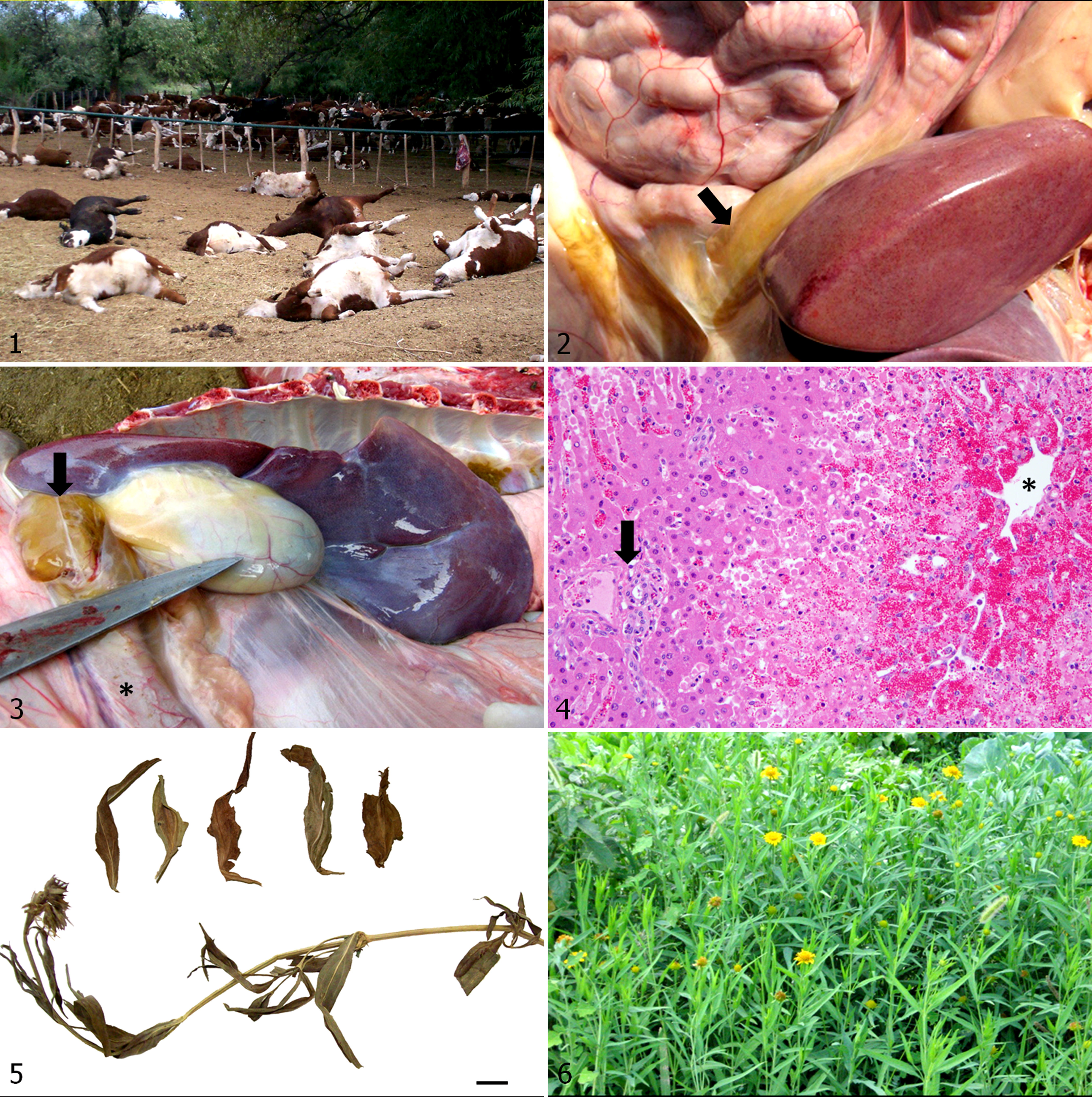
A female crossbred calf and a castrated male Polled Hereford calf were necropsied immediately after natural death. The carcasses were in good body condition. Gross findings were similar in both cases, with diffuse petechiation of the hepatic capsule and markedly enhanced reticular pattern of the liver; yellowish gelatinous mural edema in the gallbladder, as well as along the common bile duct and the duodenal serosa around the insertion of the common hepatic duct (Figs. 2 and 3); and a moderate amount of dark red (bloody) fluid in the large intestinal lumen. Histologically, both calves had diffuse, acute, severe, centrilobular (periacinar) to midzonal hepatic necrosis with loss of hepatocytes, lobular collapse with disruption of the hepatic cords, and individualization of hepatocytes with angular borders, cytoplasmic hypereosinophilia, pyknosis, karyorrhexis, or fading nuclei, with associated centrilobular hemorrhage (Fig. 4).
Whole plants and dry fragments of W. glauca were found in the remnants of the alfalfa bales being fed to the calves (Fig. 5). Patches of lush green W. glauca plants in the flowering stage, many defoliated by the mowers, were also found in the alfalfa fields at the neighboring farm where the hay bales had been produced (Fig. 6).
In the 2 samples of rumen content subjected to microscopic analysis, epidermal fragments of W. glauca were identified based on their amber color and the presence of characteristic and specific trichomes (uniseriate glandular hairs). No vegetal fragments characteristic of other acutely toxic plants in this region of Argentina, such as C. parqui or Baccharis cordifolia, 10 were observed.
Discussion
The epidemiologic characteristics of the outbreak, clinical signs, and the presence of dry W. glauca whole and fragmented plants in the alfalfa hay, as well as standing green intact and defoliated W. glauca in the alfalfa field where the hay had been baled, strongly suggested intoxication by W. glauca. Plant ingestion was confirmed with the identification of specific W. glauca epidermal trichomes by microscopic analysis of rumen contents in both calves. The toxic dose of W. glauca is considered low, with some authors reporting single lethal toxic doses for cattle and sheep of 4 to 5 g dry matter per kg body weight (g/kg BW) 2,7 and others reporting even smaller toxic doses of 1.5 g/kg BW for sheep. 6 Considering the mean body weight of the affected calves (150 kg) and the toxic dose of dry W. glauca for cattle (4–5 g/kg BW), we estimate that the total amount of W. glauca ingested to cause death in 147 calves had to be between 88.2 and 110.25 kg (22.05–27.56 kg per 400-kg bale). Given the fact that W. glauca is highly toxic and that ingestion of small quantities are enough to cause death, its identification within the rumen contents of affected animals, along with compatible gross and microscopic lesions, allows for the diagnosis of W. glauca intoxication. Microscopic analysis of gastric contents has been used to confirm W. glauca ingestion and intoxication in experimental cases in sheep 10 and in natural cases in an axis deer and a llama. 4
In addition, although not pathognomonic for this condition, the gross and microscopic lesions found in both calves, particularly the severe, diffuse, centrilobular hepatocellular necrosis and hemorrhage, supported the diagnosis of W. glauca intoxication. The lesions were similar to experimental intoxications reported by other authors in sheep, 2,6 cattle, 2,7 pigs, 6 and rats, 9 as well as natural cases in several animal species. 4,7 There were no other common local acutely hepatotoxic plants such as C. parqui, Xanthium sp, and Myoporum laetum in the feed or hay fields, nor did the cattle have access to any other type of feed. Copper intoxication due to excessive parenteral supplementation produces similar gross and microscopic hepatic lesions, 8 but this possibility was ruled out when the owner denied any recent copper supplementation. Furthermore, other typical lesions associated with acute copper intoxication, such as jaundice, hemoglobinuric nephrosis, or hemoglobinuria, were not observed in these calves. In addition, the regional edema of the choledochoduodenal junction in the calves of the current report is not reported in copper intoxication 8 but is a frequent necropsy finding in intoxications caused by several atractyloside or CAT-containing plants such as W. glauca and Xanthium sp. 1
In Argentina, naturally occurring death of livestock due to W. glauca intoxication has been diagnosed in grazing animals and in feedlot operations feeding contaminated hay. 4 The plant is the most common toxic cause of death in cattle (E. R. Odriozola, personal communication, 2012). This plant species is widely distributed in Argentina, and poisoning is likely to occur anywhere the plant is present. Therefore, we believe that natural poisoning from W. glauca consumption is underreported. This report highlights the importance of W. glauca as a major cause of significant economic losses in the agricultural sector in Argentina.
Footnotes
Declaration of Conflicting Interests
The author(s) declared no potential conflicts of interest with respect to the research, authorship, and/or publication of this article.
Funding
The author(s) received no financial support for the research, authorship, and/or publication of this article.