
Abstract
Toxicity related to consumption of Cistus sp. pl. has been described in ruminants in some countries. This report describes the clinical and pathological findings of Cistus salviifolius toxicosis in 3 beef cattle herds located in 2 different areas of Sicily, Italy. Outbreaks were observed after grazing in poor winter pasture where C. salviifolius was abundant. Mean morbidity and mortality were 29% and 21%, respectively. Most of the affected animals (6 to 36 months old) showed anorexia, weight loss, and pollakiuria culminating in recumbency and death. Occasionally, abortion and neurological signs were observed. In animals with acute signs, there was a moderate decrease of sodium and chloride concentrations in serum. Animals with chronic signs showed an increase of serum urea, creatine phosphokinase (CPK), aspartate transaminase (AST), lactate dehydrogenase (LDH), and phosphorus and a decrease in total serum protein, calcium, chloride, and magnesium concentrations. Moderate anemia and slight neutropenia, lymphocytosis, and eosinophilia were detected in all groups. At necropsy, the main lesion was severe distention of the urinary bladder with turbid hemorrhagic urine and crystalluria. Histologically, chronic cystitis, interstitial nephritis, eosinophilic enteritis, and nonsuppurative necrotizing hepatitis were observed. To our knowledge, this is the first report of C. salviifolius toxicosis in cattle in Italy.
Cistus L. (Cistaceae) is a genus of about 20 frutescent and suffrutescent species with a predominantly Mediterranean distribution except for 5 species endemic in the Canary Islands. 1 Among the white-flowered species in the subgenus Leucocistus, Cistus salviifolius L. is one of the most common in Italy. Nevertheless, the presence of this plant is reported throughout the Mediterranean basin 11 from Portugal to Greece, part of Turkey and Israel, and extending to North Africa. C. salviifolius is a bushy, nondeciduous, perennial plant, flowering between March and June in the Mediterranean area. This plant is well adapted to the xeric environments of the Mediterranean maquis and garrigues, but it is also found in degraded woodlands of many types. In Italy, its presence as a native species has been confirmed in a large part of the peninsula. 2
This plant is characterized by ovate leaves producing a fragrant oleoresin very rich in sesquiterpenes showing enzymatic inhibitory activity and antioxidant properties. 8 C. salviifolius is also very rich in phenolic compounds when compared to other species of the same genus, a feature that has contributed to the traditional use of this plant in some Mediterranean countries as an astringent and cicatrizing agent. 4
The toxic substances contained in Cistus sp. pl. are not clear. 3 However, the toxicity due to some species of Cistus may be related to their tannins, a very complex group of plant antinutritional metabolites distinguished from other polyphenolic compounds by their ability to precipitate proteins. Their main negative effects result from either direct inhibition of digestive enzymes or from formation of indigestible complexes with endogenous proteins. Finally, fermentation by ruminal bacteria converts tannins to gallic acid and pyrogallol, both of which are nephrotoxic. 12 The concentrations of tannins in Cistus may change within a single species according to seasonal variation in leaf development and moisture content and under adverse climatic conditions. Also, the consumption of saponins contained in this plant may induce hepatogenous photosensitization. Flavonoid compounds were described in some Cistus sp. pl., and some of them are certainly related to the condensed tannins and can have antinutritive properties. 3 The simultaneous presence of saponin and flavonoid compounds may lead to synergic effects, thus increasing the toxicity of each substance. 3,12
Toxicosis due to Cistus sp. pl. has been reported in sheep in Spain, France, Italy, Portugal, and Turkey. 3,5 –7,10,12 In sheep, a characteristic central nervous system syndrome accompanied by weight loss, stranguria, and urine scalding of the perineum has been observed. Occasionally, abortion and subcutaneous edema of the head (apparently correlated only to Cistus ladanifer L.) were also reported. 10
Goats reared in the same pastures sporadically showed similar signs and lesions. 10 In cattle, this disease has been described only in Israel 12 and Portugal, 7 and in both cases, the poisoning was correlated to the ingestion of C. salviifolius during the winter. Affected cattle showed weight loss, urinary tract disease (urinary retention, cystitis, pyelonephritis, and nephrosis), and death. Serological tests revealed increases in blood urea, creatinine, aspartate transaminase (AST), creatine phosphokinase (CPK), and lactate dehydrogenase (LDH), as well as decreases in alkaline phosphatase (ALP), total serum protein, albumin, potassium, sodium, and chloride concentrations. The toxicity of Cistus sp. pl. is related to their tannin content converted by ruminal bacterial fermentation to gallic acid and pyrogallol, both nephrotoxic. 7,12
The authors describe the clinical, laboratory, and pathological findings observed in 3 beef cattle herds located in 2 different areas of Sicily affected by urinary retention syndrome due to C. salviifolius toxicosis.
Materials and Methods
Three herds from different farms were involved (A, B, and C) (Suppl. Fig. S1). Herd A was composed by 70 cattle kept in an extensive pasture (about 60 hectares) reared together with a flock of 87 goats and 23 sheep in Nebrodis Park in northeast Sicily. Herd B was composed by 48 cattle kept in a semiextensive pasture (about 30 hectares) in Palermo area of northwest Sicily. The owner reared in the same pasture a flock of 55 goats and 15 sheep. Herd C was composed by 16 cattle reared in a field (about 19 hectares) close to herd B. In all herds, cattle were reared in a mixed and spontaneous pasture composed of native grasses and plants typical of a Mediterranean-type climate with warm, dry summers and cool, wet winters. The elevation of the hills and the mountain ranged from 150 to 650 m above sea level with an average annual rainfall ranging from 600 to 1200 mm. Herds B and C were supplemented with hay and straw. Herd A moved in the summer period to another area about 60 km from the winter pasture. All herds were composed by mixed-breed Charolais and Limousin beef cattle and purebred Marchigiana, with age distribution for each farm of approximatively 25% young animals (6–32 months old) and 75% adult animals (32–108 months old). Calves younger than 6 months were not considered in the at-risk group due to their different diet. All herds received antiparasitic treatments with ivermectin once a year in spring. No vaccinations were given.
Sporadic unusual clinical signs attributable to the urinary tract (ie, pollakiuria) were observed by the owners for several years, but the number of affected animals and the severity of the disease were different, mainly depending on local climate. Continuing deaths or emergency slaughters and the observation at the abattoir of cystitis and urinary bladders filled with turbid hemorrhagic urine with crystals led the breeders to refer to the unusual disease. All the herds were kept under veterinary observation from March 2016 to June 2017; information was recorded concerning date, plant exposure, onset and severity of clinical signs, and when individual animals were culled/died. Affected animals were classified in 3 groups: acute cases that died within 10 to 15 days from the beginning of the clinical signs (group 1), chronic cases that died or underwent emergency slaughter due to worsening of clinical signs from 16 days and 9 months after the beginning of the clinical signs (group 2), and surviving animals affected by moderate, nonlethal disease (group 3). Clinical, hematological, biochemical, and postmortem investigations were performed on selected cases (Suppl. Table S1). The parameters of affected animals were compared with healthy animals of the same farms.
Samples from gastrointestinal tract, liver, urinary bladder, kidney, spleen, trachea, lung, heart, brain, and muscular tissue were fixed in 10% neutral-buffered formalin and sent to the Department of Veterinary Science of the University of Turin where tissue portions were paraffin embedded, sectioned at 3 to 4 μm, and stained with hematoxylin and eosin for histological examination. Fecal exams were performed during necropsy. Fresh and Diff-Quick-stained smears of urine from dead animals were observed by light microscopy.
Examination of the pasture revealed the presence of many shrubs suspected to belong to the Cistus genus, and subsequently, botanical investigations were made in collaboration with the Department of Life Sciences and Systems Biology of the University of Turin.
Results
Clinical Findings
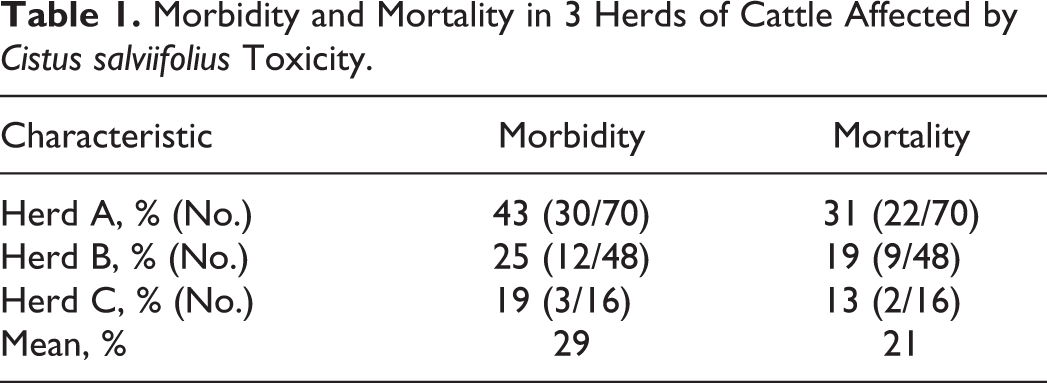
The affected animals were 6 to 36 months old. No clinical signs were detected in calves under 6 months of age. The mean morbidity and mortality observed in the 3 herds (considering all animals 6 months of age or older) were 29% and 21%, respectively (Table 1). The time from the first exposure to C. salviifolius shrubs in contaminated pasture to clinical disease was calculated to be approximately 2 to 3 months. The course of the clinical disease varied from 10-15 days to 9 months or longer and culminated with death or emergency slaughter.
Morbidity and Mortality in 3 Herds of Cattle Affected by Cistus salviifolius Toxicity.
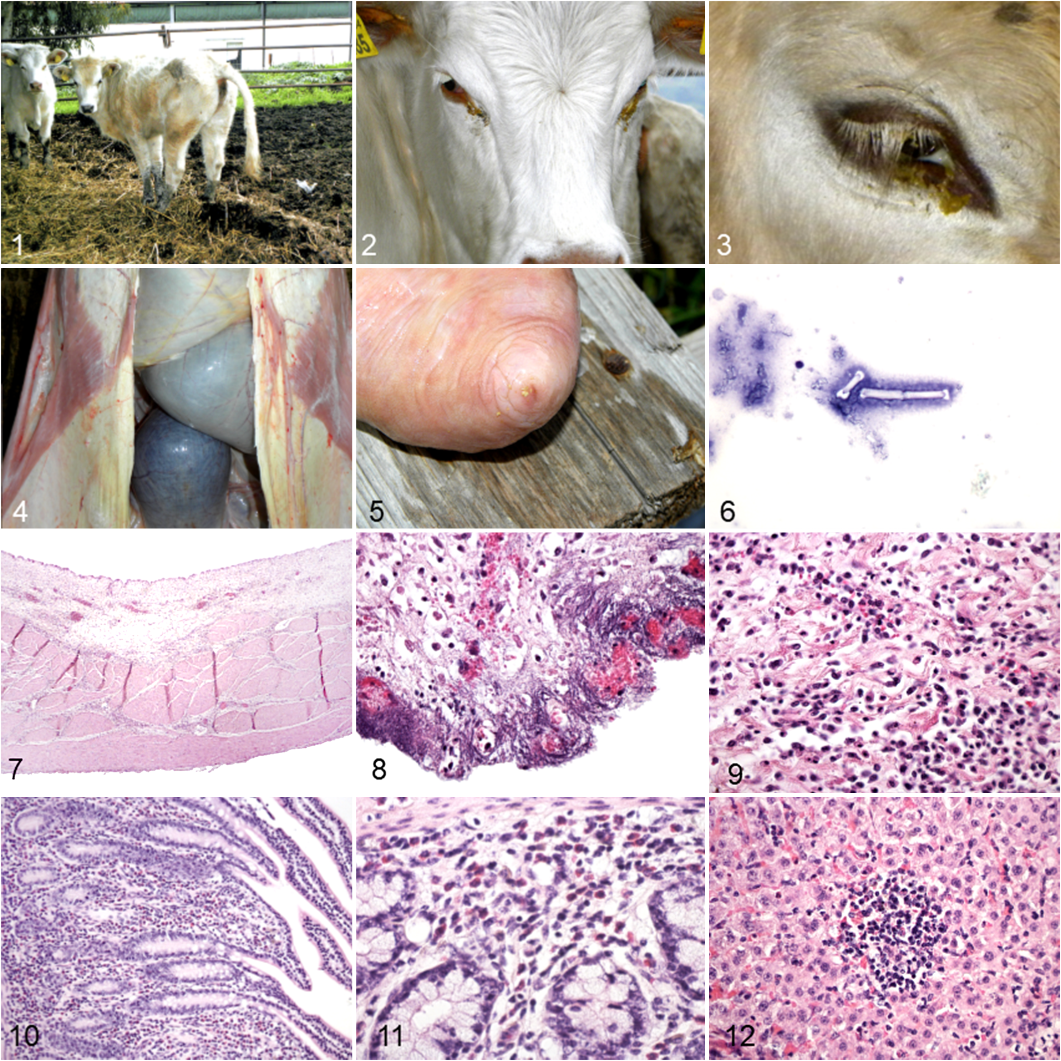
The outbreaks with acute signs were observed in late winter and early spring, but chronic cases were observed throughout the year. Apathy, anorexia, and pollakiuria were the first clinical signs observed. Unusual urination was observed several times a day and was characterized by an abnormal stance with arched back and elevation of the tail for several minutes, sometimes accompanied by dribbling of urine (Fig. 1). The animals appeared dehydrated and debilitated. The muzzle and eyelids were encrusted with dry exudate (Figs. 2, 3), and wheezing was frequent. Often photophobia and keratitis or corneal opacity were present. Some animals showed evidence of decreased vision. The motility of the rumen was decreased, and in some cases, rumination ceased. Constipation was rarely observed. In groups 1 (acute cases) and 2 (chronic cases), heart and respiratory rate were moderately higher (90–106 bpm and 39–48 bpm, respectively) than normal (40–80 bpm and 12–36 bpm, respectively), 9 especially in group 2. The severity of clinical signs increased with the progression of the disease. Cattle with marked urinary difficulties suffered from rapid weight loss, culminating in recumbency, bruxism, and death. Sporadically, abortion and neurological signs such as tremors and stagger were present. One bull in herd A also had several episodes of syncope. The majority of males also had testicular hypoplasia and occasionally focal to multifocal necrosis of the scrotum. The rectal temperature always remained normal. Animals affected by less severe forms (group 3) mainly showed intermittent urinary disorders and poor body condition. In these animals, heart and respiratory rates were normal, and ruminal motility was within the normal range. At rectal examination, all cases had distention of the bladder. Less affected animals partly recovered after removal from pasture containing C. salviifolius. However, the recovery was slow and the animals remained pollakiuric and with poor body condition for several months. In many cases, the owners preferred to slaughter the sick animals.
Farmer of both herds A and B also described neurological signs in sheep and photophobia and keratitis in goats grazing in the same pastures, but no clinical or postmortem examinations were made on these animals.
Laboratory Findings
Hematological and serum biochemical parameters are reported in Table 2. Moderate anemia, with decrease of hematocrit and hemoglobin, slight neutropenia, lymphocytosis, and eosinophilia were detected in all groups. There were increased serum concentrations of urea, AST, CPK, and LDH in group 2 and phosphorus in both chronic groups (groups 2 and 3). There was decreased serum total protein and magnesium in group 2, decreased chloride in all groups, and decreased calcium in both chronic groups. Sodium was decreased in group 1 and at the low end of the normal range in both chronic groups. Creatinine concentrations were normal in all groups, except for a decrease in 1 animal of group 2.
Hematological and Biochemical Findings in Cattle Affected by Cistus salviifolius Toxicity.a
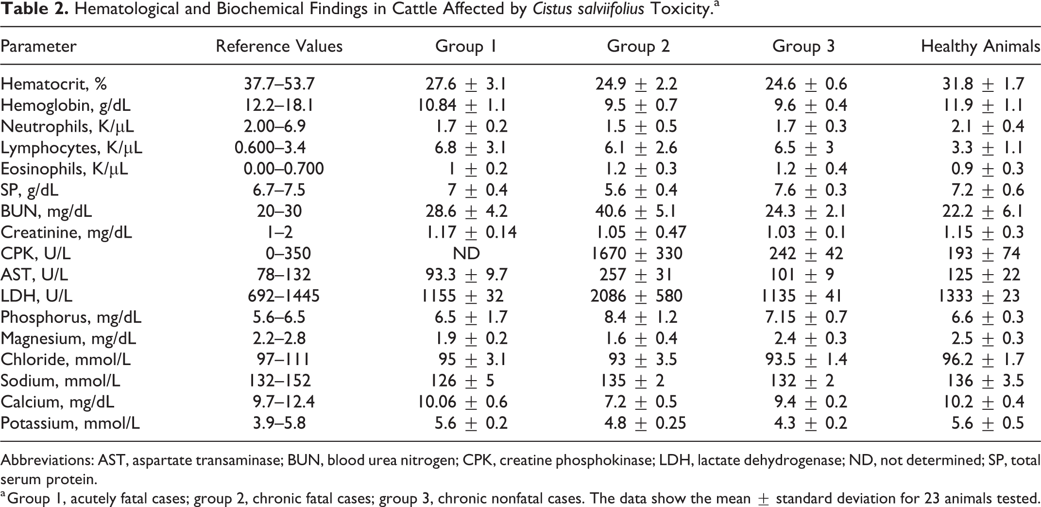
Abbreviations: AST, aspartate transaminase; BUN, blood urea nitrogen; CPK, creatine phosphokinase; LDH, lactate dehydrogenase; ND, not determined; SP, total serum protein.
a Group 1, acutely fatal cases; group 2, chronic fatal cases; group 3, chronic nonfatal cases. The data show the mean ± standard deviation for 23 animals tested.
Pathological Findings
Complete postmortem investigations, including histological evaluations, were performed on case Nos. 1, 2, and 3. Coprological examinations did not reveal any parasites.
Case No. 1 was a 13-month-old crossbred beef heifer from farm A that died 14 days after the observed onset of clinical signs. At postmortem, there was severe distention of the urinary bladder with turbid hemorrhagic urine containing small calculi and thickening/hyperemia of the wall (Figs. 4, 5). Diffuse reddening throughout the small intestine with enlargement of mesenteric lymph nodes was also present. Abundant crystals were observed in the smears of urine, and they were elongated, variable in size from 20–25 µm to 100–120 μm, and colorless (Fig. 6).
Histologically, there was severe multifocal to disseminated cystitis (Fig. 7). The transitional epithelium of the urinary bladder was absent in most areas, and the mucosa and submucosa were edematous and diffusely infiltrated (particularly in the ulcerated areas) by lymphocytes, plasma cells, macrophages, and a few neutrophils (Figs. 8, 9). The submucosa contained disseminated hemorrhages, neovascularization, vascular ectasia, and fibrosis. Multifocally, chronic inflammation and fibrosis also affected the muscularis and focally extended to the serosa. The small intestine had diffuse infiltration of the mucosa with eosinophils and expansion of lymphoid follicles (Figs. 10, 11). The mesenteric lymph nodes contained diffuse infiltrates of eosinophils. The kidneys had mild to moderate, multifocal, interstitial infiltrates of mononuclear cells.
Case No. 2 was a 28-month-old crossbred beef heifer from farm B that died 3.5 months after the observed onset of clinical signs, with severe emaciation, atrophy of the visceral fat deposits, and moderate accumulation of serous fluid in the body cavities. The reported gross lesions were turbid hemorrhagic urine, enteritis, and yellow discoloration of kidneys with moderate hydronephrosis. Histologic lesions were similar to case No. 1, with severe multifocal chronic-active cystitis, moderate to severe diffuse chronic eosinophilic enteritis, moderate lymphoid hyperplasia of the mesenteric lymph nodes, and moderate multifocal nonsuppurative interstitial nephritis. There were disseminated small areas of vacuolation in the white matter of the brain, multifocal neuronal satellitosis in the cerebral cortex, and vascular proliferation.
Case No. 3 was a 16-month-old Marchigiana beef heifer from farm B that died 1.5 months after the observed onset of clinical signs. At necropsy, the urinary bladder was greatly distended with turbid urine. Moderate multifocal chronic-active cystitis and slight to moderate, multifocal nonsuppurative interstitial nephritis were present as described above. The liver had multifocal aggregates of lymphocytes and rare macrophages mainly in the periportal areas, as well as disseminated randomly distributed foci of inflammation and necrosis (Fig. 12).
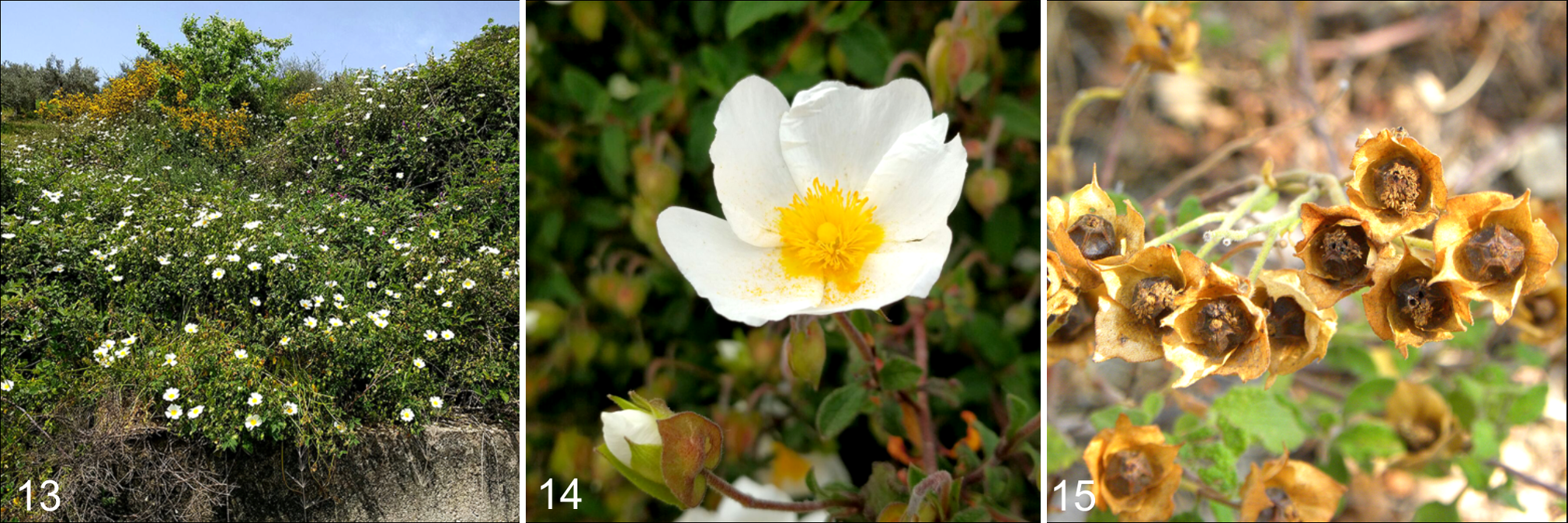
Pasture Inspection
A severe infestation of partially eaten C. salviifolius shrubs was present in all pastures, covering 10% to 30% of the areas where the owners reported cattle grazing. Based on leaf and flower morphology, all the plants examined were confirmed as C. salviifolius (Figs. 13–15).
Discussion
In the present study, the authors observed acute cases of toxicosis in spring, following the consumption of C. salviifolius during the winter, where the animals were forced to ingest this shrub in sparse pasture due to the insufficient feed supplementation. This would explain why, in the absence of supplemental feeding during the poor season, the authors observed higher morbidity and mortality in herd A compared to the other 2 herds where animals received supplementation of their diet. Significant differences in levels of tannins, phenols, and flavonoids between leaves and flower buds have been documented in plants of C. salviifolius collected in Tunisia during the spring. 4 In another study, rapid fluctuations in these levels were documented at the onset of flowering. 7,12 Therefore, it is possible that the outbreak here described also coincided with an increase of these secondary compounds due to seasonal variation of the plant growth. However, the possibility that C. salviifolius growing in the Nebrodi Park is more toxic compared to the plants growing in Palermo or other areas cannot be ruled out, considering also that a consistent degree of phenotypic plasticity has been found in this species elsewhere.
It is not clear whether the animals’ age affects susceptibility to Cistus sp. pl. toxicosis. In the present case, disease occurred in animals younger than 36 months of age, while others 12 reported a mortality rate of 59% in the first and second calving cows and only of 5% in adult cows. It remains unclear whether the apparent increased susceptibility of young cattle reflects age-related metabolic effects or a tendency of adults to avoid the most toxic plants.
Pollakiuria with abnormal urination posture was the main clinical sign observed and was associated with urinary retention. This clinical sign could be caused by failure of contractile function, inappropriate outlet resistance (inhibition of the relaxation of the external and/or internal urethral sphincters mechanism), or both. Phenolic compounds, and therefore tannins, may block pudendal nerves and produce urinary retention. 12 In Cistus toxicosis, severe and prolonged retention is suggested to cause bladder distention as a result of tearing and disruption of the tight junctions between smooth muscle cells of the urinary bladder wall. 7,12 Indeed, interstitial edema, hydropic degeneration, and necrosis in the smooth muscle of the urinary bladder were identified, and many cases also had chronic or subacute vasculitis. Bladder distension and resulting lesions could cause detrusor atony that closely mimics the autonomous bladder syndrome observed in affected animals. The pudendal nerves were not examined in this study, so damage to these nerves cannot be ruled out. Furthermore, the crystals observed in the urine, sometimes voluminous, may have mechanically resulted in inflammation of the urinary bladder and potentially, in the most severe cases, might be responsible for urinary stasis. Yeruham et al 12 pointed out that in Cistus sp. pl. exposure, the oliguria might be caused by renal damage due to the nephrotoxic nature of tannins and their metabolites or secondary to the pathologic accumulation of urine. Renal damage, paralysis of the urinary bladder, and uremia are considered the cause of death in toxicosis by Cistus sp. pl. In the present study, despite a severe chronic active cystitis, the damage observed in the kidneys was limited and insufficient to cause the alterations of biochemical parameters. Even though Yeruham et al 12 described elevation of blood urea nitrogen and creatinine levels associated with nephrosis, in the present cases, the levels of creatinine were within the normal range. Similar unchanged values of creatinine were observed by Lima et al. 7
Despite the presence of severe chronic clinical signs similar to the literature, 7,12 the lesions of the urinary bladder and kidney in this study were less severe. Moreover, chronic enteritis was mainly characterized by severe and diffuse eosinophilic infiltration in the absence of parasites and despite receiving antiparasitic drugs once a year. Chronic eosinophilic enteritis was also observed by Yeruham et al, 12 but no explanation of this feature was given. We speculate that the presence of eosinophilic infiltrates in intestine and mesenteric lymph nodes might result from an allergic reaction related to ingestion of C. salviifolius, but further investigations should be performed.
Regarding the nervous system degeneration, Lima et al 7 described in 1 animal a mild edema and mononuclear infiltrates in the cauda equina that might have been due to recumbency or perhaps related to chronic distention of the urinary bladder. In the present research, only 1 animal had moderate and multifocal to disseminated cerebral degenerative changes. It is unknown if this lesion resulted from the toxicosis, but the authors do not exclude a correlation with renal lesions or endotoxicosis secondary to poor body condition. Ligios et al 6 reported neurological signs in small ruminants intoxicated by different species of Cistus and observed the accumulation of brown periodic acid–Schiff (PAS)–positive pigments in the perikarya of larger neurons. No such pigment accumulation was detected in the present study.
Based on epidemiology, clinical signs, pathological findings, and botanical investigations, a diagnosis of C. salviifolius toxicosis was made.
Because there is no specific medical therapy, the authors only advised the owners to remove their animals from the pastures infested with C. salviifolius during the at-risk season (from October to March), especially the animals under 3 years old (considered the at-risk category), and provide forage when removal is impossible. Further studies are needed to determine the pathogenesis of the urine retention. These findings underline the importance of plant poisoning in domestic livestock and its dependence on specific environmental variables such as changes in weather and plant growth rates, as well as the importance of a complete environmental anamnesis in obtaining a definitive diagnosis.
Supplemental Material
Supplemental Material, Combined_supplemental_materials-Mignacca_et_al - Cistus salviifolius Toxicity in Cattle
Supplemental Material, Combined_supplemental_materials-Mignacca_et_al for Cistus salviifolius Toxicity in Cattle by Sebastian Alessandro Mignacca, Marco Mucciarelli, Elena Colombino, Elena Biasibetti, Salvatore Muscia, Benedetta Amato, Vincenzo Di Marco Lo Presti, Irene Vazzana, Andrea Galbo and Maria Teresa Capucchio in Veterinary Pathology
Footnotes
Acknowledgements
We thank Dr. Buttitta Rosalinda and Dr. Gagliardo Antonino of the Ambulatorio Veterinario Gagliardo (Bagheria, Palermo, Italy) for fresh observation and staining of the urine smears.
Declaration of Conflicting Interests
The author(s) declared no potential conflicts of interest with respect to the research, authorship, and/or publication of this article.
Funding
The author(s) received no financial support for the research, authorship, and/or publication of this article.
Supplemental material for this article is available online.
References
Supplementary Material
Please find the following supplemental material available below.
For Open Access articles published under a Creative Commons License, all supplemental material carries the same license as the article it is associated with.
For non-Open Access articles published, all supplemental material carries a non-exclusive license, and permission requests for re-use of supplemental material or any part of supplemental material shall be sent directly to the copyright owner as specified in the copyright notice associated with the article.