
Abstract
Tumor protein 53 (TP53) is a tumor suppressor gene that is frequently mutated in urinary bladder tumors in both humans and animals. In cattle, urinary bladder tumors have been reported as occurring spontaneously as well as in conjunction with bracken fern consumption-induced bovine enzootic hematuria (BEH). The goal of this study was to evaluate various types of bovine urinary bladder neoplasms for the presence of TP53 alterations, using the polymerase chain reaction–single-strand conformation polymorphism (PCR-SSCP) method. DNA was extracted from both epithelial and mesenchymal urinary bladder tumor samples in cattle, associated with the chronic consumption of bracken fern. PCR was performed using primers targeted to exons 5 to 8, following electrophoresis and isolation, and the products were assessed by SSCP. Tumors in which alterations in the electrophoresis patterns were noted included hemangiomas, papillomas, and carcinomas in situ. Exemplars of these tumor types were selected for sequencing, and although no changes were noted in the 5 to 8 exon range, on either side of the designed primers for exon 6, there was some portion of intron 6 in which sequencing demonstrated a deletion of the thyamine nucleotide at position 9332. In summary, although mutations were not observed within exons 5 to 8, this represents the first report of an intronic mutation in the TP53 gene in association with bovine urinary bladder tumors. Mutations within introns can predispose tissues to the development of cancer, and therefore, a possible association between mutations of the introns of TP53 and the development of urinary bladder tumors in cattle with BEH should be further investigated.
The TP53 protein is a phosphoprotein with a molecular weight of 53 KDa consisting of 378 amino acids. This protein is coded by a 20-Kbp gene that contains 11 exons and 10 introns. In cattle, this gene is located on chromosome 19 (19q15) with the same properties of human TP53. 12,20,22 TP53 is a transcriptional factor and has been described as the guardian of the genome or as the cellular gatekeeper of growth and cell division. Significantly, this gene has been demonstrated to be a tumor suppressor gene with prominent roles in the cell cycle, gene transcription, DNA repair, and apoptosis. 14,23,25,26
The transcriptional activity of the TP53 gene can be neutralized by a selective process during tumorogenesis. 9 In fact, inactivation of TP53 allows neoplastic cells to escape from apoptosis. 17 Therefore, inactivation of the TP53 gene represents a key event in carcinogenesis. 39 Genetic mutations are considered to be the most frequent causes of TP53 inactivation 36 and include mutations that prevent TP53 activation, resulting from protein conformational changes, as well as microRNA and epigenetic changes.
Acquired mutations of the TP53 gene have been identified in most types of human neoplasia, and development of almost half of human tumors is associated with mutations that resulted in inactivation of the TP53 gene. 3
Chronic sublethal feeding of cattle with different species of bracken fern such as Pteridium aquilinum subsp aquilinum and subsp caudatum, as well as mulga fern or rock fern (Cheilanthes sieberi), 28 has been demonstrated to cause the neoplastic syndrome associated with bovine enzootic hematuria (BEH). A large number of toxic, carcinogenic, mutagenic, and immunosuppressive substances such as thiaminase, quercetin, shikimic acid, prunasin, ptaquiloside or braxin C, ptaquiloside Z and aquiline A, and a bleeding factor with an unknown structure have been recognized in bracken fern. 28,29 Cattle affected with BEH frequently develop urinary bladder tumors of both epithelial and mesenchymal origins. 4,8,11,28,30 Furthermore, a potent relationship or co-carcinogenesis has been shown to exist between bovine papillomavirus type 2 (BPV-2) and bracken fern in urinary bladder carcinogenesis in cattle, 10,14,15,28,35 including tumors associated with BEH.
The goal of this study was to evaluate different types of bovine urinary bladder tumors (related to BEH) for the presence of the TP53 mutation, using polymerase chain reaction–single-strand conformation polymorphism (PCR-SSCP) and direct sequencing methods.
Materials and Methods
Tissue Samples
Fifteen paraffin-embedded samples of urinary bladder neoplasms were selected from cows with different types of urinary bladder lesions. Each of the 15 samples had a single neoplastic lesion and no inflammatory histopathologic lesions. All of the samples originated from the northern regions of Iran in which cattle are susceptible to chronic consumption of bracken fern throughout the year. The selected neoplastic samples included transitional cell carcinomas (n = 3), carcinomas in situ (n = 3), hemangiomas (n = 3), squamous cell carcinomas (n = 2), papillomas (n = 2), and single cases of fibroma (n = 1) and leiomyoma (n = 1). Collected tissues had been fixed for 48 to 72 hours in 10% neutral buffered formalin, embedded in paraffin blocks, sectioned at 5 microns, and finally stained by standard hematoxylin and eosin (H&E) methodology for initial histopathologic diagnosis.
DNA Extraction
DNA was extracted from paraffin-embedded tissues. Ten sections of 5 μm thickness were cut by microtome from each block and were transferred into a 1.5-ml microtube. Between samples, the microtome was washed with xylene and ethanol prior to each sectioning, and new disposable microtome blades were used for each sample. Then, 1600 μl of 1% sodium dodecyl sulfate (SDS) and 0.1M NaOH solution (pH 12.7) and 10 beads of Chelex 20 were added to each microtube. The microtubes were heated at 100°C in a water bath for 45 minutes and subsequently cooled for 5 minutes, after which the solidified paraffin cap was pierced by a sterile micropipette tip and the underlying solution was transferred into a sterile 1.5-ml microtube. Further steps of extraction and purification of DNA were performed according to the method by Shi et al. 34
Control tissue
Samples of normal urinary bladder tissue derived from abattoir specimens of healthy cattle were collected, and these were evaluated macroscopically and histopathologically to preclude any concurrent disease. Then the samples were fixed in 10% neutral buffered formalin and embedded in paraffin blocks that were subsequently used for DNA extraction as according to the methodology described previously.
Primers
All primers were obtained and employed in accordance with the methodology described in Dequiedt et al. 13
PCR Method
PCR reaction for exons 5 through 8 was performed with 3 μl of DNA suspension (200 ng) as the template, 2.5 μl of 10× buffer (50 mM KCl, 10 mM Tris-HCl [pH 8.4], AMS buffer with 20 mM ammonium sulfate, 75 mM Tris-HCl [pH 8.8]), 1.5 mM MgCl2, 0.2 mM dNTP, and, from each primer, 0.5 pM and 1U Taq DNA polymerase (CinnaGen, Tehran, Iran) in a final volume of 25 μl. The amplification process included an initial denaturation at 94°C for 2 minutes, followed by 35 cycles of denaturation at 94°C for 20 seconds, annealing at 57.8°C for 45 seconds for exon 5, 68°C for 30 seconds for exon 6, 57°C for 30 seconds for exon 7, and 66°C for 30 seconds for exon 8 with extension for all 4 exons at 72°C for 45 to 50 seconds and a final extension at 72°C for 5 to 10 minutes. Then, 10 μl of each sample was loaded onto the 1.5% agarose gel and electrophoresis was performed. The gels were then stained with ethidium bromide and analyzed under ultraviolet light.
SSCP Method
Aliquots (5 μl) of each PCR product were mixed with 5 μl of formamide loading dye (formamide 95%, NaOH 10 mM, xylene cyanol 0.5%, bromophenol blue 0.5%). The samples were heated to 95°C for 5 minutes, until the double-stranded DNA was denaturated and converted to single strands. Samples were then immediately cooled on ice and after 5 minutes run onto the polyacrylamide 10% gel.
Electrophoresis was performed at 100 V for 20 hours at 20°C with 1× Tris-borate-EDTA (TBE) buffer in the buffer chamber. Gels were stained using a silver nitrate method. The samples with the alterations in their electrophoretic patterns were selected for direct sequencing, using Macrogen (Seoul, South Korea) in an ABI 3730 XL automatic DNA sequencer.
Results
The PCR products of exons 5 through 8 in the TP53 gene were analyzed by SSCP and direct sequencing methods. PCR products from exons 5, 7, and 8 did not demonstrate any significant changes in electrophoretic pattern motility in SSCP. However, alterations in electrophoretic patterns were identified in exon 6 from samples derived from hemangiomas, papillomas, and carcinomas in situ (Fig. 1) but not in the other neoplasm variants evaluated as part of this study.
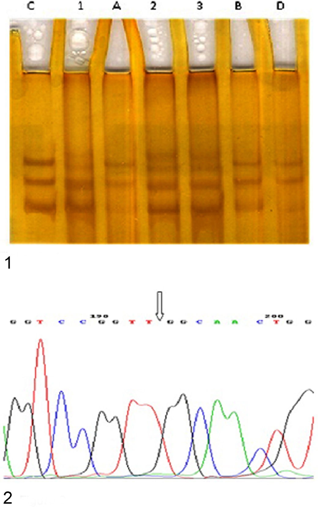
Interpretation of the sequencing results suggests that although no mutation was observed in exon 6, there was a single base mutation in intron 6. This mutation was found to be in close proximity to exon 6, with a deletion of a thymine (T) nucleotide (9332) (GGTCCGGTT/-GGCAACTGG) (Fig. 2). In comparison, no mutations were observed in the control tissues.
Discussion
The literature describing the etiopathogenesis of the BEH syndrome has demonstrated a co-carcinogenesis between bracken fern consumption and BPV-2, which creates an environment permissive for cellular transformation and development of urinary bladder tumors in affected cattle. 2,10
Increases in telomerase activity 5 and of H-RAS oncogene expression, 32 upregulation of cyclooxygenase-2 (COX-2) expression, 6 tumor suppressor gene expression, 4 activation of platelet-derived growth factor β receptor (PDGFβR), 7 and increases of ferritin heavy chain (FHC) expression 31 as well as downregulation of fragile histidine tetrads (FHIT) have all been reported in the neoplastic cells of the urinary bladder tumors in cattle nourished sublethally with bracken fern.
Furthermore, mutation and deletion of the TP53 tumor suppressor gene have been described as the most frequent genetic alterations of epithelial urinary bladder cancers in humans and are theorized to be closely related with disease progression. 14,15,35
In addition, TP53 mutations have been documented in the veterinary literature in association with the pathogenesis of neoplastic transformation in canine urinary bladder tumors. 28
The TP53 protein has been shown immunohistochemically in aggressive hemangiosarcomas of the bovine urinary bladder, and it has been suggested that the positive immunoreactivity of the p53 protein in those tumors was correlated with increased tumor aggression. 11
Although no mutations were observed in exons 5 through 8, a mutation in intron 6 in 3 tumors samples was noted. Introns are noncoding sequences between exons that are deleted by exonucleases from mRNA, during splicing but prior to translation. They are major components of eukaryotic genomes that perform important tasks and participate actively in gene development. A number of regulatory elements in gene expression are found in intron sequences. They have an important role in the transfer of critical signals for mRNA extrusion from the nucleus. 16
Splicing is performed with extreme precision, as a mistake in a single nucleotide (deletion or addition of a nucleotide) can result in alterations in codons. These changes can then cause the formation of new sequences, which finally results in the synthesis of novel amino acid structures. 18 Splicing occurs from the 5′ to 3′ side, and intronic base alterations of the 5′ side typically result in defects in mRNA splicing and often result in the formation of defective mRNA molecules. Mutations that influence mRNA splicing usually contain 3′ and 5′ splicing locations. 40
This study demonstrated a deletion of a thymine (T) nucleotide (number 9332) in samples of urinary bladder hemangiomas, papillomas, and carcinomas in situ derived from cattle fed with sublethal amounts of bracken fern. This nucleotide is located on the 5′ side of intron 6 of the bovine TP53 gene, and its mutation converts a splice sequence from 5′-GTCCGGTTT-3′ to 5′-GTCCGGTTG-3′. Nucleotide alterations of intron 6 of the TP53 gene have been reported to be associated with a syndrome of predisposition for various types of cancer, as well as resulting in an accumulation of the p53 protein and in vitro reduction of apoptosis induction with chemotherapy in diffuse large B-cell lymphoma in humans. 24 Intronic mutations have also been shown to have important roles in the stability of the wild-type p53 protein, which result in abnormal accumulation of mutated TP53 and a predisposition for cancer.
In the medical literature, a correlation between cancer phenotypes and the inheritance of intronic polymorphisms of the TP53 gene has been observed in studies of epithelial neoplasms affecting the ovaries, breasts, colon, stomach, nasopharynx, thyroid glands, urinary bladder, and lungs in humans. 37 In addition, intronic alterations in the TP53 gene have been described in a malignant schwannoma, 33 hepatocellular carcinoma, 21 pheochromocytoma, 38 diffuse large B-cell lymphoma, 24 and acute T-cell lymphoblastic leukemia 19 in humans.
In the veterinary literature, a 23-bp deletion involving the splicing junction between intron 5 and exon 6 has been described in a feline pleomorphic sarcoma, and a 4-bp deletion of intron 7 was observed in a feline fibrosarcoma. 1,27
Further analysis of intronic mutations of the TP53 gene is required to determine the potential roles of TP53 intronic mutations in the development or progression of urinary bladder tumors in cattle that are fed chronic, sublethal amounts of bracken fern.
Footnotes
Acknowledgement
We thank Reza Samani and Mahmood Khormali for their technical and laboratory assistance.
Declaration of Conflicting Interests
The author(s) declared no potential conflicts of interest with respect to the research, authorship, and/or publication of this article.
Funding
The author(s) disclosed receipt of the following financial support for the research, authorship, and/or publication of this article: The research was supported by Research Assistance of the University of Tehran.