
Abstract
A 21-week-old male untreated control SHR/NCrlNarl rat was found dead during an experiment. Grossly, pulmonary lesions were characterized by multifocal to coalescing firm gray-white nodules randomly scattered on the surface. Microscopically, bronchopneumonia was found with pyogranulomas containing neutrophils, macrophages, and numerous thick-walled yeast cells. Yeast cells, 5 to 25 μm in diameter, with no branching of hyphae were observed by staining with hematoxylin and eosin, Diff-Quik, and periodic acid-Schiff. Furthermore, polymerase chain reaction (PCR) using panfungal and nested PCR primers were used for detection of Blastomyces dermatitidis DNA in the lung tissue. After sequencing and matching with DNA sequences in the GenBank, the sample showed a similarity of 94.6% and 97% to Ajellomyces dermatitidis (B. dermatitidis), respectively. On the basis of these results, probable pulmonary blastomycosis was diagnosed. The origin of the infection in the colony rat is undetermined.
Blastomycosis occurs in many countries, including the American continent, Africa, the Middle East, and occasionally in Europe.6,8 Blastomyces dermatitidis is a dimorphic fungus (mycelia-yeast) seen mainly in young dogs and occasionally in cats. 6 The fungal cells appear as round to oval shape with thick, sharply defined and refractile cell walls. Generally, yeast cells range from 6 to 15 μm in diameter but also can be seen as small as 2 to 4 μm or as large as 20 to 30 μm. In hematoxylin and eosin (HE) staining, the protoplasm is separated from the rigid unstained cell wall by a clear space that is readily stained with basophilic or amphophilic color. When found in tissue, these yeast cells reproduce through single budding, with the bud attached to the parent cell via a very wide base.4–6,8
This fungus is present in soil, and inhalation of spores is considered the principal route of infection; thus, it frequently affects outdoor and hunting dogs. 9 In the dermis and subcutis of dogs, intraepithelial abscesses with an epithelioid reaction are observed, and a slow-healing epidermis with ulceration is also found. Primary infection in the lung will cause hematogenous dissemination and may spread to other organs, mainly bone, skin, brain, and eyes. Canine pulmonary lesions are characterized by multifocal to pyogranulomatous pneumonia, generally with firm nodules scattered throughout the lung. Nodules are granulomas with numerous macrophages (epithelioid cells), some neutrophils, multinucleated giant cells, and thick-walled yeasts.5,6 Clinical signs of B. dermatitidis infection reflect the inflammatory and multisystemic nature of the disease, such as anorexia, weight loss, and fever, with about 40% to 60% of affected dogs having a fever over 39.4°C (103°F). Pulmonary lesions occur in 65% to 85% of cases and may be clinically silent. However, lesions are more often associated with respiratory signs, including exercise intolerance, tachypnea, and cough. 7
Forty-five 5-week-old male rats (spontaneous hypertension rat, SHR/NCrlNarl) were purchased from the National Laboratory Animal Center, Taipei, Taiwan, for a hypotensive experiment at a university. They were kept in polycarbonate cages with bedding (Andersons, Maumee, Ohio) and supplied with a standard nonpurified diet (Lab Diet 5001 Rodent diet; PMI Nutrition International, LLC, St Louis, Missouri) and ion-reverse water (Millipore, Billerica, Massachusetts) freely in a conventional animal room (temperature 22–25°C, relative humidity 50%–70%, and on a 12-hour light-dark cycle). Animals were kept until 14 weeks of age, when the experiment began. A male rat in the control group was incidentally found dead after 7 weeks of treatment. Prior to death, this rat had shown signs of nasal discharge, dyspnea, anorexia, depression, and loss of body weight. Tissue samples from the aorta, heart, kidney, liver, and lung were fixed in 10% neutral buffered formalin and sent to the Animal Disease Diagnostic Center of National Chung-Hsing University for histopathological examination.
Grossly, pulmonary lesions were characterized by multifocal to coalescing firm gray-white nodules. Nodules of various sizes ranged from less than 0.1 × 0.1 × 0.1 cm to greater than 0.2 × 0.2 × 0.2 cm and were observed on the lung lobes (Fig. 1). Microscopically, multifocal, severe, subacute to chronic pyogranulomas with bronchopneumonia extending into alveoli were found in the lung (Fig. 2). The bronchioles and alveoli were infiltrated by many neutrophils and macrophages. The pyogranulomas were composed of numerous inflammatory cells, including neutrophils, epithelioid macrophages, and amorphous plant cells (foreign body) adjacent to thick-walled yeast cells (Fig. 3). Yeast cells were 5 to 25 μm in diameter and hyphae with no branching. They were also observed with Diff-Quik (DQ) or periodic acid-Schiff (PAS) staining in imprint smears and paraffin tissue sections (data not shown). The bronchus-associated lymph tissue (BALT) and plasma cells of the lung were less than normal. In addition, no significant lesions were observed in the aorta, heart, kidney, and liver. Unfortunately, neither spleen nor thymus tissues were included in this case. Slides of lungs were further stained for immunohistochemistry for Aspergillus sp. (Abcam, Cambridge, UK; rabbit polyclonal antibody, ab20419, at 200× dilution) and Candida albicans (Abcam; rabbit polyclonal antibody, ab20005, at 200× dilution), with an immunohistochemical detection substrate (Dako REAL EnVision Detection System, Peroxidase/DAB+, Rabbit/Mouse; Dako, Glostrup, Denmark) to differentiate the possible pathogen involved. However, no immunoreaction for Aspergillus sp. and C. albicans was found in the lungs (data not shown).
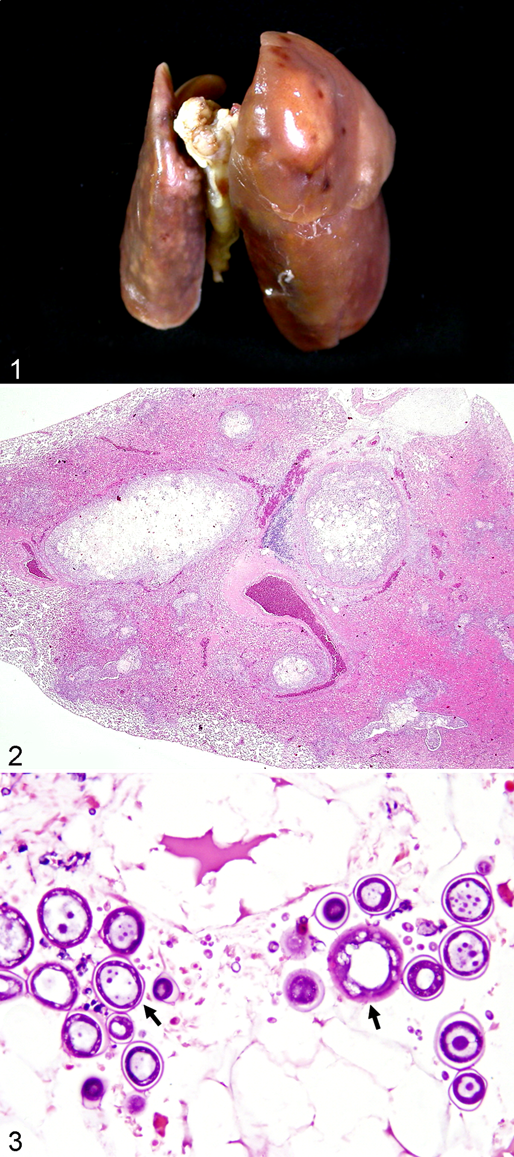
Lung; spontaneous hypertension, rat. Pulmonary lesions were characterized by multifocal to coalescing firm gray-white nodules randomly scattered throughout the lung lobes.
Furthermore, DNA was extracted from the formalin-fixed lung tissue according to the method reported by Burik et al. 3 Briefly, DNA extraction was conducted by using the QIAamp tissue kit (Qiagen, Hilden, Germany). A panfungal polymerase chain reaction (PCR) assay that detects the small-subunit rRNA gene sequence of the fungal organisms in the paraffin-embedded lung tissue was performed using extracted DNA, PCR buffer (PCR Master Mix kit, cat. RP02; GeneMark, Tainan, Taiwan), and panfungal primer (the forward primer 999 [5′-GATACCGTCGTAGTCTTA-3′] and the reverse primer 1574c [5′-ATTCCTCGTTGAAGAGC-3′]). Amplification was done by the GeneAmp PCR System 9700 thermocycler (Applied Biosystems, Singapore). After denaturation at 94°C for 5 minutes, the cycling conditions were 35 cycles at 94°C for 30 seconds, 52°C for 1 minute, and 72°C for 1 minute, followed by a final elongation period at 72°C for 7 minutes. Each PCR product was electrophoresed through a 2% agarose gel for 30 minutes at 100 V and 50 mA in 0.5X Tris-Acetate-EDTA (TAE) buffer, and the gel was stained with ethidium bromide. A known Aspergillus sp.–infected chicken lung was used as the positive control. Results revealed that PCR using panfungal universal primers could successfully amplify the predicted products (580 bp) of both the Aspergillus sp.–infected chicken and the infected rat lung. The negative control was doubly distilled water. After sequencing the products of the rat lung and matching them with DNA sequences in the GenBank, the similarities were up to 94.6% of Ajellomyces dermatitidis (B. dermatitidis).
Furthermore, we have investigated the possible pathogen of B. dermatitidis by using a Blastomyces-specific PCR method with specific detection probes that has been reported by Bialek et al. 2 A Histoplasma capsulatum 18S rDNA PCR was used. Outer primers fungus I (5′-GTT AAA AAG CTC GTA GTT G-3′) and fungus II (5′-TCC CTA GTC GGC ATA GTT TA-3′) are complementary to a highly conserved region of the small subunit rRNA gene of H. capsulatum (GenBank accession number X58572) and B. dermatitidis (GenBank accession number AF320010) and amplify a 429-bp sequence of several fungi pathogenic for humans. Inner primers histo I (5′-GCC GGA CCT TTC CTC CTG GGG AGC-3′) and histo II (5′-CAA GAA TTT CAC CTC TGA CAG CCG A-3′) are complementary to positions 643 to 666 and 873 to 849 of the 18S rDNA, respectively, and amplify a specific 231-nucleotide sequence. The nested PCR product of paraffin-embedded lung tissues was also submitted to GenBank. Sequencing results revealed that this product is 97% homologous to that of B. dermatitidis.
Because of the ability of pulmonary blastomycosis to cause severe, life-threatening disease, early and accurate diagnosis is critical to improve survival rates. Although not always clearly visible, each yeast cell contains several nuclei. Stains for bound glycogen (PAS, Gomori methenamine silver) can identify the outer wall of organisms selectively.8,10 A diagnosis can be made by microscopic examination of pus, tissue, or autopsy material in unstained wet film. The specimen can be cleared in 10% potassium or sodium hydroxide under a cover glass. Recently, Blastomyces yeast forms were easily identified with DQ staining by their negative image. 11 This technique can be used as a quick and cost-effective cytological diagnostic characteristic for confirming these specimens. Another method for identification includes cultivation of the organism in a suitable medium such as Sabouraud dextrose agar, potato dextrose agar, potato flake agar, or inhibitory mold agar at 22°C or 37°C, for 1 to 3 weeks, with creamy or waxy colonies formed on blood agar at 37°C and consisting of budding yeast cells. 4 For detection of B. dermatitidis in tissue, a panfungal PCR assay that detects the small-subunit rRNA gene sequence of the fungal organism was used in this test. The 580-bp PCR product was identified after being amplified by panfungal primers and hybridization to a 245-bp digoxygenin-labeled probe. 3 In addition, the PCR targeting a gene encoding the unique WI-1 adhesin is sensitive and more specific than the PCR targeting the 18S rDNA. 2 Our results revealed that the panfungal and nested PCR assays can also be applied to detecting multiple fungal genera and may be used as an adjunct to conventional methods for the detection of fungal infection.2,3 Because of our inability to culture the possible fungi from the formalin-fixed lung tissue, we were forced to rely on the sequencing percent match of 94.6% in panfungal primers as well as the nested PCR, which was 97% homologous to that of B. dermatitidis. Finally, probable B. dermatitidis infection was diagnosed according to the fungal morphology, pathology, and molecular identifications in this case.
Blastomycosis also occurs in dogs in endemic areas, and humans and dogs are often ill with the disease at the same time while experiencing the same exposure. 9 For the pathogenesis of B. dermatitidis, the conidia produced in the environmental mold phase cause infection when aerosolized and inhaled into the alveoli, where conversion to the yeast phase occurs. Inflammatory cells, including neutrophils and macrophages, can phagocytize the yeast phase organisms, leading to pyogenic and granulomatous lesions that are seen in the infected tissues. Several local host factors are triggered after inhalation of the conidia. 8 Monkeys are suggested to have high levels of resistance to B. dermatitidis infection. However, a disseminated blastomycosis of extrapulmonary lesions, including the lung, tracheobronchial lymph node, brain, spleen, and liver, has been reported in a rhesus monkey (Macaca mulatta) following a left ventricle catheterization experimental procedure. 10
No pulmonary blastomycosis has been reported in experimental rats. However, subcutaneous Blastomyces lesions have been induced by using subcutaneous injections of yeasts. 1 Subcutaneous nodules first appeared 3 to 7 days after injection and reached 2 to 15 mm in diameter by 7 to 28 days after inoculation. The lesions were characterized by a layer of viable yeast and granulomatous inflammation. Live yeast organisms were recovered from all lesions. No systemic spread of Blastomyces organisms was observed in this experimental study. In contrast, our case showed pyogranulomatous/granulomatous inflammatory reaction in the lung, resembling fungal infections, such as Aspergillosis. However, histological lesions were characterized by a granulomatous inflammation with necrotic centers surrounded by the viable thick-walled yeasts with budding but no branching of hyphae. It is known that dimorphic fungi can undergo a temperature-dependent morphological change from conidium or mycelium at environmental temperatures (22°C) to pathogenic yeast at normal body temperature (37°C). 4 The oxygen-rich environment of the lung along with possible hypothermia might have provided conditions helpful for the growth of B. dermatitidis in this case. The yeasts were further identified by using the panfungal PCR assay.
In addition, B. dermatitidis with amorphous plant cells were also found in this lung tissue. The possible sources of infection might be related to accidental inhalation of the foreign materials into the lungs, such as food particles or bedding, via inappropriate oral gavage administration of the test substance. Otherwise, the BALT and plasma cells of the lung were less than normal, suggesting immunosuppression occurred in this rat. The origin of the infection in a colony rat is undetermined. No illnesses were noted in the animal facility staff members. It has been suggested that B. dermatitidis can be disseminated to immunocompromised hosts who have AIDS or patients receiving tumor necrosis factor–α inhibitor therapy. 8 The opportunistic characteristics of B. dermatitidis in a mold-contaminated environment may pose a potential risk of infection, especially to animals confined to closed living conditions.
Footnotes
Acknowledgements
We thank Dr J. M. Ward for his review of the manuscript and Mr K. C. Cheng, Ian Cochrane-Lusk at GIVP, NCHU, for technical assistance.
Declaration of Conflicting Interests
The author(s) declared no potential conflicts of interest with respect to the research, authorship, and/or publication of this article.
Funding
The author(s) disclosed receipt of the following financial support for the research, authorship, and/or publication of this article: This work is supported in part by the Ministry of Education, Taiwan, ROC, under the ATU plan.