
Abstract
Atresia ani, a congenital anomaly of the anus, can be associated with other types of malformation. Two female Holstein Friesian calves had imperforate anus, rectovaginal fistula, and perineal choristomas. In one case, the choristoma was composed of mature adipose and fibrous tissue with nephrogenic rests. In the other calf, the choristoma consisted of fragments of trabecular bone coated by cartilage and containing marrow, mixed with mature adipose and fibrous tissue, striated muscle fibers, nerves, and vessels. This combination of malformations resembles the association of anorectal malformations and perineal masses in children.
Atresia ani is the most common congenital defect of the lower gastrointestinal tract in mammals and is reported mainly in ruminants and pigs. The defect may consist of a failure of perforation of the fetal anal membrane that separates the endodermal hindgut from the ectodermal part of the anus, or both rectum and anus may be absent. 2 Atresia ani is classified into 4 types. Type 1 is characterized by a normal rectum and a patent but stenotic anus. In type 2 atresia ani, or imperforate anus, the rectum ends in a blind pouch without development of the anus. Type 3 is an imperforate anus associated with a blind pouch in the orad part of the rectum. Type 4 is characterized by atresia of the orad rectum with normal development of terminal rectum and anus. 6 Hereditary factors may contribute to the pathogenesis of atresia ani of cattle, pigs, and humans.2 In calves, atresia ani has been associated with urogenital malformations (rectovaginal fistula, renal agenesis, horseshoe kidney, cryptorchidism, scrotal duplication, 3 persistent cloaca, 4 diphallia, 6 ureteral fusion, 5 urinary bladder malformation, 3 freemartinism 3 ), with skeletal anomalies (spinal dysraphism and sacral or coccygeal agenesis, 2 pubic bone malformation,3 partial agenesis of the rib plate, deformed scapula, shortened humerus, scoliosis) 7 and with intestinal atresia or agenesis of the colon. 2 A case of atresia ani associated with fibrolipomatous hamartoma has also been reported in a calf. 10 In the current report, a choristoma was associated with type II atresia ani in 2 calves.
Case Descriptions
Case No. 1
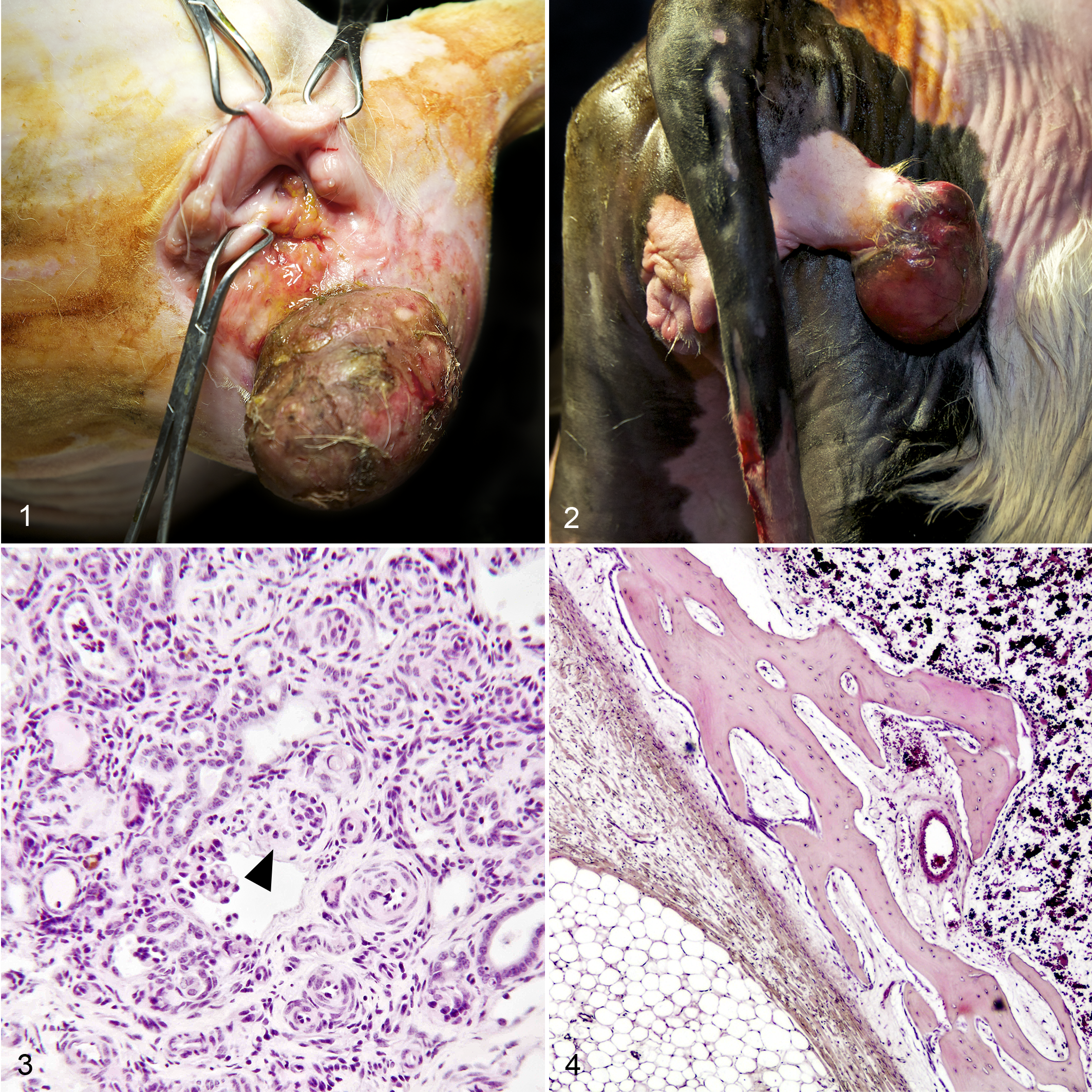
A 5-day-old female Holstein Friesian calf had a 4-day history of abdominal pain and anorexia. Type 2 atresia ani (imperforate anus), a rectovaginal fistula of 1-cm diameter, a 4-cm-diameter reducible umbilical hernia, and a perineal mass were diagnosed upon physical examination. The ulcerated, sessile perineal mass (20 × 15 × 15 cm) was to the right of the vulva (Fig. 1). The atresia ani, the rectovaginal fistula, and the umbilical hernia were surgically corrected; the perineal mass was excised. The calf was discharged 11 days after surgery.
Case No. 2
A 1-day-old female Holstein Friesian calf with imperforate anus had an ulcerated, pedunculated mass (5 cm in diameter) that protruded from the right perineum 5 cm ventral to the tailhead (Fig. 2). Feces leaked from the vagina due to the presence of a 0.5-cm-long rectovaginal fistula. The atresia ani and the rectovaginal fistula were surgically corrected; the perineal mass was excised. Ten days after surgery, the calf developed cyanosis, coughing, and a heart murmur. It died on postsurgery day 11. At necropsy, cardiac ventricular and atrial septal defects with pulmonary edema were observed. No gross abnormalities were detected in other organs.
Histologic Findings
In calf No. 1, the perineal mass was mainly composed of mature adipose tissue in abundant fibrovascular tissue. Blood vessels within the mass were prominent and dilated. A well-demarcated central area of the mass (4 × 6 mm) contained nephrogenic rests composed of renal tubules and glomeruli in fibrous stroma (Fig. 3). Some glomeruli were well developed with a urinary space and tufts of epithelial cells with well-defined capillaries; other glomeruli had a fetal appearance. In another area of the mass, a cystic structure lined by stratified urothelium and ductal structures lined by a layer of cuboidal epithelium were interpreted as Mullerian duct remnants. The diagnosis was perineal choristoma with nephrogenic rests.
In calf No. 2, the perineal mass was composed predominantly of mature adipose tissue with scattered blood and lymphatic vessels, striated muscle fibers, and nerves. In its center, fragments of trabecular bone (0.5–1.4 cm long) contained marrow and were coated by cartilage (Fig. 4). The diagnosis was perineal choristoma with heterotopic bone.
Discussion
Choristoma is defined as a mass of well-organized mature tissue at an ectopic site. It differs from hamartoma, which consists of disorganized mature mesenchymal or epithelial tissue in its normal anatomic location.1,2 Both hamartomas and choristomas have been associated with anorectal malformations in humans, 9 whereas only 1 case of concurrent atresia ani and perineal mass has been reported in veterinary medicine. In that 3-day-old male Holstein Friesian calf, the mass was composed of disorganized fibrous tissue, adipose tissue, and blood vessels. 10 Because these tissues are normally present in the perineum, the diagnosis was hamartoma.
In the present report, the nephrogenic rests in one case and the bony tissue in the other perineal mass were the ectopic tissues that supported the diagnosis of choristoma. A retroperitoneal pulmonary choristoma has been reported in a calf, 1 but to our knowledge, this is the first report of concurrent perineal choristoma, atresia ani, and rectovaginal fistula in veterinary medicine. In humans, imperforate anus or other anorectal malformations have been associated with perineal masses, including lipomas, hamartomas, choristomas, and vascular anomalies such as hemangiomas or lymphaticovenous malformations. 9 One choristoma in a study of perineal masses in 22 patients with anorectal malformation 9 was in a female infant and included nephrogenic rests like those in calf No. 1. In calf No. 2, the perineal choristoma contained adipose tissue mixed with bony trabeculae, cartilage, and bone marrow and resembled the so-called human pseudotail, which can contain bone and cartilage. 8 In human medicine, a “true tail” is defined as a remnant of the embryonic tail, which usually regresses during the seventh and eighth weeks of gestation. True tails are covered by skin and composed of various connective tissues but lack bone or cartilage. They are always associated with the distal end of the embryonic tail. Other lumbosacrococcygeal protrusions, unrelated to the embryonic tail, are categorized as pseudotails. They resemble a vestigial tail in location only, may be composed of normal or abnormal tissues, and can develop from different pathologic conditions, such as an elongation of sacral or coccygeal vertebrae, a lipoma or fibrolipoma, a teratoma or a pygomelus. A pseudotail is characterized by the presence of bone, cartilage, notochord, or spinal cord.8 Calf No. 2 also had cardiac ventricular and atrial septal defects. The possibility that the cardiac defects were part of a more complex malformation cannot be ruled out.
In conclusion, perineal choristomas can accompany anorectal malformations in cattle. In humans, the migration and fusion of the labioscrotal folds occur simultaneously with formation of the urorectal septum. The concurrence of these developments may explain the association of perineal masses, especially those with nephrogenic tissue, and anorectal malformations. Histologic evaluation of perineal masses in neonatal animals is necessary for diagnosis because their composition and derivation varies.
Footnotes
Declaration of Conflicting Interests
The author(s) declared no potential conflicts of interest with respect to the research, authorship, and/or publication of this article.
Funding
This study has been funded by Prozoo project supported by Regione Lombardia, Fondazione Cariplo, Fondazione Banca Popolare di Lodi..