
Abstract
A 4-year-old dog was presented for acute, progressive tetraparesis and cervical hyperesthesia. Symmetrical tubular structures coursing along the lateroventral aspects of the spinal cord at the fourth and fifth cervical vertebrae were identified in magnetic resonance images. At necropsy, vertebral arteries and their spinal branches were severely ectatic bilaterally, and the cervical spinal cord was compressed. Histologically, the ectatic branches of the vertebral and ventral spinal arteries were surrounded by fibrosis with scant mononuclear cell infiltrates and hemorrhage. Spinal branches of the vertebral arteries had focally severe reduction in the tunica media. A thrombus was in an arterial branch. Smaller vessels in adjacent tissue had fibrinoid degeneration. Axonal degeneration was detected in the affected spinal cord and nerve roots. The segmental degenerative radiculomyelopathy in this dog was attributed to anomalous ectasia of the vertebral and ventral spinal arteries.
Keywords
A 4-year-old neutered male crossbred Catahoula Leopard dog was referred to the Veterinary Medical Teaching Hospital, University of Missouri, for acutely progressive, nonambulatory tetraparesis and severe cervical hyperesthesia of 48 hours’ duration. A functional deficit was localized to the C6–T2 segments of the spinal cord by neurologic examination. Markedly enlarged intervertebral foramina at C4-5 and C5-6 were identified in radiographs. The dorsal and middle portions of the C4 and C5 vertebral bodies were irregularly shaped and sclerotic. In magnetic resonance images (transverse and sagittal T1- and T2-weighted sequences), bilaterally symmetrical hypointense tubular structures coursed along the lateroventral aspects of the C4 and C5 spinal cord segments (Figs. 1, 2), compressing the ventral aspect of the spinal cord and effacing the dorsal aspect of the C4 and C5 vertebral bodies. The tubular structures communicated with a large vessel extending from the thorax. A vascular anomaly was suspected with secondary spinal cord compression and vertebral erosion. Based on the imaging results, severity of clinical signs, and rapid deterioration of the patient, the prognosis was deemed poor, and the dog was humanely euthanized.
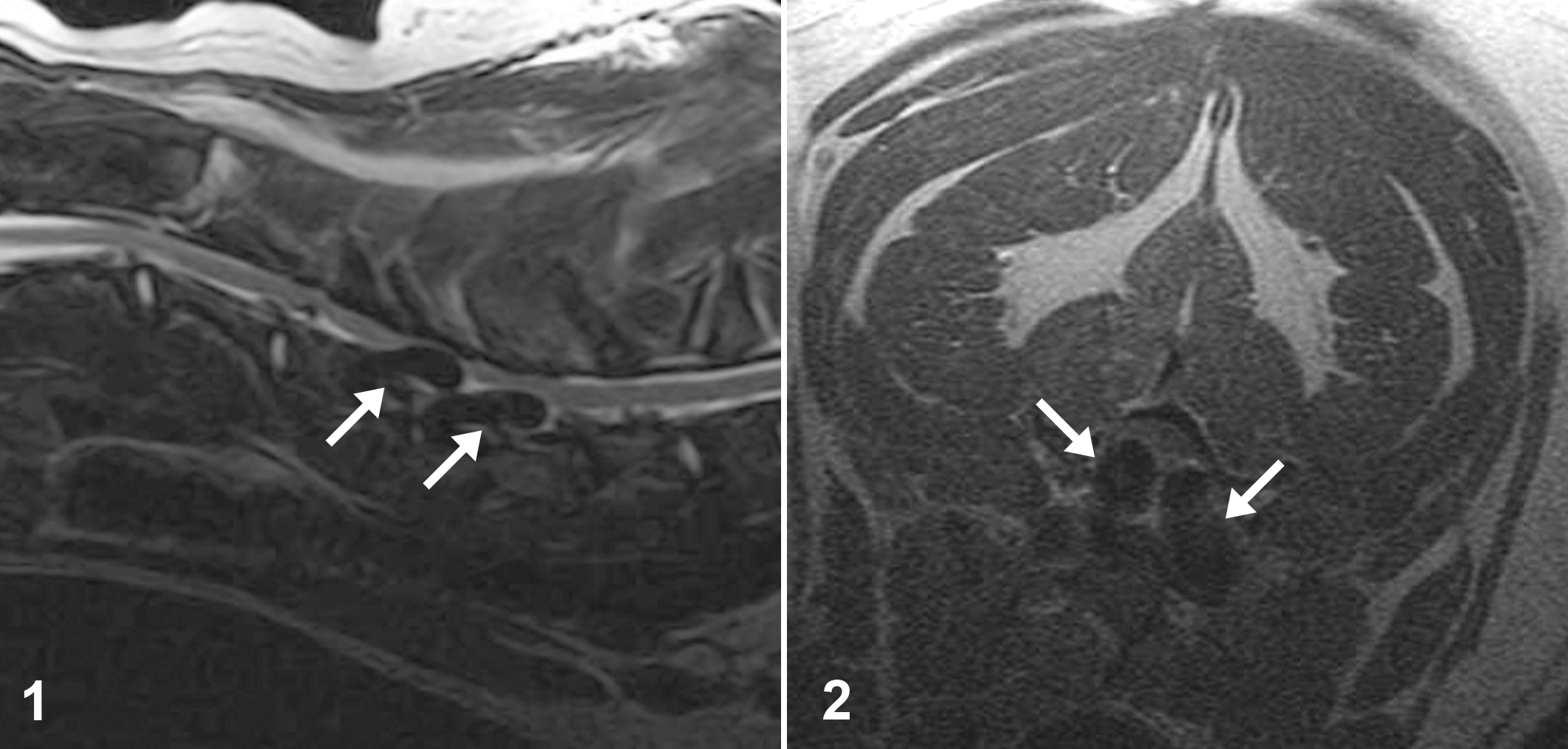
T2-weighted sagittal magnetic resonance image of the neck; dog. Hypointense tubular structures (arrows) course along the lateroventral aspects of the spinal cord, near the fourth and fifth cervical vertebrae.
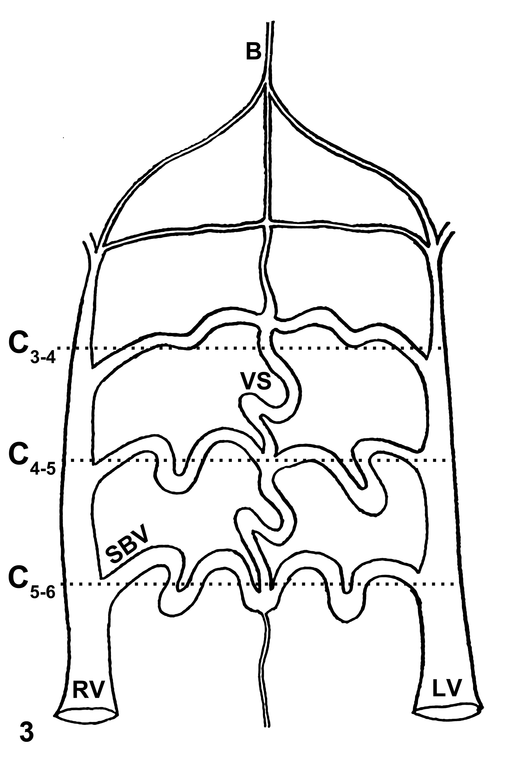
Anomalous vertebral arteries of the affected dog. C, cervical vertebra; RV, right vertebral artery; LV, left vertebral artery; SBV, spinal branches of vertebral arteries; VS, ventral spinal artery; B, basilar artery.
Pathologic Findings
Grossly, severely ectatic right and left vertebral arteries entered the vertebral column lateroventrally at C6 (Figs. 3, 4). The ectasia resulted in saccular arterial dilations up to 5.2 cm long and 1.2 cm in external diameter. A probe was easily inserted in both vessels and could be extended cranially to the C4 region. The spinal canal was enlarged from C4 to C6, and the spinal branches of the vertebral arteries at this site were tortuous and ectatic, up to 5 mm in diameter (Figs. 3, 5). The ventral spinal artery (in formalin-fixed C4-to-C5 spinal cord) was ectatic, ranging from 2 to 3 mm in diameter, whereas the artery was 0.4 to 0.6 mm in diameter in unaffected areas of the spinal cord. The ventral aspect of the spinal cord at C4 to C5 was compressed and discolored red-brown.
Ventral surface of vertebral column. Ectatic right and left vertebral arteries (arrows) enter the vertebral column lateroventrally at C6.
The only other salient finding at necropsy was an asymmetrical saccular aneurysm that caused marked mural thinning of the aortic arch. Just distal to the aneurysm, a whitish, firm, irregular plaque was on the intimal surface of the aorta.
Tissue samples were fixed in 10% buffered formalin, processed routinely, embedded in paraffin, and sectioned at 4 μm. The C4 and C5 vertebral bodies were decalcified before histologic processing with a solution of hydrochloric acid, EDTA, and water (Surgipath Decalcifier II, Surgipath Medical Industries Inc, Richmond, IL). Sections were stained with hematoxylin and eosin and, in selected sections, Verhoeff–van Gieson elastin stain.
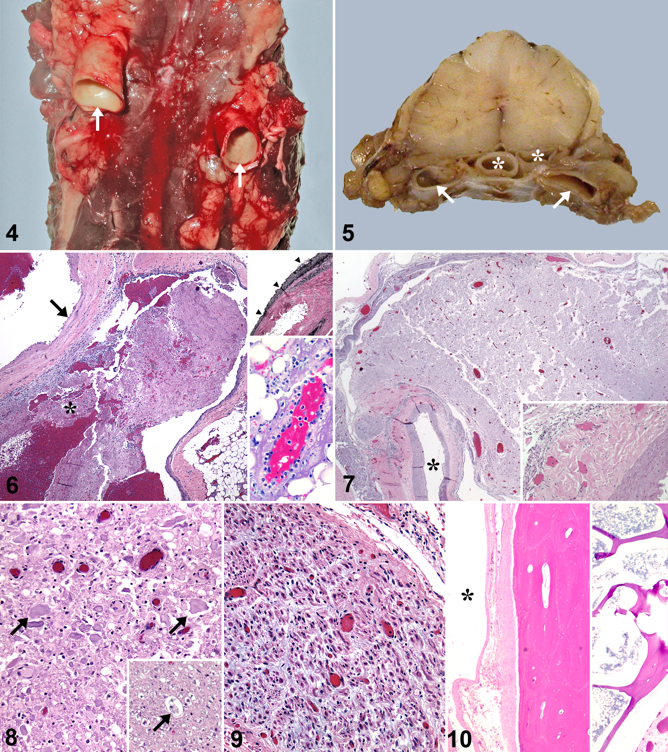
Histologically, ectatic vertebral and ventral spinal arteries had an elastic tunica media of variable thickness with adventitial and periarterial fibrosis (Figs. 6, 7), light periarterial infiltration by mononuclear leukocytes, and mild hemorrhage (Fig. 7). Spinal branches of the vertebral arteries had focally extensive reduction in the tunica media (Fig. 6). A thrombus in a branch artery (Fig. 6) had disrupted the tunica intima and tunica media. Several smaller vessels in adjacent adipose tissue had fibrinoid degeneration and were surrounded by fibrinocellular fluid (Fig. 6, inset). The ventral white matter of the overlying spinal cord was compressed by the ectatic ventral spinal arteries, with one side more severely affected; a few sections had marked rarefaction of white and gray matter (Fig. 7). In cross sections of compressed white matter, many axons were swollen or absent (Fig. 8); some dilated myelin sheaths contained macrophages (Fig. 8, inset). Congestion and spongy change of the white matter were consistent with edema. The dorsal and ventral nerve roots were edematous with scattered degenerated axons (Fig. 9) and reduced myelin staining with Luxol fast blue (not shown). Cortical and trabecular bone loss was prominent along the dorsal aspect of C4 and C5 vertebral bodies, with sclerotic thickening of remaining cortical and trabecular bone that encroached on the medullary cavity (Fig. 10). The tunica media had marked thinning in the saccular aneurysm of the aortic arch. The intimal plaque, noted grossly, was a focus of mineralized cartilage that expanded the tunica intima and inner aspect of the tunica media.
Discussion
Myelopathy associated with vascular ectasia is seldom reported in domestic animals, and most cases have involved the vasculature within the spinal cord itself (intramedullary ectasia). Intramedullary vascular anomalies have been reported in dogs, 1,6,8 2 horses, 3,7 a Hereford calf, 2 and an African warthog. 11 One case of intradural–extramedullary vascular anomaly has been reported in a dog. 9,10
Human spinal vascular malformations have been classified as capillary telangiectasias, cavernous malformations, venous malformations, arteriovenous malformations, or other vascular malformations (eg, arterial malformations). 5 Both capillary telangiectasias and cavernous malformations consist of variably dilated capillaries lined by a single layer of endothelial cells and devoid of muscular and elastic fibers. 5 Vessels in capillary telangiectasias are separated by normal spinal cord parenchyma, whereas those in cavernous malformations are not separated by nervous tissue. 5 Venous malformations consist solely of anomalous veins separated by neural parenchyma. The vascular channels are thin-walled vessels of variable diameter with walls of flattened endothelium and fibromuscular tissue but devoid of elastic tissue. 5 Arteriovenous malformations, the most commonly reported human vascular anomaly, are direct arterial–venous communications without intervening capillaries. 5 Additionally, intermediate and combined forms exist, and some anomalies only fit into the other vascular malformations category. 12 Based on the prominence of elastin fibers in the tunica media of the ectatic vessels, the vascular anomaly in this dog could be classified as an arterial malformation.
In the previously reported canine case of intradural–extramedullary vascular anomaly, the ectatic and tortuous left vertebral artery and its branches traversed and compressed the cervical spinal cord. 9,10 The anomaly in that case was attributed to the lack of the right subclavian artery, which probably resulted in increased blood flow through the left subclavian and vertebral arteries, and retrograde flow to a common right arterial vessel (ie, one functioning as the right subclavian artery) via the right vertebral artery. 9,10 In the present case, no abnormalities were observed in the subclavian arteries or other branches of the aortic arch to account for the ectasia of cervical blood vessels. The only change in the great vessels was a saccular aneurysm and focal cartilaginous metaplasia in the aortic arch, which may have resulted from intimal damage secondary to turbulent blood flow. Dogs have a more developed cephalic (ie, headward) collateral anastomotic network (including vertebral and carotid arteries) than humans, and this low-resistance network promotes vascular ectasia. 4,9 As in the previous canine case of intradural–extramedullary vascular anomaly, 9,10 both the vertebral and ventral spinal arteries were involved.
Most human vascular malformations are considered idiopathic and congenital; however, some may be inherited or secondary to trauma, irradiation, or other injury. 12 One reported canine case of spinal intramedullary cavernous malformation was also in a Catahoula Leopard dog. 6 Based on the paucity of reported vascular anomalies in dogs, their occurrence in 2 dogs of the same uncommon breed suggests the possibility of genetic predisposition. In summary, the segmental degenerative radiculomyelopathy in this dog was attributed to anomalous ectasia of vertebral and ventral spinal arteries, an uncommon extramedullary arterial malformation.
Footnotes
Acknowledgements
We thank the Veterinary Medical Diagnostic Laboratory histology personnel for technical support; Howard Wilson, University of Missouri, for assistance with the illustrations; and Dr Fred Wininger, Department of Veterinary Medicine and Surgery, for the magnetic resonance images.
Declaration of Conflicting Interests
The author(s) declared no potential conflicts of interest with respect to the research, authorship, and/or publication of this article.
Funding
The author(s) received no financial support for the research, authorship, and/or publication of this article.